

Arachidonic acid-induced expression of the organic solute and steroid transporter-beta (Ost-beta) in a cartilaginous fish cell line
- PMID: 18407792
- PMCID: PMC2471870
- DOI: 10.1016/j.cbpc.2008.03.005
Arachidonic acid-induced expression of the organic solute and steroid transporter-beta (Ost-beta) in a cartilaginous fish cell line
Abstract
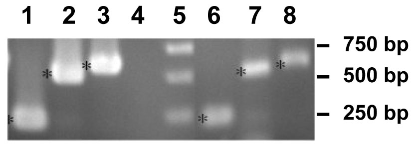
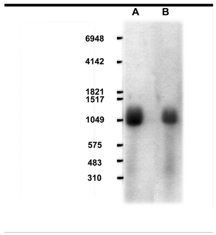
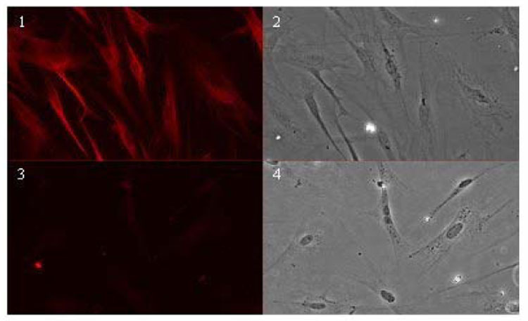
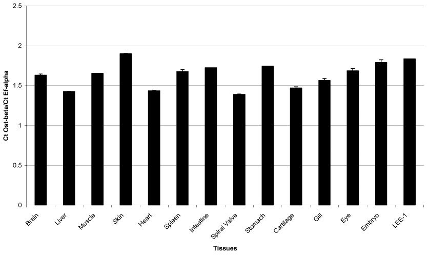
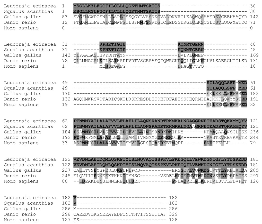
The organic solute and steroid transporter (OST/Ost) is a unique membrane transport protein heterodimer composed of subunits designated alpha and beta, that transports conjugated steroids and prostaglandin E(2) across the plasma membrane. Ost was first identified in the liver of the cartilaginous fish Leucoraja erinacea, the little skate, and subsequently was found in many other species, including humans and rodents. The present study describes the isolation of a new cell line, LEE-1, derived from an early embryo of L. erinacea, and characterizes the expression of Ost in these cells. The mRNA size and amino acid sequence of Ost-beta in LEE-1 were identical to that previously reported for Ost-beta from skate liver, and the primary structure was identical to that of the spiny dogfish shark (Squalus acanthias) with the exception of a single amino acid. Ost-beta was found both on the plasma membrane and intracellularly in LEE-1 cells, consistent with its localization in other cell types. Interestingly, arachidonic acid, the precursor to eicosanoids, strongly induced Ost-beta expression in LEE-1 cells and a lipid mixture containing arachidonic acid also induced Ost-alpha. Overall, the present study describes the isolation of a novel marine cell line, and shows that this cell line expresses relatively high levels of Ost when cultured in the presence of arachidonic acid. Although the function of this transport protein in embryo-derived cells is unknown, it may play a role in the disposition of eicosanoids or steroid-derived molecules.
Figures
Similar articles
-
OST alpha-OST beta: a key membrane transporter of bile acids and conjugated steroids.Front Biosci (Landmark Ed). 2009 Jan 1;14(8):2829-44. doi: 10.2741/3416. Front Biosci (Landmark Ed). 2009. PMID: 19273238 Free PMC article. Review.
-
Analysis and functional annotation of expressed sequence tags from in vitro cell lines of elasmobranchs: Spiny dogfish shark (Squalus acanthias) and little skate (Leucoraja erinacea).Comp Biochem Physiol Part D Genomics Proteomics. 2010 Sep;5(3):199-206. doi: 10.1016/j.cbd.2010.04.004. Epub 2010 Apr 24. Comp Biochem Physiol Part D Genomics Proteomics. 2010. PMID: 20471924 Free PMC article.
-
Homologue gene of bile acid transporters ntcp, asbt, and ost-alpha in rainbow trout Oncorhynchus mykiss: tissue expression, effect of fasting, and response to bile acid administration.Fish Physiol Biochem. 2014 Apr;40(2):511-25. doi: 10.1007/s10695-013-9862-y. Epub 2013 Sep 12. Fish Physiol Biochem. 2014. PMID: 24026769
-
Multidrug resistance-associated protein 3 (Mrp3/Abcc3/Moat-D) is expressed in the SAE Squalus acanthias shark embryo-derived cell line.Zebrafish. 2007 Winter;4(4):261-75. doi: 10.1089/zeb.2007.0520. Zebrafish. 2007. PMID: 18284333 Free PMC article.
-
Cell and molecular biology of the spiny dogfish Squalus acanthias and little skate Leucoraja erinacea: insights from in vitro cultured cells.J Fish Biol. 2012 Apr;80(5):2089-111. doi: 10.1111/j.1095-8649.2011.03205.x. Epub 2012 Feb 7. J Fish Biol. 2012. PMID: 22497417 Review.
Cited by
-
OST alpha-OST beta: a key membrane transporter of bile acids and conjugated steroids.Front Biosci (Landmark Ed). 2009 Jan 1;14(8):2829-44. doi: 10.2741/3416. Front Biosci (Landmark Ed). 2009. PMID: 19273238 Free PMC article. Review.
-
Heteromeric Solute Carriers: Function, Structure, Pathology and Pharmacology.Adv Exp Med Biol. 2021;21:13-127. doi: 10.1007/5584_2020_584. Adv Exp Med Biol. 2021. PMID: 33052588 Review.
-
Establishing primary cultures of embryonic intestinal cells from the elasmobranch, Leucoraja erinacea.In Vitro Cell Dev Biol Anim. 2012 Aug;48(7):413-7. doi: 10.1007/s11626-012-9534-8. Epub 2012 Jul 18. In Vitro Cell Dev Biol Anim. 2012. PMID: 22806972 No abstract available.
-
Pleiotropic functions of the organic solute transporter Ostα-Ostβ.Dig Dis. 2011;29(1):13-7. doi: 10.1159/000324123. Epub 2011 Jun 17. Dig Dis. 2011. PMID: 21691099 Free PMC article. Review.
-
Novel insights into the organic solute transporter alpha/beta, OSTα/β: From the bench to the bedside.Pharmacol Ther. 2020 Jul;211:107542. doi: 10.1016/j.pharmthera.2020.107542. Epub 2020 Apr 2. Pharmacol Ther. 2020. PMID: 32247663 Free PMC article. Review.
References
-
- Ballard WW, Mellinger J, Lechenault H. A series of normal stages for development of Scyliorhinus canicula, the Lesser Spotted Dogfish (Chondrichthyes: Scyliorhinidae) J Exp Zool. 1993;267:318–336.
-
- Ballatori N. Biology of a novel organic solute and steroid transporter, Ost-alpha/Ost-beta. Exp Biol Med. 2005;230:689–698. - PubMed
-
- Ballatori N, Christian WV, Lee YJ, Dawson PA, Soroka CJ, Boyer JL, Madejczyk MS, Li N. OSTalpha-OSTbeta, a major basolateral bile acid and steroid transporter in human intestinal, renal, and biliary epithelia. Hepatology. 2005;42:1270–1279. - PubMed
-
- Barnes DW, Parton A, Tomana M, Hwang J-H, Czechanski A, Collodi P. Stem cells from cartilaginous fish. Methods in Cell Biology, 2007 in press. - PubMed
Publication types
MeSH terms
Substances
Grants and funding
- ES03828/ES/NIEHS NIH HHS/United States
- R01-DK067214/DK/NIDDK NIH HHS/United States
- T32-ES07026/ES/NIEHS NIH HHS/United States
- ES01247/ES/NIEHS NIH HHS/United States
- P30 ES001247/ES/NIEHS NIH HHS/United States
- P20-RR016463/RR/NCRR NIH HHS/United States
- T32 ES007026/ES/NIEHS NIH HHS/United States
- P30 ES003828/ES/NIEHS NIH HHS/United States
- R01 RR019732/RR/NCRR NIH HHS/United States
- P20 RR016463/RR/NCRR NIH HHS/United States
- R01 DK067214/DK/NIDDK NIH HHS/United States
- R01-RR019732/RR/NCRR NIH HHS/United States
LinkOut - more resources
Full Text Sources
Molecular Biology Databases
Research Materials
