. 2008 Jun 27;283(26):18344-54.
doi: 10.1074/jbc.M801192200. Epub 2008 Apr 11.

A p120 catenin isoform switch affects Rho activity, induces tumor cell invasion, and predicts metastatic disease

Affiliations

A p120 catenin isoform switch affects Rho activity, induces tumor cell invasion, and predicts metastatic disease

Masahiro Yanagisawa et al. J Biol Chem. .

Abstract

p120 catenin is a cadherin-associated protein that regulates Rho GTPases and promotes the invasiveness of E-cadherin-deficient cancer cells. Multiple p120 isoforms are expressed in cells via alternative splicing, and all of them are essential for HGF signaling to Rac1. However, only full-length p120 (isoform 1) promotes invasiveness. This selective ability of p120 isoform 1 is mediated by reduced RhoA activity, both under basal conditions and following HGF treatment. All p120 isoforms can bind RhoA in vitro, via a central RhoA binding site. However, only the cooperative binding of RhoA to the central p120 domain and to the alternatively spliced p120 N terminus stabilizes RhoA binding and inhibits RhoA activity. Consistent with this, increased expression of p120 isoform 1, when compared with other p120 isoforms, is predictive of renal tumor micrometastasis and systemic progression, following nephrectomy. Furthermore, ectopic expression of the RhoA-binding, N-terminal domain of p120 is sufficient to block the ability of p120 isoform 1 to inhibit RhoA and to promote invasiveness. The data indicate that the increased expression of p120 isoform 1 during tumor progression contributes to the invasive phenotype of cadherin-deficient carcinomas and that the N-terminal domain of p120 is a valid therapeutic target.


Figures
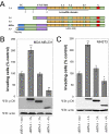
FIGURE 1.
FIGURE 1.
Differential effects of p120 isoforms on cell invasiveness. A, p120 isoforms tested in this study include the naturally occurring isoforms 1A, 3A, and 4A. Murine p120 isoform 1A contains a coiled-coil domain and a regulatory phosphorylation domain in its N terminus, a central armadillo domain, and the alternatively spliced exon A in the C terminus. Murine p120 isoforms 3A and 4A lack the coiled-coil or the entire N-terminal domain, respectively. The ΔRho p120 mutant is identical to p120 isoform 1A with an in-frame deletion of amino acids 622-628. Finally, N1 is a FLAG-tagged N-terminal mutant of p120 lacking the central and C-terminal domains. B, MDA-MB-231 cells with knocked down expression of endogenous p120 (shRNA) were infected with retroviruses expressing neomycin resistance alone (neo), or together with murine p120 isoforms 1A, 3A, or 4A. After G418 selection, the invasiveness of p120-reexpressing cells was tested in vitro toward a gradient of HGF, as described under “Experimental Procedures.” After a 20-h incubation, cells on the underside of a Matrigel-coated transwell membrane were counted under a ×20 objective. Data are expressed as percentage of control and represent the means ± S.E. of three independent determinations performed in duplicate. **, p < 0.001 as compared with the neomycin control (Student's t test). Reexpression of murine p120 isoform 1A promotes cell invasiveness, whereas expression of p120 isoforms 3A or 4A has no effect or suppresses invasiveness, respectively (n = 6). Lower two panels, lysates from all cell lines were subjected to SDS-PAGE and Western blotted (WB) for expression of p120 (mAb pp120) and actin. C, NIH3T3 fibroblasts were infected with retroviruses expressing neomycin resistance alone or in combination with p120 isoform 1A, p120 isoform 3A, or p120 isoform 4A. After G418 selection, stable polyclonal cell lines were assayed for invasiveness in transwell assays as above, with the exception that 5% fetal bovine serum was added to the bottom chamber as the chemoattractant, since NIH3T3 cells lack c-Met. Results represent the mean ± S.E. of three independent determinations performed in duplicate. Lower two panels, lysates from all cell lines were subjected to SDS-PAGE and Western blotted for expression of p120 (using mAb 8D11) and actin.
FIGURE 2.
FIGURE 2.
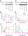
Differential effect of p120 isoforms on Rho GTPase activities. A, levels of total and GTP-bound Rac1 were determined in serum-starved MDA-MB-231 cells infected with control pRS and LZRS-neo viruses (pRS + neo), p120-depleted cells (shRNA + neo), or cells reexpressing p120 isoform 1A (shRNA + p120 1A) or 4A (shRNA + p120 4A). p120-depleted cells exhibited lower Rac1 activity under these basal conditions. Expression of either murine p120 isoform 1A or 4A rescued Rac1 activation (n = 3). B, levels of total and GTP-bound Rac1 were determined in the same serum-starved cell lines as above, following incubation with 20 ng/ml HGF for the indicated times. Following Western blotting and densitometry, mean ± S.E. activity data from at least three independent determinations was expressed as percentage of total (active versus total × 100). p120-depleted cells exhibited no Rac1 activation at any time following HGF addition, whereas control cells and cells reexpressing p120 isoform 1A or 4A exhibited HGF-induced Rac1 activation with a peak at 10 min (p < 0.01; ANOVA). C, the above data were also expressed as percentage of control (active Rac1 at time xversus active at time 0 × 100). As can be seen in the graph, both p120 isoforms 1A and 4A rescue Rac1 activation. D, similar to Rac1, levels of total and active RhoA were also measured in control serum-starved MDA-MB-231 cells (pRS + neo), p120-depleted cells (shRNA + neo), or cells reexpressing p120 isoform 1A (shRNA + p120 1A) or 4A (shRNA + p120 4A). p120-depleted cells exhibited higher RhoA activity under these basal conditions, as reported previously (11). Expression of p120 isoform 1A blocked Rho activation, whereas expression of the N-terminally truncated p120 isoform 4A was unable to block RhoA activation, despite its ability to induce the activation of Rac1 (n = 6). E, levels of total and GTP-bound RhoA were also determined in HGF-treated cells. When mean ± S.E. RhoA activity data were expressed as percentage of total, both basal and HGF-induced RhoA activities were much higher (2-10-fold) in shRNA-expressing cells than in control cells (pRS) or cells expressing p120 isoform 1A (p120-1A). Cells expressing p120 isoform 4A exhibited 10-20-fold higher levels of activated RhoA than control cells (n ≥ 3). F, when the same data were expressed as percentage of control, a biphasic response to HGF was uncovered. p120-depleted cells lacked both an initial activation phase and a late decrease in RhoA activity. Cells expressing murine p120 1A were unable to induce RhoA activation in response to HGF but retained the long term inhibition phase (p < 0.05; ANOVA), whereas cells expressing p120 isoform 4A induced RhoA within 5 min (p < 0.05; ANOVA) but failed to inhibit RhoA in the long term (n ≥ 3). Representative raw Western blot data for B and E are shown in supplemental Fig. 1.
FIGURE 3.
FIGURE 3.
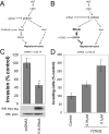
RhoA-ROCK signaling mediates the differential effects of p120 isoforms on cell invasion. A, effect of p120 isoform 1 on HGF-mediated migration/invasiveness. HGF-mediated cell scattering, migration, and invasiveness require the up-regulation of Rac1 signaling (right) and down-regulation of cell contractility, promoted by RhoA-ROCK signaling (left). p120 depletion causes the up-regulation of basal RhoA activity and bocks HGF-mediated Rac1 activation. Our data suggest that p120 isoform 1 mediates both the activation of Rac1 in response to HGF and maintains RhoA signaling at a low level, thus promoting cell motility. This model explains why p120 depletion suppresses migration and invasiveness. It also predicts that activation of RhoA signaling in cells expressing p120 isoform 1 would suppress cell invasion. B, effect of p120 isoform 4 on HGF-mediated migration/invasiveness. Our data indicate that cells expressing p120 isoform 4 can activate Rac1 in response to HGF. However, these cells have very high basal RhoA activity, which is further increased by HGF treatment. This model suggests that the increased RhoA-ROCK signaling in p120 isoform 4-expressing cells is able to suppress overall cell invasiveness. It also predicts that inhibition of RhoA-ROCK signaling would promote motility and invasiveness in cells expressing p120 isoform 4. C, p120-depleted MDA-MB-231 cells stably expressing murine p120 isoform 1A were electroporated and transfected transiently with pcDNA3-RhoA-V14-Myc (V14-RhoA) or pcDNA control. Control immunofluorescence experiments verified that more than 60% of cells expressed CA-RhoA. One day after transfection, cells were tested in vitro for invasion toward HGF, as described in the legend to Fig. 1. In agreement with our hypothesis (A), overexpression of CA-RhoA significantly suppressed the invasiveness of p120 isoform 1A-expressing cells (n = 6). D, the in vitro invasiveness of p120-depleted cells stably expressing murine p120 isoform 4A was tested under control conditions or following treatment with the ROCK inhibitor Y27632 (0.01-0.1 μm). In agreement with our hypothesis (B), HGF-induced invasiveness of p120 isoform 4-expressing cells was significantly increased in cells treated for 24 h with Y27632. WB, Western blot.
FIGURE 4.
FIGURE 4.
N-terminal and central regions of p120 cooperate to promote RhoA binding and inhibit RhoA activity. A, immunoblot analysis of GDP-RhoA association with various p120 isoforms and deletion mutants. His-tagged RhoA and GST-p120 proteins were bacterially expressed and purified. The extent of GDP-RhoA association with recombinant p120 isoforms 1 versus 4, with or without the 622-628 (ΔRho) deletion, was determined in binding assays, in vitro. Both the central ΔRho p120 region and the N-terminal domain are required for efficient RhoA association. Deletion of both domains abrogates RhoA binding. Data shown are representative of at least four independent determinations. B, 2 μg of [3H] GDP bound RhoA (in the presence of GST; GST) were incubated alone, with ∼4 μg of GST-p120 isoform 1A, GST-p120 isoform 4A, or GST-p120 isoform 1-ΔRho (p120-ΔRho) or with 0.5 μg of His-TrioC, an RhoA-specific GEF. Incubations were carried out at ambient temperature in a GEF assay buffer (15). The amount of RhoA-bound radionucleotide at time 0 was taken as 100%. As previously shown, p120 isoform 1 inhibited the intrinsic guanine nucleotide exchange activity of RhoA. p120 proteins lacking the N-terminal domain (p120-4A) or ΔRho region (p120-ΔRho) had no effect on RhoA exchange activity. Identical results were obtained in at least three independent determinations. C, the extent of GDP-RhoA association with a recombinant p120 N-terminal mutant containing amino acids (a.a.) 1-232 was determined in binding assays, in vitro. The N-terminal 232 amino acids of p120 associate with RhoA-GDP (top). The bottom panel shows the expression of different GST proteins. D, model of p120-RhoA interactions. Our data suggest that naturally occurring p120 isoforms, including those lacking the N-terminal domain (like isoform 4), can associate with RhoA via their central ΔRho region. The N-terminal p120 domain is required for stabilization of RhoA binding (increased affinity interaction) and for inhibiting the dissociation of GDP from RhoA. Therefore, interaction of p120 isoform 1 with RhoA results in local inhibition of RhoA activity, whereas p120 isoform 4 fails to inhibit RhoA but may promote the recruitment of RhoA at sites where it can be activated by upstream signaling events (i.e. HGF signaling).
FIGURE 5.
FIGURE 5.
An N-terminal p120 fragment suppresses invasiveness. A, effect of p120 N-terminal fragment overexpression on RhoA and Rac1 activities. p120-depleted MDA-MB-231 cells reexpressing murine p120 isoform 1A were infected with retroviruses expressing zeocin resistance alone (zeo) or together with a FLAG-tagged N-terminal p120 truncation mutant containing amino acids 1-323 (N1). The relative levels of active RhoA and Rac1 were determined in stable polyclonal cell lines using pull-down assays and Western blots as described earlier. Expression of N1 did not affect endogenous levels of cadherin 11 or Rac1 activation but caused a significant increase in basal RhoA activity. B, NIH3T3 fibroblasts and MDA-MB-231 cells stably expressing zeocin resistance alone or together with the N1 p120 fragment were subjected to invasion assays to determine the effect of N1 on cell invasiveness. Results are the mean ± S.E. of three independent determinations performed in duplicate. **, p < 0.001 as compared with the zeocin resistance alone control (Student's t test). Bottom two panels, lysates from all cell lines were subjected to SDS-PAGE and Western blotted (WB) for expression of N1 (using the anti-FLAG tag mAb M2) and actin.
FIGURE 6.
FIGURE 6.
p120 isoforms and ccRCC. A, immunohistochemical analysis of a ccRCC sample compared with patient-matched normal kidney. E-cadherin expression is lost in this ccRCC tumor sample, as it is in the majority of ccRCC cases. Expression of p120 is maintained but largely mislocalized to the cytoplasm. Expression of the HGF receptor c-Met is absent in normal tissue but becomes highly induced in the tumor. B, to determine whether endogenous p120 isoforms correlate with invasiveness in ccRCC, we initially determined the relative p120 isoform expression in 36 tumor and patient-matched normal freshly frozen kidney samples (see “Experimental Procedures”). The ratio of p120 isoform 1 (which induces invasion in vitro) to the shorter p120 isoform 3 (lower band; which does not induce invasion in vitro) was calculated in both normal (N) and ccRCC tumor (T) samples. p120 isoform 1 and 3 expression in MDA-MB-231 cells is also shown as an additional control (C). Normal kidney exhibited a p120 isoform 1A/3A ratio of ∼0.4. p120 isoform switch is indicated by a higher relative expression of p120 isoform 1 in tumor samples (ratio larger than 1). According to this analysis, the second tumor (T2) exhibits a p120 isoform switch, whereas the first one does not. C, when p120 isoform switch data were compared with disease progression, a significant correlation was uncovered between p120 isoform switch and the incidence of metastatic ccRCC disease. 89.2% of ccRCC tumors that metastasized following nephrectomy within a 5-year follow-up exhibited a p120 isoform switch versus only 25% of localized tumors.

References

    1. Berx, G., and Van Roy, F. (2001) Breast Cancer Res. 3 289-293 - PMC - PubMed
    1. Conacci-Sorrell, M., Zhurinsky, J., and Ben-Ze'ev, A. (2002) J. Clin. Invest. 109 987-991 - PMC - PubMed
    1. Thiery, J. P. (2002) Nat. Rev Cancer 2 442-454 - PubMed
    1. Batlle, E., Sancho, E., Franci, C., Dominguez, D., Monfar, M., Baulida, J., and Garcia De Herreros, A. (2000) Nat. Cell Biol. 2 84-89 - PubMed
    1. Cano, A., Perez-Moreno, M. A., Rodrigo, I., Locascio, A., Blanco, M. J., del Barrio, M. G., Portillo, F., and Nieto, M. A. (2000) Nat. Cell Biol. 2 76-83 - PubMed

Publication types