. 2008 Jun;190(12):4360-6.
doi: 10.1128/JB.00239-08. Epub 2008 Apr 11.

The peptidoglycan of stationary-phase Mycobacterium tuberculosis predominantly contains cross-links generated by L,D-transpeptidation

Affiliations

The peptidoglycan of stationary-phase Mycobacterium tuberculosis predominantly contains cross-links generated by L,D-transpeptidation

Marie Lavollay et al. J Bacteriol. 2008 Jun.

Abstract

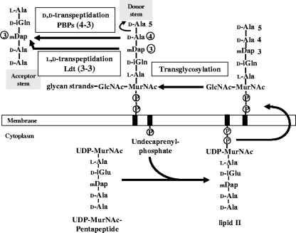
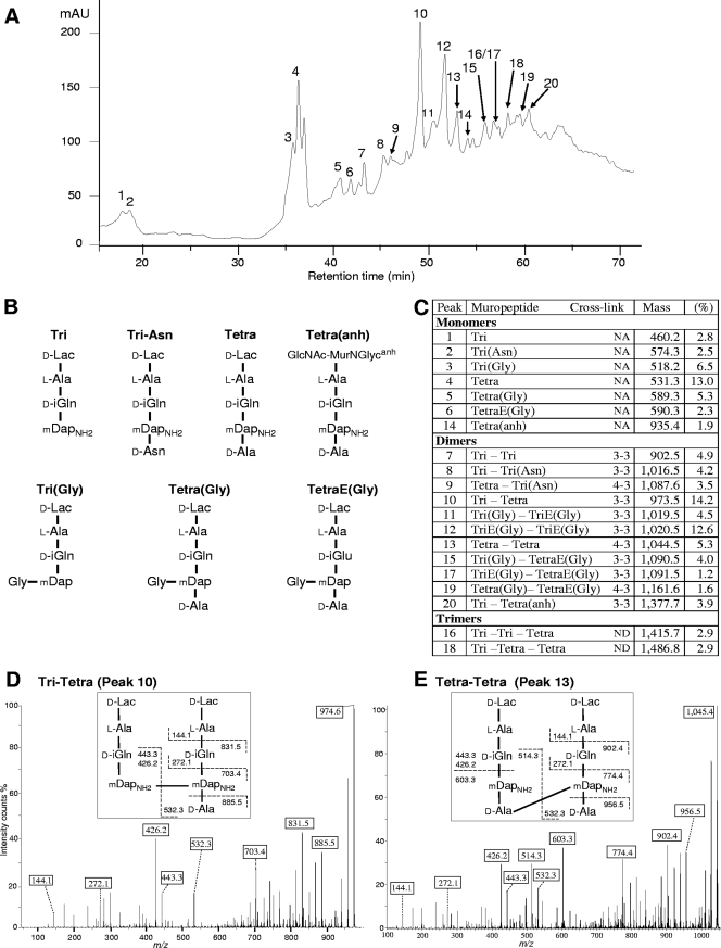
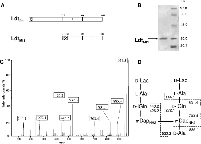
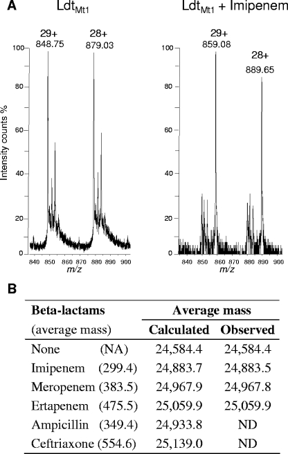
Our understanding of the mechanisms used by Mycobacterium tuberculosis to persist in a "dormant" state is essential to the development of therapies effective in sterilizing tissues. Gene expression profiling in model systems has revealed a complex adaptive response thought to endow M. tuberculosis with the capacity to survive several months of combinatorial antibiotic treatment. We show here that this adaptive response may involve remodeling of the peptidoglycan network by substitution of 4-->3 cross-links generated by the D,D-transpeptidase activity of penicillin-binding proteins by 3-->3 cross-links generated by a transpeptidase of L,D specificity. A candidate gene, previously shown to be upregulated upon nutrient starvation, was found to encode an L,D-transpeptidase active in the formation of 3-->3 cross-links. The enzyme, Ldt(Mt1), was inactivated by carbapenems, a class of beta-lactam antibiotics that are poorly hydrolyzed by the M. tuberculosis beta-lactamases. Ldt(Mt1) and carbapenems may therefore represent a target and a drug family relevant to the eradication of persistent M. tuberculosis.


Figures

FIG. 1.
FIG. 1.
Schematic representation of peptidoglycan synthesis. The nucleotide UDP-MurNAc-pentapeptide is formed in the cytoplasm by sequential addition of l-Ala, d-Glu, meso-diaminopimelic acid (mDap), and the dipeptide d-Ala-d-Ala. The membrane steps of peptidoglycan synthesis involve the transfer of the phospho-MurNAc-pentapeptide moiety of the nucleotide to the lipid carrier (undecaprenyl-phosphate) and the addition of the second sugar (GlcNAc). The complete precursor linked to the lipid carrier (lipid II) is translocated through the membrane and polymerized by glycosyltransferases (formation of glycan strands) and by the d,d-transpeptidase activity of PBPs (formation of the cross-links). These enzymes cleave the d-Ala4-d-Ala5 bond of a pentapeptide donor and link the carbonyl of d-Ala4 to the side chain amine of mDap at the third position of an acceptor stem (4→3 cross-links). β-Lactam antibiotics are structural analogues of the d-Ala4-d-Ala5 extremity of the precursors and act as suicide substrates of the d,d-transpeptidases. l,d-Transpeptidases cleave the mDap3-d-Ala4 bond of a tetrapeptide donor and link the carbonyl of mDap3 to the acceptor stem (3→3 cross-links).
FIG. 2.
FIG. 2.
Structure of the peptidoglycan of M. tuberculosis. (A) rp-HPLC profile of peptidoglycan fragments obtained by digestion of the peptidoglycan of strain H37Rv with muramidases and treatment with ammonium hydroxide. mAU, absorbance unit × 103 at 210 nm. (B) Structure of monomers. d-iGln, d-iso-glutamine; d-iGlu, d-iso-glutamic acid; d-Lac, d-lactate; GlcNAc-MurNGlycanh, N-acetylglucosamine-anhydro-N-glycolylmuramic acid; mDap, meso-diaminopimelic acid; mDapNH2, mDap with an amidated ɛ carboxyl group. (C) Peptidoglycan composition. The relative abundance (%) was calculated by integration of the absorbance. “Mass” refers to observed monoisotopic mass. NA, not applicable; ND, not determined. E, d-iGlu. (D) Sequencing of a dimer generated by l,d-transpeptidation. Tandem mass spectrometry was performed on the [M+H]+ ion at m/z 974.6 (peak 10). Boxes indicate ions generated by cleavage at single peptide bonds. (E) Sequencing of a dimer generated by d,d-transpeptidation (peak 13; [M+H]+ ion at m/z 1,045.4).
FIG. 3.
FIG. 3.
Characterization of LdtMt1 from M. tuberculosis. (A) Domain composition of l,d-transpeptidases from E. faecium (Ldtfm) and M. tuberculosis (LdtMt1). Residues 121 to 251 of LdtMt1 are related (29% identity) to the catalytic domain of Ldtfm (domain II, positions 338 to 466). Hatched boxes represent hydrophobic regions that could act as a membrane anchor for Ldtfm and as a signal peptide for LdtMt1. (B) Purification of a soluble fragment of LdtMt1 produced in E. coli. (C) Analysis of a dimer formed in vitro by LdtMt1. Fragmentation was performed on the [M+H]+ ion at m/z 974.5. Boxes indicate ions generated by cleavage at single peptide bonds. (D) Structure of the dimer and inferred fragmentation pattern.
FIG. 4.
FIG. 4.
Formation of adducts between LdtMt1 and β-lactams. (A) LdtMt1 (12.5 μM) was incubated without antibiotic (left) or with 125 μM imipenem (right). Proteins and adducts were detected by electrospray mass spectrometry. Peaks at m/z 848.75 and 879.03 correspond to the [M+29H]29+ and [M+28H]28+ ions of the native protein, respectively (deduced average mass of 24,584.4). Peaks at m/z 859.08 and 889.65 correspond to the [M+29H]29+ and [M+28H]28+ ions of the LdtMt1-imipenem adduct (deduced average mass of 24,883.5). (B) Formation of adducts between LdtMt1 and various β-lactams. NA, not applicable; ND, not detected.

Similar articles

Cited by

References

    1. Amrein, K. E., B. Takacs, M. Stieger, J. Molnos, N. A. Flint, and P. Burn. 1995. Purification and characterization of recombinant human p50csk protein-tyrosine kinase from an Escherichia coli expression system overproducing the bacterial chaperones GroES and GroEL. Proc. Natl. Acad. Sci. USA 921048-1052. - PMC - PubMed
    1. Arbeloa, A., J. E. Hugonnet, A. C. Sentilhes, N. Josseaume, L. Dubost, C. Monsempes, D. Blanot, J. P. Brouard, and M. Arthur. 2004. Synthesis of mosaic peptidoglycan cross-bridges by hybrid peptidoglycan assembly pathways in gram-positive bacteria. J. Biol. Chem. 27941546-41556. - PubMed
    1. Auger, G., J. van Heijenoort, D. Mengin-Lecreulx, and D. Blanot. 2003. A MurG assay which utilizes a synthetic analogue of lipid I. FEMS Microbiol. Lett. 219115-119. - PubMed
    1. Betts, J. C., P. T. Lukey, L. C. Robb, R. A. McAdam, and K. Duncan. 2002. Evaluation of a nutrient starvation model of Mycobacterium tuberculosis persistence by gene and protein expression profiling. Mol. Microbiol. 43717-731. - PubMed
    1. Biarrotte-Sorin, S., J. E. Hugonnet, V. Delfosse, J. L. Mainardi, L. Gutmann, M. Arthur, and C. Mayer. 2006. Crystal structure of a novel beta-lactam-insensitive peptidoglycan transpeptidase. J. Mol. Biol. 359533-538. - PubMed

Publication types

MeSH terms

LinkOut - more resources