. 2008 Jun;190(12):4342-50.
doi: 10.1128/JB.00047-08. Epub 2008 Apr 11.

Investigating early events in receptor binding and translocation of colicin E9 using synchronized cell killing and proteolytic cleavage

Affiliations

Investigating early events in receptor binding and translocation of colicin E9 using synchronized cell killing and proteolytic cleavage

Ying Zhang et al. J Bacteriol. 2008 Jun.

Abstract

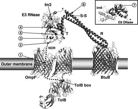
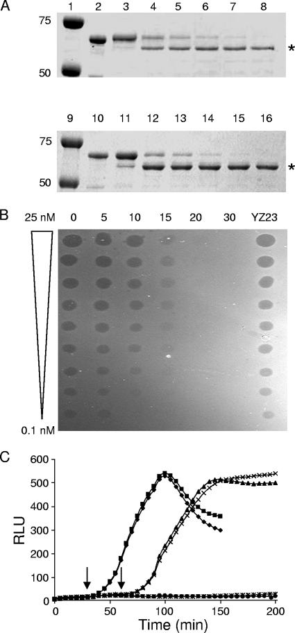
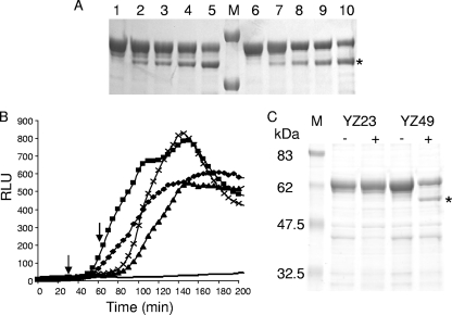
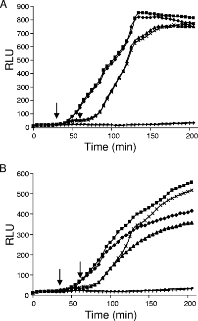
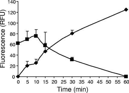
Enzymatic colicins such as colicin E9 (ColE9) bind to BtuB on the cell surface of Escherichia coli and rapidly recruit a second coreceptor, either OmpF or OmpC, through which the N-terminal natively disordered region (NDR) of their translocation domain gains entry into the cell periplasm and interacts with TolB. Previously, we constructed an inactive disulfide-locked mutant ColE9 (ColE9(s-s)) that binds to BtuB and can be reduced with dithiothreitol (DTT) to synchronize cell killing. By introducing unique enterokinase (EK) cleavage sites in ColE9(s-s), we showed that the first 61 residues of the NDR were inaccessible to cleavage when bound to BtuB, whereas an EK cleavage site inserted at residue 82 of the NDR remained accessible. This suggests that most of the NDR is occluded by OmpF shortly after binding to BtuB, whereas the extreme distal region of the NDR is surface exposed before unfolding of the receptor-binding domain occurs. EK cleavage of unique cleavage sites located in the ordered region of the translocation domain or in the distal region of the receptor-binding domain confirmed that these regions of ColE9 remained accessible at the E. coli cell surface. Lack of EK cleavage of the DNase domain of the cell-bound, oxidized ColE9/Im9 complex, and the rapid detection of Alexa Fluor 594-labeled Im9 (Im9(AF)) in the cell supernatant following treatment of cells with DTT, suggested that immunity release occurred immediately after unfolding of the colicin and was not driven by binding to BtuB.


Figures

FIG. 1.
FIG. 1.
Structural model of the assembly of the ColE9S-S translocon shortly after binding to the BtuB receptor. The model is based on the structure of ColE3/Im3 modeled on that of the BtuB-ColE3 R domain complex (26, 39). The figure highlights the position of the disulfide bond (S-S) within the R domain (R), the NDR, and the TolB box relative to the ordered T domain (T), and the E3 RNase bound to Im3 in relation to the BtuB, OmpF outer membrane receptors, and periplasmic TolB. The locations of the EK cleavage sites are shown by numbered arrows 1 to 7, where arrow 1 indicates an insertion of DDDK after residue 61; arrow 2 indicates an insertion of DDDK after residue 82; arrow 3 indicates an insertion of DDDK after G101; arrow 4 indicates a mutation of P161-ADDI-T166 to P161-DDDK-T166; arrow 5 indicates a mutation of S287-VSDV-L292 to S287-DDDK-L292; arrow 6 indicates a mutation of K441-DAKDK-L447 to K441-ADDDK-L447; and arrow 7 indicates an insertion of DDDK after S478. The asterisk shows the position of attachment of the ColE9 DNase/Im9 domains (inset) on the full-length colicin. For clarity, OmpF is shown as a dimer rather than the physiologically relevant trimeric arrangement. (Adapted from reference with permission of the publisher.)
FIG. 2.
FIG. 2.
Proteolytic cleavage of an EK cleavage site at residue 61 of ColE9 (YZ23). (A) SDS-PAGE of free YZ23 (lanes 2 to 8) and YZ23/Im9 (lanes 10 to 16) containing an engineered EK cleavage site at residue 61, incubated with EK in vitro for 0 min (lanes 3 and 11), 5 min (lanes 4 and 12), 10 min (lanes 5 and 13), 15 min (lanes 6 and 14), 20 min (lanes 7 and 15), and 30 min (lanes 8 and 16). Untreated controls are shown in lanes 2 and 10, and protein molecular mass markers of 50 and 75 kDa in lanes 1 and 9 are indicated. The asterisk marks the cleavage products. (B) Spot test activity assay of the EK cleaved YZ23. Twofold doubling dilutions of 25 nM YZ23 cleaved with EK for 0, 5, 10, 15, 20, or 30 min were spotted as 2-μl aliquots onto a freshly grown lawn of E. coli DH5α cells. Untreated YZ23 were spotted as a control. (C) Luminescence (RLU) generated by YZ23 in the lux reporter assay showing protection of YZ23 from EK cleavage. E. coli DPD1718 cells were incubated with 4 nM oxidized YZ23, followed by incubation with EK for 30 min (⧫) or 60 min (▴) before being reduced with 1 mM DTT. E. coli DPD1718 cells that were not treated with EK but reduced with 1 mM DTT after 30 min (▪) or 60 min (×) were used as positive controls for DNA damage, while E. coli DPD1718 cells that were treated with trypsin for 30 min prior to 1 mM DTT treatment (✳) or untreated DPD1718 (•) were grown as negative controls of DNA damage. Arrows indicate the addition of DTT at 30 and 60 min, respectively.
FIG. 3.
FIG. 3.
Proteolytic cleavage of an EK cleavage site at residue 82 of ColE9 (YZ49). (A) Im9-free YZ49 (lanes 1 to 5) and YZ49/Im9 (lanes 6 to 10) were incubated with EK in vitro for 10 min (lanes 2 and 7), 15 min (lanes 3 and 8), 20 min (lanes 4 and 9), or 30 min (lanes 5 and 10). Untreated controls are shown in lanes 1 and 6. The asterisk marks the cleavage products, and protein molecular mass markers of 50 and 75 kDa are shown in lane M. (B) Luminescence (RLU) generated by YZ49 after incubation with EK in the lux reporter assay. E. coli DPD1718 cells were treated with 4 nM oxidized YZ49, followed by incubation with EK for 30 min (⧫) or 60 min (▴) before being reduced with 1 mM DTT. E. coli DPD1718 cells incubated with YZ49 that were not treated with EK but reduced with 1 mM DTT after 30 (▪) or 60 min (×) were used as positive controls for DNA damage, while untreated DPD1718 (−) was grown as negative controls for DNA damage. The arrows indicate the addition of DTT at 30 or 60 min, respectively. (C) Capture of colicin proteins in vivo showing specific cleavage of YZ49 at the EK cleavage site. E. coli DPD1718 cells were treated with oxidized free YZ49 or YZ23 and then incubated with (+) or without (−) EK for 60 min. Bound colicin was rescued from heat-inactivated cells after binding to exogenously added Im9 using nickel-treated magnetic beads, concentrated, and subjected to SDS-12% PAGE. Molecular mass markers (M) are indicated on the left in kilodaltons. The asterisk marks the specific cleavage product of 54.2 kDa.
FIG. 4.
FIG. 4.
Protection from proteolytic cleavage of the DNase by Im9. The lux induction assay was used to show that Im9-complexed YZ25 was not cleaved (A), whereas Im9-free YZ25 was cleaved by EK after binding to BtuB (B). The luminescence of cultures of E. coli DPD1718 treated with oxidized YZ25/Im9 (A) or free YZ25 (B), followed by EK treatment for 30 min (⧫) or 60 min (▴), and then immediately reduced with 1 mM DTT was compared to that of E. coli DPD1718 treated with oxidized YZ25/Im9 or free YZ25 without EK treatment but reduced with 1 mM DTT after 30 min (▪) or 60 min (×). The luminescence of E. coli DPD1718 cells treated with oxidized YZ25/Im9 (A) or YZ25 (B) and then trypsin treatment for 30 min, followed by reduction with 1 mM DTT (+), and cells with no addition (−) is shown. Arrows indicate the addition of DTT at 30 and 60 min, respectively.
FIG. 5.
FIG. 5.
Immunity protein is released from the colicin after reduction of the disulfide bond. DH5α(pET21a) cells were preincubated with 10 nM BH29-Im9AF. After the removal of unbound BH29-Im9AF, activity was initiated by the addition of 2 mM DTT. The release of fluorescently labeled Im9 in the cell supernatant (RFU) is shown at set time intervals over a 60-min period as values plotted after subtraction of background release from samples that were not reduced with DTT (⧫). The coinciding reduction in cell-bound fluorescence (RFU) over the same period is also shown (▪). The means ± the standard errors of the mean are shown for two independent experiments.

Similar articles

Cited by

References

    1. Bassford, P. J., R. J. Kadner, and C. A. Schnaitman. 1977. Biosynthesis of the outer membrane receptor for vitamin B12, E colicins, and bacteriophage BF23 by Escherichia coli: kinetics of phenotypic expression after the introduction of bfe+ and bfe alleles. J. Bacteriol. 129265-275. - PMC - PubMed
    1. Bénédetti, H., R. Lloubès, C. Lazdunski, and L. Letellier. 1992. Colicin A unfolds during its translocation in Escherichia coli cells and spans the whole-cell envelope when its pore has formed. EMBO J. 11441-447. - PMC - PubMed
    1. Bonsor, D. A., I. Grishkovskaya, E. J. Dodson, and C. Kleanthous. 2007. Molecular mimicry enables competitive recruitment by a natively disordered protein. J. Am. Chem. Soc. 1294800-4807. - PubMed
    1. Bourdineaud, J. P., P. Boulanger, C. Lazdunski, and L. Letellier. 1990. In vivo properties of colicin A: channel activity is voltage dependent but translocation may be voltage independent. Proc. Natl. Acad. Sci. USA 871037-1041. - PMC - PubMed
    1. Bouveret, E., L. Journet, A. Walburger, E. Cascales, H. Bénédetti, and R. Lloubès. 2002. Analysis of the Escherichia coli Tol-Pal and TonB systems by periplasmic production of Tol, TonB, colicin, or phage capsid soluble domains. Biochimie 84413-421. - PubMed

Publication types

MeSH terms

LinkOut - more resources