. 2008 Apr 22;105(16):6033-8.
doi: 10.1073/pnas.0708354105. Epub 2008 Apr 11.

Membrane damage by human islet amyloid polypeptide through fibril growth at the membrane

Affiliations

Membrane damage by human islet amyloid polypeptide through fibril growth at the membrane

Maarten F M Engel et al. Proc Natl Acad Sci U S A. .

Abstract

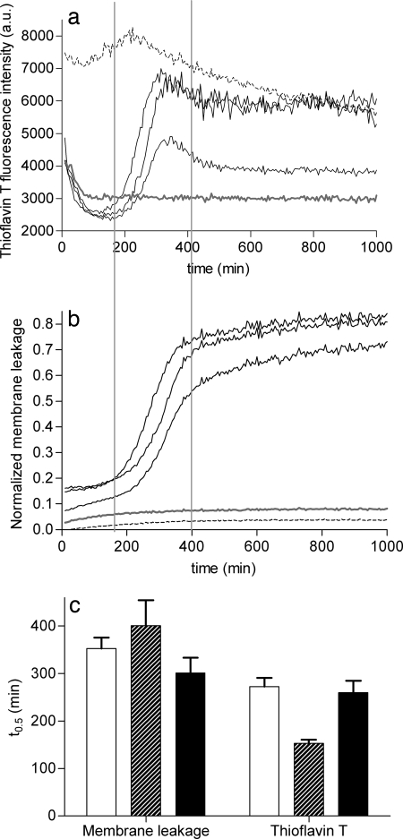
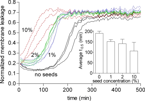
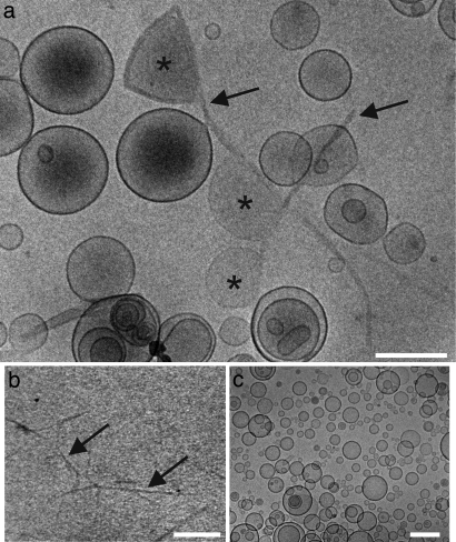
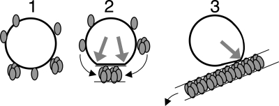
Fibrillar protein deposits (amyloid) in the pancreatic islets of Langerhans are thought to be involved in death of the insulin-producing islet beta cells in type 2 diabetes mellitus. It has been suggested that the mechanism of this beta cell death involves membrane disruption by human islet amyloid polypeptide (hIAPP), the major constituent of islet amyloid. However, the molecular mechanism of hIAPP-induced membrane disruption is not known. Here, we propose a hypothesis that growth of hIAPP fibrils at the membrane causes membrane damage. We studied the kinetics of hIAPP-induced membrane damage in relation to hIAPP fibril growth and found that the kinetic profile of hIAPP-induced membrane damage is characterized by a lag phase and a sigmoidal transition, which matches the kinetic profile of hIAPP fibril growth. The observation that seeding accelerates membrane damage supports the hypothesis. In addition, variables that are well known to affect hIAPP fibril formation, i.e., the presence of a fibril formation inhibitor, hIAPP concentration, and lipid composition, were found to have the same effect on hIAPP-induced membrane damage. Furthermore, electron microscopy analysis showed that hIAPP fibrils line the surface of distorted phospholipid vesicles, in agreement with the notion that hIAPP fibril growth at the membrane and membrane damage are physically connected. Together, these observations point toward a mechanism in which growth of hIAPP fibrils, rather than a particular hIAPP species, is responsible for the observed membrane damage. This hypothesis provides an additional mechanism next to the previously proposed role of oligomers as the main cytotoxic species of amyloidogenic proteins.


Conflict of interest statement

The authors declare no conflict of interest.

Figures

Fig. 1.
Fig. 1.
Comparison of the kinetics of hIAPP fibril growth and hIAPP-induced membrane leakage. (a and b) ThT fluorescence (a) and membrane leakage (b) were measured independently after the addition of hIAPP at a final concentration of 5 μM to DOPC/DOPS 7:3 LUVs (43 μM lipids) at time t = 0. The three solid black lines represent three hIAPP samples measured at the same time, in three adjacent wells on the same microtiter plate, with the same stock solutions. Representative traces are shown for mIAPP (gray lines) and preformed hIAPP fibrils (dashed lines). The two vertical lines are shown to facilitate comparison of the kinetic traces in a and b. (c) The average midpoints (t0.5) of the sigmoidal transitions for both ThT fluorescence and membrane leakage were obtained by fitting the curves to a standard sigmoidal function (see Experimental Procedures) and are shown for three independent experiments, each performed in triplicate, on different days, with different hIAPP stock solutions. The black bars correspond to the two experiments shown in a and b. The error bars indicate the standard deviation.
Fig. 2.
Fig. 2.
Effect of seeding on hIAPP-induced membrane leakage. Kinetic curves of membrane leakage without (black lines) and with 1% (dotted blue lines), 2% (green lines), and 10% (dashed red lines) hIAPP seeds. The three lines of the same color represent three samples measured at the same time in three adjacent wells on the same microtiter plate with the same stock solutions. (Inset) Comparison of the average t0.5 of three independent experiments, each performed in triplicate. The error bars indicate the standard deviation.
Fig. 3.
Fig. 3.
Cryo-TEM of the interaction of hIAPP and mIAPP with LUVs. (a) hIAPP in the presence of LUVs. (b) hIAPP fibrils in the absence of LUVs. (c) mIAPP in the presence of LUVs. Distorted LUVs (asterisks) are in contact with hIAPP fibrils (black arrows). All samples contain 5% DMSO. LUVs are composed of DOPC/DOPS 7:3 with a phospholipid concentration of 7 mM. IAPP concentration is 1 mM. (Scale bars, 200 nm.)
Fig. 4.
Fig. 4.
Simplified schematic representation of the different stages of the proposed membrane-associated hIAPP fibril growth that results in membrane damage. Starting from a situation with an intact membrane (black circle) and monomeric hIAPP (gray ellipsoids), hIAPP monomers or oligomers adsorb on or insert into the membrane (Left). Next, membrane-located hIAPP participates in initiation and propagation of fibril growth at the membrane leading to a forced change in membrane curvature and concomitant temporal membrane leakage, at the locations where fibrils and membrane separate (gray arrows) (Center). Finally, mature hIAPP fibrils that line the surface of a distorted membrane start detaching, initiating recovery of vesicle shape (Right). Oligomers have been depicted arbitrarily as a cluster of four hIAPP monomers. Black arrows indicate the movement of hIAPP species.

References

    1. Höppener JWM, Ahrén B, Lips CJM. Islet amyloid and type 2 diabetes mellitus. N Engl J Med. 2000;343:411–419. - PubMed
    1. Mirzabekov TA, Lin MC, Kagan BL. Pore formation by the cytotoxic islet amyloid peptide amylin. J Biol Chem. 1996;271:1988–1992. - PubMed
    1. Quist A, et al. Amyloid ion channels: A common structural link for protein-misfolding disease. Proc Natl Acad Sci USA. 2005;102:10427–10432. - PMC - PubMed
    1. Anguiano M, Nowak RJ, Lansbury PT., Jr Protofibrillar islet amyloid polypeptide permeabilizes synthetic vesicles by a pore-like mechanism that may be relevant to type II diabetes. Biochemistry. 2002;41:11338–11343. - PubMed
    1. Porat Y, Kolusheva S, Jelinek R, Gazit E. The human islet amyloid polypeptide forms transient membrane-active prefibrillar assemblies. Biochemistry. 2003;42:10971–10977. - PubMed

Publication types