Review
. 2009 Sep;35(5):959-72.
doi: 10.1093/schbul/sbn022. Epub 2008 Apr 11.

A review of the fetal brain cytokine imbalance hypothesis of schizophrenia

Affiliations
Review

A review of the fetal brain cytokine imbalance hypothesis of schizophrenia

Urs Meyer et al. Schizophr Bull. 2009 Sep.

Abstract

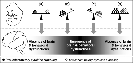
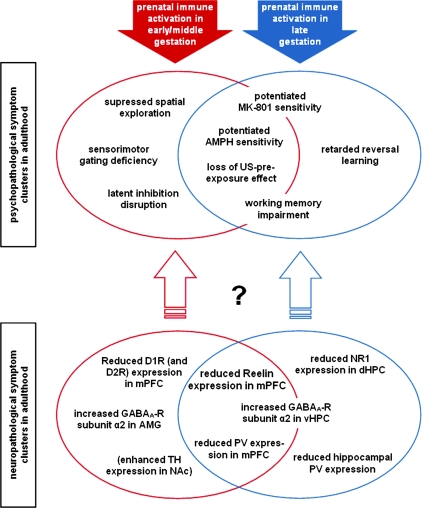
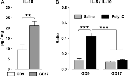
Maternal infection during pregnancy increases the risk of schizophrenia and other brain disorders of neurodevelopmental origin in the offspring. A multitude of infectious agents seem to be involved in this association. Therefore, it has been proposed that factors common to the immune response to a wide variety of bacterial and viral pathogens may be the critical link between prenatal infection and postnatal brain and behavioral pathology. More specifically, it has been suggested that the maternal induction of pro-inflammatory cytokines may mediate the neurodevelopmental effects of maternal infections. Here, we review recent findings from in vitro and in vivo investigations supporting this hypothesis and further emphasize the influence of enhanced anti-inflammatory cytokine signaling on early brain development. Disruption of the fetal brain balance between pro- and anti-inflammatory cytokine signaling may thus represent a key mechanism involved in the precipitation of schizophrenia-related pathology following prenatal maternal infection and innate immune imbalances.


Figures

Fig. 1.
Fig. 1.
Hypothesized Model of the Modulation of Adult Brain and Behavioral Functions by Imbalances in Fetal Brain Cytokines. The pro-inflammatory and anti-inflammatory classes of cytokines are represented by black dots and white diamonds, respectively. (a) Brain and behavioral development is under the influence of a balance between pro- and anti-inflammatory cytokines in prenatal life. (b) A shift toward excess levels of pro-inflammatory cytokines (eg, by in utero exposure to infection) leads to the emergence of severe brain and behavioral abnormalities in adult life. (c) A shift toward enhanced anti-inflammatory signaling alone (eg, by genetically determined increases in IL-10 production) also precipitates adult brain and behavioral dysfunctions. (d) The concomitant induction of both pro- and anti-inflammatory cytokines (eg, after in utero infections in individuals with enhanced IL-10–mediated anti-inflammatory signaling) largely nullifies each other's long-term negative influences on brain and behavioral functions because the balance between the 2 cytokine classes is maintained. For a detailed discussion of the model see Meyer et al.
Fig. 2.
Fig. 2.
Emergence of Distinct Psychopathological and Neuropathological Symptom Clusters in Adulthood After Prenatal Polyriboinosinic-Polyribocytidilic Acid (PolyI:C)–Induced Immune Activation in Early/Middle (Red) and Late (Blue) Gestation in the Mouse. The diagram illustrates the identified structural and functional brain abnormalities that are characteristic of the symptom profiles associated with prenatal PolyI:C exposure in early/middle and late gestation. Some of the psychopathological and neuropathological traits are clearly restricted to the symptom cluster associated with prenatal immune activation in early/middle or late gestation, whereas others are common to both symptom clusters. The exact correspondence between the distinct neuropathological and psychopathological symptom clusters remains to be determined. The neuropathological effects presented in brackets denote that they were studied only after prenatal PolyI:C–induced immune activation in early/middle gestation so far. AMG, amygdala; AMPH, amphetamine; D1R/D2R, dopamine D1/D2 receptor; dHPC, dorsal hippocampus; GABAA-R, gamma-amino-butyric acid (A) receptor; mPFC, medial prefrontal cortex; MK-801, dizocilpine; NAc, nucleus accumbens; PV, Parvalbumin; TH, tyrosine hydroxylase; US, unconditioned stimulus; vHPC, ventral hippocampus.
Fig. 3.
Fig. 3.
(A) Basal Protein Levels (pg/mg) of the Anti-Inflammatory Cytokine IL-10 Are Increased in the Fetal Brains of Mice Derived From Pregnant Mothers in Late Gestation (Gestation Day 17, GD17) Compared With Fetuses of Mothers in Early/Middle Gestation (GD9). Pregnant mice were killed by decapitation, and the fetal brains were dissected as described in Meyer et al., Levels of IL-10 were measured using a multiplexed particle–based flow cytometric cytokine as described previously., **P < .01, based on one-way analysis of variance (ANOVA) [F(1,21) = 13.8]; N(GD9 fetuses) = 10, N(GD17 fetuses) = 13. Fetuses derived from 4 mothers in each gestation period. (B) Acute maternal immunological stimulation by the viral mimic polyriboinosinic-polyribocytidilic acid (PolyI:C) (2 mg/kg, intravenously) increases the fetal brain ratio of IL-6/IL-10 in fetuses derived from mothers in early/middle gestation (GD9), but not in fetuses of mothers in late gestation (GD17). The former effect indicates a shift in the fetal brain balance toward increased pro-inflammatory cytokines in early/middle gestation. Pregnant mice were killed by decapitation 2 h after maternal vehicle (saline) or PolyI:C treatment, and the fetal brains were dissected as described previously., Levels of IL-6 and IL-10 were measured using a multiplexed particle–based flow cytometric cytokine assay as described in Meyer et al., ***P < .01, based on Fisher's post hoc comparisons following a significant gestation day × maternal treatment interaction in the 2 × 2 ANOVA [F(1,36) = 17.1, P < .001]; N(GD9-saline fetuses) = 8, N(GD9-PolyI:C fetuses) = 7, N(GD17-saline fetuses) = 13, N(GD17-PolyI:C fetuses) = 12. Fetuses derived from 3 mothers in each gestation period/treatment condition.

References

    1. Rees S, Harding R. Brain development during fetal life: influences of the intra-uterine environment. Neurosci Lett. 2004;361:111–114. - PubMed
    1. Rees S, Inder T. Fetal and neonatal origins of altered brain development. Early Hum Dev. 2005;81:753–761. - PubMed
    1. Weinberger DR. Implications of normal brain development for the pathogenesis of schizophrenia. Arch Gen Psychiatry. 1987;44:660–669. - PubMed
    1. Rapoport JL, Addington AM, Frangou S, Psych MR. The neurodevelopmental model of schizophrenia: update 2005. Mol Psychiatry. 2005;10:434–449. - PubMed
    1. Ross CA, Margolis RL, Reading SA, Pletnikov M, Coyle JT. Neurobiology of schizophrenia. Neuron. 2006;52:139–153. - PubMed

Publication types