. 2008 May 29;453(7195):657-61.
doi: 10.1038/nature06928. Epub 2008 Apr 13.

Functional genomic screen reveals genes involved in lipid-droplet formation and utilization

Affiliations

Functional genomic screen reveals genes involved in lipid-droplet formation and utilization

Yi Guo et al. Nature. .

Abstract

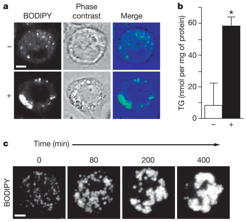
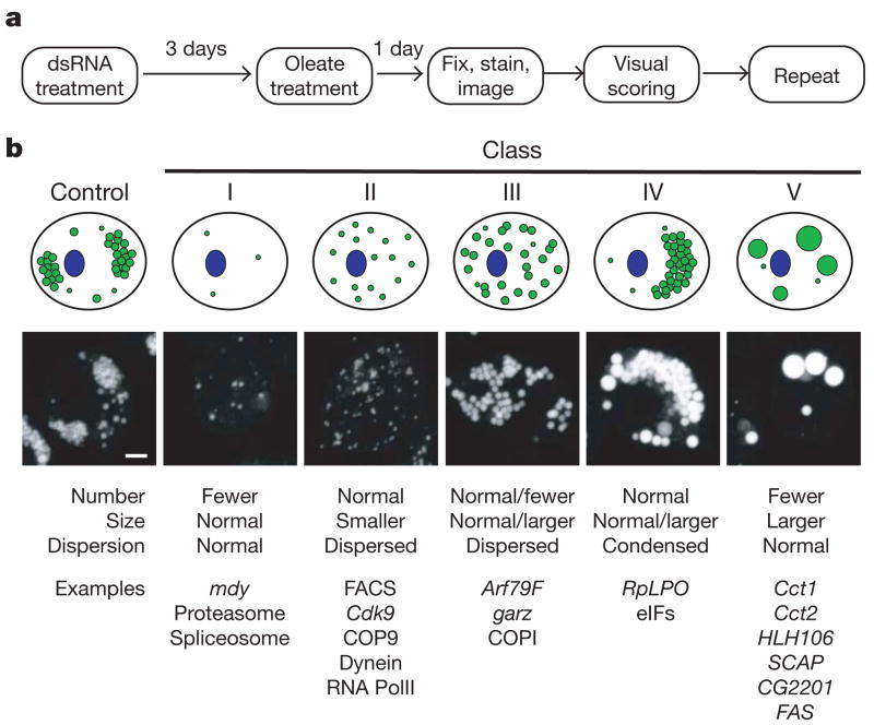
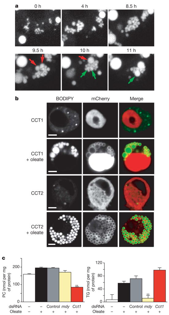
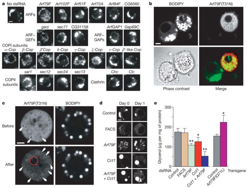
Eukaryotic cells store neutral lipids in cytoplasmic lipid droplets enclosed in a monolayer of phospholipids and associated proteins. These dynamic organelles serve as the principal reservoirs for storing cellular energy and for the building blocks for membrane lipids. Excessive lipid accumulation in cells is a central feature of obesity, diabetes and atherosclerosis, yet remarkably little is known about lipid-droplet cell biology. Here we show, by means of a genome-wide RNA interference (RNAi) screen in Drosophila S2 cells that about 1.5% of all genes function in lipid-droplet formation and regulation. The phenotypes of the gene knockdowns sorted into five distinct phenotypic classes. Genes encoding enzymes of phospholipid biosynthesis proved to be determinants of lipid-droplet size and number, suggesting that the phospholipid composition of the monolayer profoundly affects droplet morphology and lipid utilization. A subset of the Arf1-COPI vesicular transport proteins also regulated droplet morphology and lipid utilization, thereby identifying a previously unrecognized function for this machinery. These phenotypes are conserved in mammalian cells, suggesting that insights from these studies are likely to be central to our understanding of human diseases involving excessive lipid storage.


Figures

Figure 1
Figure 1. Oleate increases the formation of lipid droplets in Drosophila S2 cells
a, S2 cells incubated for 24 h without (upper) or with (lower) 1 mM oleate. Staining with BODIPY, phase-contrast image, and overlay are shown. b, Oleate-loaded cells have an increased triacylglycerol (TG) content. Cells were incubated as in a and their TG contents were measured. Results are means and s.d. for three experiments; P <0.05. c, Lipid-droplet formation occurs in steps. Cells treated as in a were stained with BODIPY. Single cells were followed by four-dimensional confocal time-lapse microscopy. Representative maximum projections of three-dimensional stacks at the indicated times are shown. Scale bars, 3 μm.
Figure 2
Figure 2. Genome-wide screen identified genes regulating the formation of lipid droplets
a, Outline for strategy to screen for genes involved in lipid-droplet biogenesis. See the text for details. b, Genes involved in the screen for lipid-droplet biogenesis fall into distinct phenotypic classes. The 132 most striking phenotypes were classified according to lipid-droplet number, size and dispersion. From this classification, five major classes emerged; a graphic representation (top), an example image (middle) and some gene examples (bottom) are shown. Scale bar, 3 μm.
Figure 3
Figure 3. Phosphatidylcholine content regulates the size and abundance of lipid droplets
a, Lipid-droplet formation induced by oleate in Cct1 knockdown cells (Fig. 1a). Single cells were followed by time-lapse confocal microscopy. Representative projections revealed that droplets first proliferate normally (upper) and then fuse (lower). Examples of fusion are indicated (red and green arrows). b, CCT enzymes localize to the surface of droplets after induction with oleate. Cct1 and Cct2 were transiently expressed in S2 cells as amino-terminal mCherry-tagged fusion proteins and were stained and imaged before or after induction. BODIPY staining, mCherry fluorescence, and merge are shown. Scale bar, 3 μm. c, Cct1 knockdown cells have less phosphatidylcholine (PC) and more triacylglycerol (TG). S2 cells were treated with dsRNAs as indicated, loaded with oleate (as in Fig. 1a) and lysed. PC (upper) and TG (lower) levels in the extract were measured. Results are means and s.d. for three independent experiments. **, P <0.01 versus control RNAi.
Figure 4
Figure 4. ArfI–COPI complex members function in the formation of lipid droplets
a, Knockdowns of Arf79F, the GEF gene gartenzwerg (garz) and specific subunits of the COPI coat affected lipid-droplet formation similarly. Representative images are shown. For descriptions of controls see Supplementary Information. GAP, GTPase-activating protein. b, Arf79F(T31N) localizes to the droplet surface and causes a similar phenotype to Arf79F knockdown. Arf79F(T31N) expressed as a carboxy-terminal mCherry-tagged fusion protein in S2 cells was observed by confocal microscopy after loading with oleate and staining with BODIPY. A representative confocal midsection is shown for BODIPY (top left), mCherry fluorescence (top right) and a merge (bottom right). c, Arf79F(T31N)–mCherry localizes to the droplet surface. The photobleached region (6 min) is indicated by a red circle. Arrows indicate the association of Arf79F(T31N) with the surface of droplets. d, Arf79F, Cct1 and double knockdowns lead to decreased lipolysis. S2 cells were treated with dsRNAs for three days as indicated, loaded with 1 mM oleate for one day, and imaged by confocal microscopy after staining with BODIPY (day 0, left panels). Representative confocal midsections are shown. Oleate was removed from the medium and the cells were starved for one day in serum-free medium to induce lipolysis (day 1, right). Scale bars, 3 μm. FACS, gene encoding a long-chain-fatty-acid-CoA ligase (CG8732). e, Arf79F, Cct1 and double knockdowns lead to decreased glycerol release to the medium. A transgene encoding Arf79F(Q71L) leads to increased release of glycerol. Experiments were as in d, and the glycerol released was measured. Results are means and s.d. for three independent experiments. *, P <0.05, ** P <0.01 versus control RNAi (left) and versus control transgene (right).

References

    1. Bartz R, et al. Lipidomics reveals that adiposomes store ether lipids and mediate phospholipid traffic. J Lipid Res. 2007;48:837–847. - PubMed
    1. Brown DA. Lipid droplets: proteins floating on a pool of fat. Curr Biol. 2001;11:R446–R449. - PubMed
    1. Miura S, et al. Functional conservation for lipid storage droplet association among Perilipin, ADRP, and TIP47 (PAT)-related proteins in mammals, Drosophila, and Dictyostelium. J Biol Chem. 2002;277:32253–32257. - PubMed
    1. Bartz R, et al. Dynamic activity of lipid droplets: protein phosphorylation and GTP-mediated protein translocation. J Proteome Res. 2007;6:3256–3265. - PubMed
    1. Martin S, Parton RG. Lipid droplets: a unified view of a dynamic organelle. Nature Rev Mol Cell Biol. 2006;7:373–378. - PubMed

Publication types