. 2008 May;4(5):313-21.
doi: 10.1038/nchembio.83.

Identification of RIP1 kinase as a specific cellular target of necrostatins

Affiliations

Identification of RIP1 kinase as a specific cellular target of necrostatins

Alexei Degterev et al. Nat Chem Biol. 2008 May.

Abstract

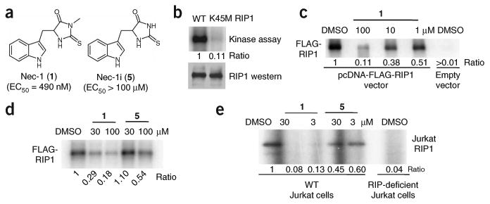
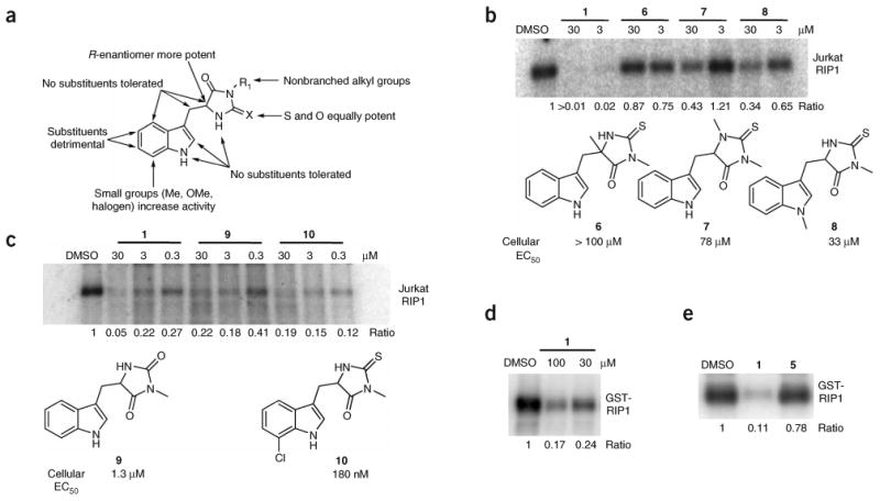
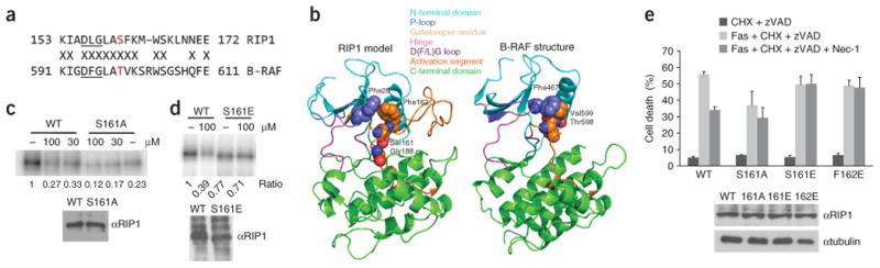
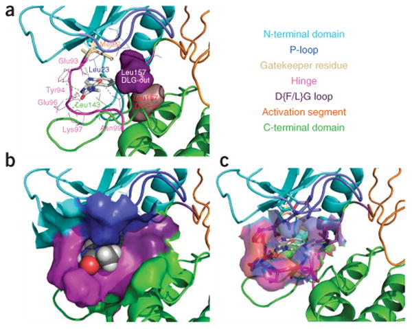
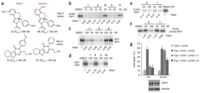
Necroptosis is a cellular mechanism of necrotic cell death induced by apoptotic stimuli in the form of death domain receptor engagement by their respective ligands under conditions where apoptotic execution is prevented. Although it occurs under regulated conditions, necroptotic cell death is characterized by the same morphological features as unregulated necrotic death. Here we report that necrostatin-1, a previously identified small-molecule inhibitor of necroptosis, is a selective allosteric inhibitor of the death domain receptor-associated adaptor kinase RIP1 in vitro. We show that RIP1 is the primary cellular target responsible for the antinecroptosis activity of necrostatin-1. In addition, we show that two other necrostatins, necrostatin-3 and necrostatin-5, also target the RIP1 kinase step in the necroptosis pathway, but through mechanisms distinct from that of necrostatin-1. Overall, our data establish necrostatins as the first-in-class inhibitors of RIP1 kinase, the key upstream kinase involved in the activation of necroptosis.


Figures

Figure 1
Figure 1
Nec-1 (1) is an inhibitor of RIP1 kinase. (a) Structures of 1 and 5. EC50 values for inhibition of cellular necrosis in TNFα-treated FADD-deficient Jurkat cells were determined as described in the Methods and were previously reported. (b) Phosphorylation of RIP1 requires its kinase activity. Expression constructs of FLAG-tagged wild-type (WT) or a kinase-inactive point mutant of RIP1 (K45M) were transfected into 293T cells and RIP1 kinase assay was performed as described in the Methods in the presence of [γ-32P]ATP for 30 min at 30 °C. Samples were subjected to SDS-PAGE and RIP1 band was visualized by autoradiography. Relative intensities of radioactive bands were quantified and are shown (ratio) in this and all other autoradiographs. In parallel to kinase reactions, a sample of beads was subjected to western blot analysis using anti-RIP1 antibody to ensure equal protein amounts in kinase reactions. (c) 1 inhibits the autophosphorylation of overexpressed RIP1 in vitro in a dose-dependent fashion. Assay was performed as in b, except different amounts of 1 were added 15 min before ATP. (d) Inactive analog of 1 (5) displays substantially reduced activity against RIP1 kinase in vitro. Assay was performed as in c with indicated concentrations of 1 and 5. (e) 1 inhibits the autophosphorylation of endogenous RIP1 in vitro. RIP1 was immunoprecipitated from lysates of WT and RIP1-deficient Jurkat cells using agarose-conjugated anti-RIP1 antibody, and kinase reactions were performed as in c in the presence of the indicated amounts of 1 or 5. Assays were performed at least two or three times, and similar results were obtained each time. The representative images are shown.
Figure 2
Figure 2
SAR analysis of RIP1 inhibition by necrostatins. (a) Summary of cellular SAR of 1 based on ref. . (b,c) In vitro activity of select inactive (b) and active (c) 1 analogs. Endogenous RIP1 kinase assays were performed as in Figure 1e in the presence of indicated amounts of 1 analogs. Structures of the derivatives are shown. EC50 values for inhibition of cellular necrosis in TNFα-treated FADD-deficient Jurkat cells by necrostatins were determined as described in the Methods and were previously reported. (d,e) 1 inhibits kinase activity of recombinant RIP1 expressed in Sf9 cells. Recombinant RIP1, expressed in Sf9 cells, was subjected to in vitro kinase assay in the presence of indicated amounts (d) or 100 μM (e) of 1 (d,e) or 5 (e). Assays were performed at least two or three times, and similar results were obtained each time. The representative images are shown.
Figure 3
Figure 3
Effect of Ser161 and Phe162 mutations on necroptosis and inhibition by 1. (a) Sequence alignment of the magnesium binding and activation segments of B-RAF and RIP1. Thr598 of B-RAF and Ser161 of RIP1 are shown in red. Kinase-conserved DFG motif is underlined. (b) Homology model of RIP1. (c,d) S161A (c) and S161E (d) mutations attenuate RIP1 kinase sensitivity to 1 in vitro. Kinase assays of RIP1 mutants overexpressed in 293T cells were performed as described in Figure 1. “–” indicates DMSO. In parallel to kinase reactions, a sample of beads was subjected to western blot analysis using anti-RIP1 antibody to ensure equal protein amounts in kinase reactions. Assays were performed at least two or three times, and similar results were obtained each time. The representative images are shown. (e) Mutations of Ser161 and Phe162 attenuate inhibition of necroptosis by 1. Corresponding pcDNA-FLAG-RIP1 vectors were transiently electroporated into RIP1-deficient Jurkat cells along with pEGFP. Cells were allowed to recover for 48 h, treated with anti-FAS antibody, cycloheximide and zVAD-fmk to induce necroptosis and 30 μM 1, followed by analysis by FACS as described in the Methods. The data were obtained in a single experiment performed in triplicate and represent mean values ± s.d. Experiment was repeated multiple times, and similar results were obtained each time. Equal expression of RIP1 mutants was confirmed by western blot using anti-RIP1 and anti-tubulin antibodies.
Figure 4
Figure 4
Molecular model of the RIP1–11 complex. (a) RIP1 is shown in ribbon representation with the binding site amino acids shown in wireframe representation, and with the gatekeeper residue and 11 shown in stick representation. Solid surfaces of DLG leucine in its predicted DFG-in and DFG-out conformations are highlighted in purple and brown, respectively. (b) RIP1 is shown in ribbon representation with the binding site solid surface displayed, and 11 is shown in spacefill representation. (c) RIP1 is shown in ribbon representation with the binding site transparent surface displayed, and binding site amino acids and 11 are shown in stick representation. Different structural features of RIP1 kinase are shown in different colors and are annotated on the included text panel.
Figure 5
Figure 5
Necrostatin-3 and necrostatin-5 inhibit RIP1 kinase activity. (a) Structures of necrostatin-3 and necrostatin-5 analogs. Cellular EC50 in TNFα-treated FADD-deficient Jurkat cells are shown. (b) Additional Necs inhibit activity of Jurkat cell RIP1. In vitro kinase reaction in the presence of the indicated concentrations of necrostatins was performed as in Figure 1e. (c,d) 2, but not 16, specifically inhibits recombinant RIP1 kinase, expressed in Sf9 cells. In vitro kinase reactions in the presence of the indicated concentrations of necrostatins were performed as in Figure 2d. (e,f) 2 still inhibits activity of S161E mutant of RIP1 in vitro. WT FLAG-RIP1 (e) and S161E FLAG-RIP1 (f) were expressed in 293T cells and subjected to in vitro kinase assay in the presence of the indicated concentrations of necrostatins as in Figure 1c. Assays were performed at least two or three times, and similar results were obtained each time. The representative images are shown. (g) 2 attenuates necroptosis mediated by S161E mutant of RIP1. Recapitulation of WT and S161E mutants in RIP-deficient Jurkat cells and necroptosis viability assay were performed as described in Figure 3e. Compounds were used at 30 μM. The data were obtained in a single experiment performed in triplicate and represent mean values ± s.d. Experiment was repeated multiple times, and similar results were obtained each time. Equal expression of RIP1 mutants was confirmed by western blot using anti-RIP1 and anti-tubulin antibodies.

Similar articles

Cited by

References

    1. Thompson CB. Apoptosis in the pathogenesis and treatment of disease. Science. 1995;267:1456–1462. - PubMed
    1. Lindsten T, Thompson CB. Cell death in the absence of Bax and Bak. Cell Death Differ. 2006;13:1272–1276. - PubMed
    1. Los M, Wesselborg S, Schulze-Osthoff K. The role of caspases in development, immunity, and apoptotic signal transduction: lessons from knockout mice. Immunity. 1999;10:629–639. - PubMed
    1. Chautan M, Chazal G, Cecconi F, Gruss P, Golstein P. Interdigital cell death can occur through a necrotic and caspase-independent pathway. Curr Biol. 1999;9:967–970. - PubMed
    1. Oppenheim RW, et al. Programmed cell death of developing mammalian neurons after genetic deletion of caspases. J Neurosci. 2001;21:4752–4760. - PMC - PubMed

Publication types

Associated data