. 2008 May;15(5):469-76.
doi: 10.1038/nsmb.1403. Epub 2008 Apr 13.

The HSA domain binds nuclear actin-related proteins to regulate chromatin-remodeling ATPases

Affiliations

The HSA domain binds nuclear actin-related proteins to regulate chromatin-remodeling ATPases

Heather Szerlong et al. Nat Struct Mol Biol. 2008 May.

Abstract

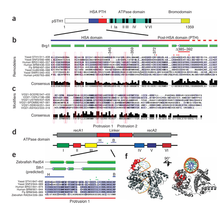
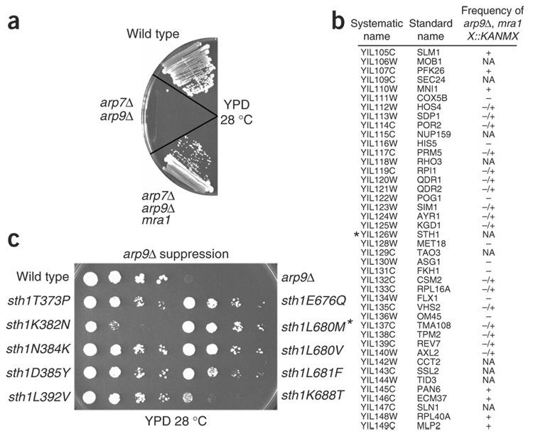
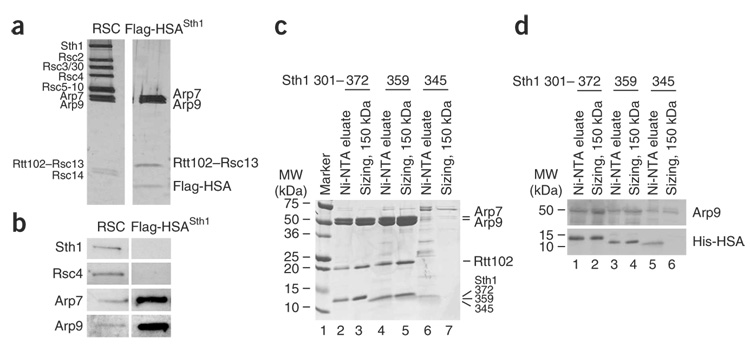
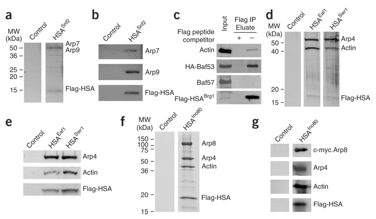
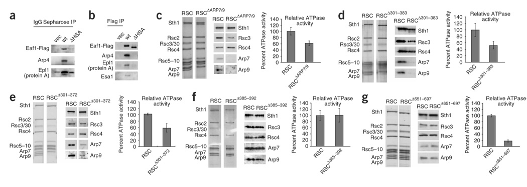
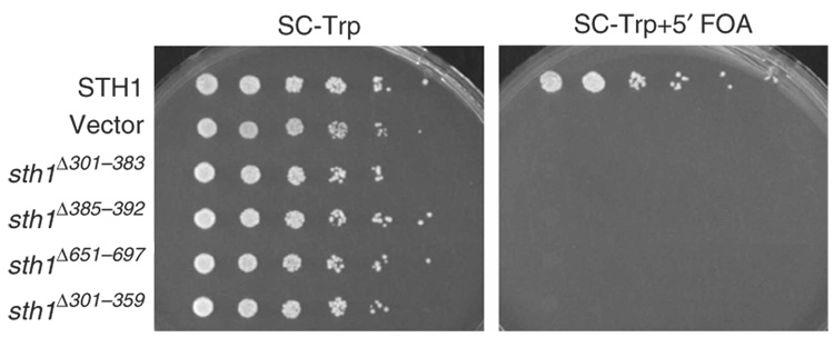
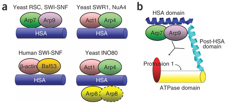
We identify the helicase-SANT-associated (HSA) domain as the primary binding platform for nuclear actin-related proteins (ARPs) and actin. Individual HSA domains from chromatin remodelers (RSC, yeast SWI-SNF, human SWI-SNF, SWR1 and INO80) or modifiers (NuA4) reconstitute their respective ARP-ARP or ARP-actin modules. In RSC, the HSA domain resides on the catalytic ATPase subunit Sth1. The Sth1 HSA is essential in vivo, and its omission causes the specific loss of ARPs and a moderate reduction in ATPase activity. Genetic selections for arp suppressors yielded specific gain-of-function mutations in two new domains in Sth1, the post-HSA domain and protrusion 1, which are essential for RSC function in vivo but not ARP association. Taken together, we define the role of the HSA domain and provide evidence for a regulatory relationship involving the ARP-HSA module and two new functional domains conserved in remodeler ATPases that contain ARPs.


Figures

Figure 1
Figure 1
HSA, post-HSA (PTH) and protrusion 1 domains in ARP-containing remodeling complexes are conserved; arpΔ suppressors cluster in Sth1. (a) A schematic depicting the domains of Sth1. Clusters of arpΔ suppressor mutations are depicted with asterisks. (b) Alignment of HSA and post-HSA domains of remodelers. Secondary-structure predictions used PSIPRED. Locations of mutations that suppress arpΔ lethality are indicated with asterisks (red, within the HSA; green, within protrusion 1). (c) Alignment of HSA domains of Eaf1 (also known as Vid21)–related proteins, with residues conserved within remodelers extended to b and boxed in red. The post-HSA domain is not present in Eaf1-related proteins from Saccharomyces cerevisiae (SCERE), Yarrowia lipolytica (YARL), Ustilago maydis (USTMA), Schizosaccharomyces pombe (SPOMBE), Debaryomyces hansenii (DEBHA), Ashbya gossypii (ASHGO). (d) Schematic of SNF2 family ATPase domains, which bear a unique insertion between domains III and IV, containing two protrusions and two highly conserved subdomains (H and B). sth1 ATPase mutations that suppress arpΔ lethality cluster between these regions (green asterisk). recA1 and recA2 refer to the regions in helicases/translocases that share homology to recA. (e) Mutations that suppress arpΔ mutations (green asterisks) cluster tightly within protrusion 1, between the H-B region. (f) Structural depiction of protrusion 1 of Sulfolobus solfataricus Rad54 (in red). DNA and ATP binding regions are not localized to protrusion 1.
Figure 2
Figure 2
Particular sth1 mutations suppress arpΔ mutations. (a) Suppression of the arp7Δ arp9Δ lethality by mra1-1. (b) Linkage analysis maps arpΔ suppressor mutations to the STH1 locus. Genes flanking STH1 (marked with an asterisk) on chromosome IX are depicted, and the scoring reflects the frequency of obtaining triple-mutant spores (mra1 arp9Δ XD::KANMX): +, spore patches; −/+, infrequent punctate spore colonies; –, no spores; NA; not applicable (essential genes). Shown is the data for mra1-1, and linkage to STH1 was also demonstrated for the nine other mra mutations (not shown). (c) arp9Δ suppression by sth1 alleles. Reference strains were wild type and arp9Δ. An asterisk indicates the arp9Δ arp7Δ background. All strains are the original spontaneous suppressors.
Figure 3
Figure 3
The HSA domain of Sth1 is sufficient to bind Arp7 and Arp9. (a) Tagged Sth1 HSA (amino acids 301–383) copurifies Arp7, Arp9 and Rtt102 (Methods). Proteins were revealed by silver staining and identified by MS sequencing (see text). (b) Western analysis of RSC and the HSA–Arp7–Arp9–Rtt102 complexes. (c) Reconstitution of the complex and determination of the minimal ARP binding HSA domain. Sth1 HSA truncation derivatives (Sth1301–372, Sth1301–359 or Sth1301–345 tagged with 10 × His) were expressed along with untagged Arp7, Arp9 and Rtt102. Extracts were purified using Ni-NTA agarose and subjected to gel filtration for sizing (Methods). (d) Western analysis of recombinant complexes.
Figure 4
Figure 4
Individual HSA domains are sufficient to bind actin and particular ARPs. (a) The yeast Snf2 HSA domain nucleates Arp7 and Arp9. The HSA region of Snf2 (578–659, Flag-tagged and 10 × His-tagged) was expressed in a wild-type strain (YBC928). Complexes were purified with Ni-NTA, then anti-Flag beads (Methods) and visualized by silver staining. (b) Western analysis confirms the protein identities in a. (c) The BRG1 HSA associates with BAF53 and actin. The BRG1 HSA (462–531, Flag-tagged) and HA-tagged BAF53 were coexpressed by transient transfection in RKO cells. Immunoprecipitation analysis with anti-Flag antibody followed by western blotting reveals specific associations with BAF53 and actin. (d) The Swr1 and Eaf1 HSA domains nucleate Arp4 and actin. The HSA regions (Flag-tagged and 10 × His–tagged) of Swr1 or Eaf1 (p1871 and p1870) were expressed, along with untagged Arp4 and actin (p1883) in a wild-type strain (YBC928). Complexes were purified using Ni-NTA and anti-Flag beads (Methods) and visualized by silver staining. (e) Western analysis confirms protein identities in d. (f) The Ino80 HSA nucleates Arp4, Arp8 and actin. The HSA region of Ino80 (462–598, Flag-tagged and His-tagged, (p2441)), along with untagged Arp4 and actin (p1883), V5-Arp5 and c-myc–Arp8 (p2436), were expressed in a wild-type strain. Complexes were purified with Ni-NTA, then anti-Flag beads (Methods) and visualized by Coomassie. (g) Western analysis confirms the protein identities shown in f.
Figure 5
Figure 5
The HSA domains of Sth1 and NuA4 are required for Arp and actin binding, and, in RSC, full ATPase activity. (a) The Eaf1 HSA region is necessary for Epl1 association with Eaf1, Arp4 and actin. Flag-tagged Eaf1 (p2345), or a derivative lacking the HSA domain (Eaf1ΔHSA (p2348)) was expressed in an eaf1Δ strain (YBC3068) that also expressed TAP-tagged Epl1 from its genomic locus. Proteins associated with Epl1 (by pull-down with IgG sepharose) were examined by western analysis. (b) The Eaf1 HSA region is necessary for association of Arp4 and actin with Eaf1. Flag-tagged Eaf1 (p2345), or a derivative lacking the HSA domain (Eaf1ΔHSA (p2348)) was expressed in an eaf1Δ strain. Eaf1 was pulled down with anti-Flag agarose, and associated proteins were examined by western analysis. (c–g) RSC complex derivatives containing Sth1 domain mutations, or arp mutations, were purified and examined for composition by silver staining (left) and western analysis (middle), and then monitored for ATPase activity, relative to a wild-type derivative purified alongside the mutant complex (right). For each, the error bar indicates s.d. (c) RSC complex lacking Arp7 and Arp9. (d) RSC complex lacking the Sth1 HSA region (RSCΔ301–383). (e) RSC complex lacking the Sth1 region sufficient for ARP binding (RSCΔ301–372). (f) RSC complex lacking the Sth1 post-HSA domain (RSCΔ385–392). (g) RSC complex lacking the Sth1 protrusion 1 domain (RSCΔ651–697).
Figure 6
Figure 6
The HSA, post-HSA and protrusion 1 domains are essential for viability. TRP1-marked plasmids bearing wild-type STH1, sth1Δ301–383, sth1Δ385–392, sth1Δ651–697 or sth1Δ301–359 (p976, p1724, p1683, p2500 or p2465, respectively) were transformed into an sth1Δ strain (YBC943) covered by wild-type STH1 on a URA3-marked plasmid (p114). Loss of the URA3-marked plasmid is enforced on medium containing 5′ fluoroorotic acid (FOA), which prevents the growth of Ura3+ cells. Inability to lose the URA3-marked plasmid bearing wild-type STH1 demonstrates the lack of complementation by the sth1 domain deletion derivative. Vector, empty vector control.
Figure 7
Figure 7
Summary of HSA domain complexes and a model for HSA–ARP regulation of ATPase activity via the adjacent post-HSA domain and protrusion 1. (a) The HSA domains from yeast modifying complexes RSC (Sth1), SWI-SNF (Snf2), SWR1 (Swr1), NuA4 (Eaf1), INO80 (Ino80) and human SWI-SNF (BRG1) nucleate binding of their respective ARP–ARP or ARP–actin members. (b) The HSA–ARPs, post-HSA and protrusion 1 domains interact to regulate the function of the ATPase. Actin-related proteins bind strongly to the HSA, with a weak interaction with the post-HSA also detected. The ARPs and protrusion 1 positively regulate ATPase activity, and the ability of the ARPs to influence (or bind) protrusion 1 may be regulated by the post-HSA domain.

Comment in

Similar articles

Cited by

References

    1. Havas K, Whitehouse I, Owen-Hughes T. ATP-dependent chromatin remodeling activities. Cell. Mol. Life Sci. 2001;58:673–682. - PMC - PubMed
    1. Narlikar GJ, Fan HY, Kingston RE. Cooperation between complexes that regulate chromatin structure and transcription. Cell. 2002;108:475–487. - PubMed
    1. Vignali M, Hassan AH, Neely KE, Workman JL. ATP-dependent chromatin-remodeling complexes. Mol. Cell. Biol. 2000;20:1899–1910. - PMC - PubMed
    1. Saha A, Wittmeyer J, Cairns BR. Chromatin remodeling by RSC involves ATP-dependent DNA translocation. Genes Dev. 2002;16:2120–2134. - PMC - PubMed
    1. Brown CE, Lechner T, Howe L, Workman JL. The many HATs of transcription coactivators. Trends Biochem. Sci. 2000;25:15–19. - PubMed

Publication types

MeSH terms