. 2008 Jul 31;27(33):4521-31.
doi: 10.1038/onc.2008.103. Epub 2008 Apr 14.

Mycoplasma infection suppresses p53, activates NF-kappaB and cooperates with oncogenic Ras in rodent fibroblast transformation

Affiliations

Mycoplasma infection suppresses p53, activates NF-kappaB and cooperates with oncogenic Ras in rodent fibroblast transformation

D Y Logunov et al. Oncogene. .

Abstract

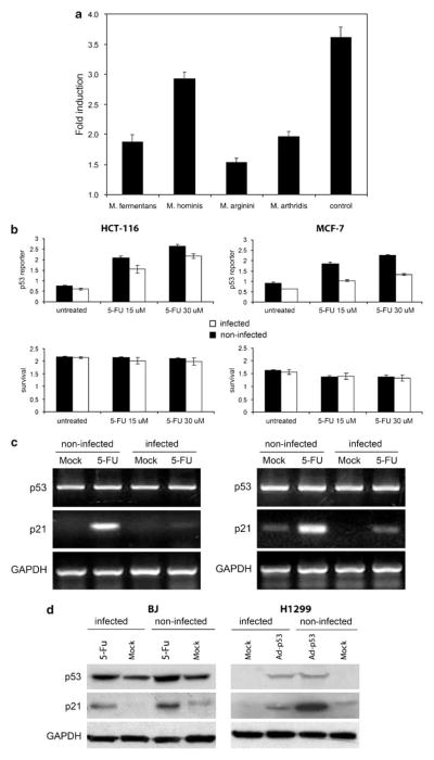
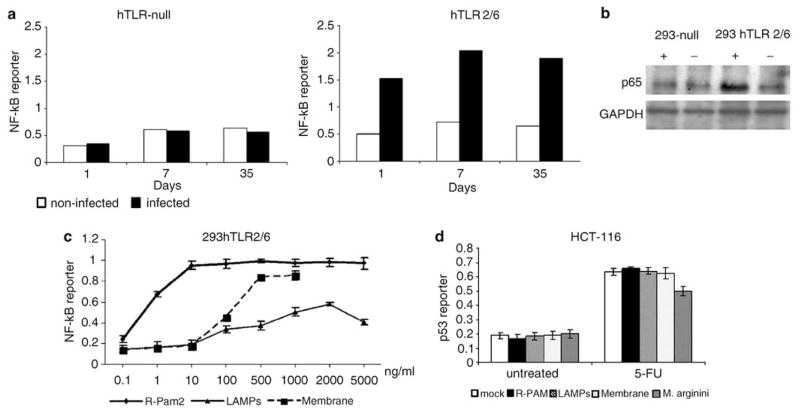
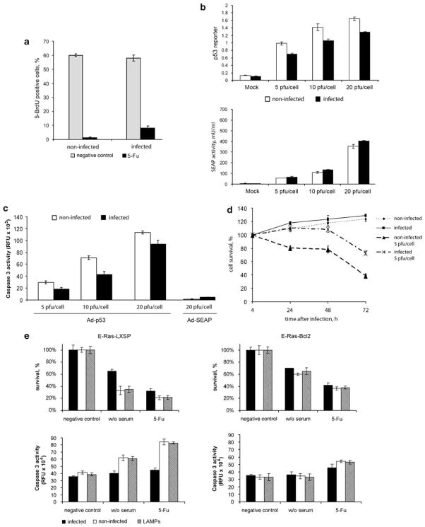
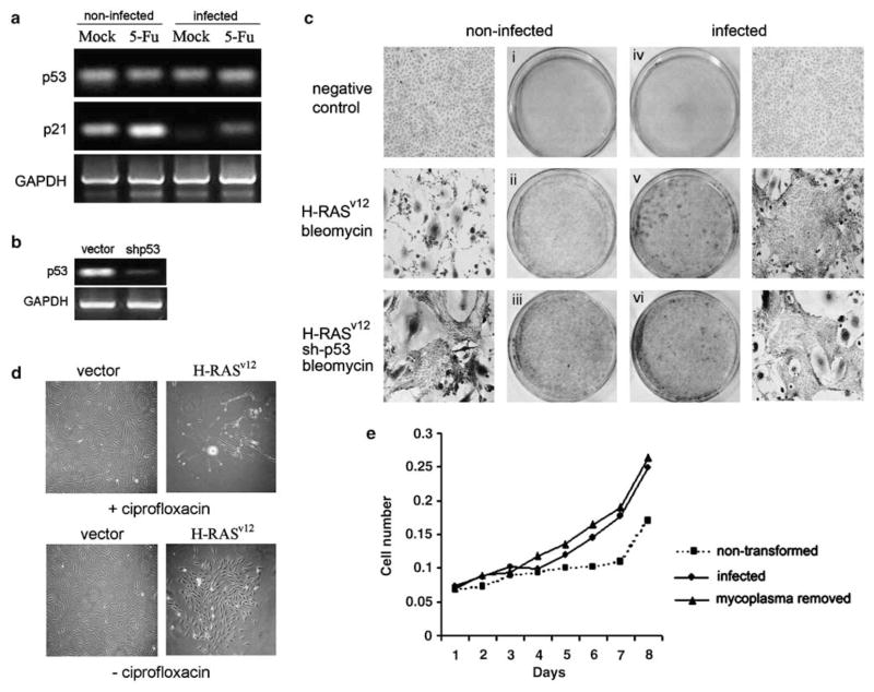
Prokaryotes of the genus Mycoplasma are the smallest cellular organisms that persist as obligate extracellular parasites. Although mycoplasma infection is known to be associated with chromosomal instability and can promote malignant transformation, the mechanisms underlying these phenomena remain unknown. Since persistence of many cellular parasites requires suppression of apoptosis in host cells, we tested the effect of mycoplasma infection on the activity of the p53 and nuclear factor (NF)-kappaB pathways, major mechanisms controlling programmed cell death. To monitor the activity of p53 and NF-kappaB in mycoplasma-infected cells, we used a panel of reporter cell lines expressing the bacterial beta-galactosidase gene under the control of p53- or NF-kappaB-responsive promoters. Cells incubated with media conditioned with different species of mycoplasma showed constitutive activation of NF-kappaB and reduced activation of p53, common characteristics of the majority of human tumor cells, with M. arginini having the strongest effect among the species tested. Moreover, mycoplasma infection reduced the expression level and inducibility of an endogenous p53-responsive gene, p21(waf1), and inhibited apoptosis induced by genotoxic stress. Infection with M. arginini made rat and mouse embryo fibroblasts susceptible to transformation with oncogenic H-Ras, whereas mycoplasma-free cells underwent irreversible p53-dependent growth arrest. Mycoplasma infection was as effective as shRNA-mediated knockdown of p53 expression in making rodent fibroblasts permissive to Ras-induced transformation. These observations indicate that mycoplasma infection plays the role of a p53-suppressing oncogene that cooperates with Ras in cell transformation and suggest that the carcinogenic and mutagenic effects of mycoplasma might be due to inhibition of p53 tumor suppressor function by this common human parasite.


Figures

Figure 1
Figure 1
Suppression of p53 transcriptional activity by mycoplasmas. (a) ConA-3T3 cells carrying a p53-dependent β-galactosidase (β-gal) reporter (ConALacZ) were treated overnight with 5% conditioned broths collected from cell-free cultures of the mycoplasma species indicated or with regular medium (‘control’). Doxorubicin was then added to a final concentration of 1 μg/ml and cells were cultured for an additional 24 h. The induction of β-gal activity in total cell lysates was determined by o-nitrophenyl β-D-galactopyranoside staining measured spectrophotometrically at 414 nm. The values plotted indicate the ratio of the β-gal activity in doxorubicin-treated cells infected with a given mycoplasma species to the β-gal activity in untreated cells infected with the same mycoplasma species. (b) p53-responsive reporter activity in control and M. arginini-infected HCT-116 and MCF-7 cells. HCT-116 and MCF-7 cells stably transfected with the ConALacZ reporter construct were plated into 96-well plates and incubated in medium containing the indicated concentration of 5-FU for 16 h. The cells were infected with M. arginini at least 2 weeks before the experiment. Presence of M. arginini in cell culture was confirmed by semiquantitative PCR vis-à-vis nuclear gene sequences. β-gal activity was measured by o-nitrophenyl β-D-galactopyranoside (ONPG) staining (upper panels) and cell survival was measured by methylene blue staining (bottom panels). Infected (M. arginini) cells (black bars), noninfected cells (white bars). (c) Reverse transcription (RT)–PCR analysis of p53 and p21 mRNA expression in control and M. arginini-infected HCT-116 and MCF-7 cells. Cells were left uninfected or were infected by growth in 5% M. arginini-conditioned medium overnight. To induce p53, cells were then treated with 5-FU (30 μM) for 24 h. Levels of p53 and p21 expression were measured by RT–PCR. Expression of glyceraldehyde-3-phosphate dehydrogenase (GAPDH) was used as a positive control to confirm the quality and amount of cDNA in each sample. HCT-116 cells (left panel), MCF-7 cells (right panel). (d) Effect of M. arginini infection on p53 and the p53-transcriptional target, p21. Lysates from control (noninfected) and M. arginini-infected BJ and H1299 cells were used for western blot analysis of p53 and p21 protein levels. GAPDH expression was used to control for protein concentration between lanes. To induce p53 activity, BJ cells were treated with 30 μM 5-FU for 16 h and H1299 cells were treated with 10 PFU (plaque-forming units) of Ad-p53 per cell.
Figure 2
Figure 2
Activation of nuclear factor (NF)-κB-dependent transcription in cells infected with M. arginini. (a) N F-κB-responsive reporter activity in control and M. arginini-infected 293hTLR-null cells (not expressing any Toll-like receptors (TLRs)) and 239hTLR2/TLR6 cells (expressing hTLR2 and TLR6 but not any other TLRs). 293hTLR-null and 239hTLR2/TLR6 cells bearing the NF-κB-LacZ reporter construct were infected with M. arginini. Next, cells were plated into 96-well plates and β-galactosidase (β-gal) activity was measured by o-nitrophenyl β-D-galactopyranoside (ONPG) staining 1, 7 and 35 days later. Infected cells (black bars), noninfected cells (white bars). (b) Influence of M. arginini infection on the nuclear localization of the p65 subunit of NF-κB. 293hTLR-null and 239hTLR2/TLR6 cells infected (+) or noninfected (−) with mycoplasma were collected 40 days post-infection and lysed for extraction of nuclear proteins. p65 and glyceraldehyde-3-phosphate dehydrogenase (GAPDH) (control for equal protein loading) were detected by western blotting. (c) Effect of different mycoplasma components on NF-κB activity. N F-κB-responsive reporter activity in 293 cells expressing hTLR2/hTLR6 treated with different concentrations of mycoplasmal membranes, lipid-associated mycoplasmal proteins (LAMPs) and synthetic analogs of mycoplasmal MALP-2 lipopeptides (R-PAM2). (d) Induction of p53-responsive reporter activity in HCT-116 ConALacZ cells left untreated (‘mock’), treated with mycoplasmal membranes (‘membrane,’ 1μg/ml), lipid-associated membrane proteins (‘LAMPs,’ 2 μg/ml) or synthetic analogs of mycoplasmal MALP-2 lipopeptides (‘R-PAM,’ 0.1 μg/ml) or infected with M. arginini. p53 reporter activity was determined in cells grown in 15 μM 5-FU for 24 h and in cells grown without 5-FU (‘negative control’).
Figure 3
Figure 3
M. arginini infection inhibits p53-mediated checkpoint control and apoptosis. (a) Effect of M. arginini infection on the ability of REF52 cells synchronized at G0/G1 by serum starvation (72 h) to enter S phase (incorporate 5-bromodeoxyuridine (BrdU) into DNA) following serum stimulation in the presence or absence of DNA-damaging treatment (30 μM 5-FU). After 7h of cell incubation with medium containing 10% fetal bovine serum (FBS) with 5-BrdU (‘negative control’, light gray bars) or 5-BrdU+5-FU (‘5-FU’, black bars), incorporation of 5-BrDU was detected by immunofluorescent staining with anti-BrdU antibodies. For each treatment, no fewer than 500 cells were analysed. (b) Infection of cells with M. arginini reduces the activity of ectopically expressed p53. Mycoplasma-infected (black bars) or -noninfected (white bars) p53-null H1299 cells containing the p53-responsive lacZ reporter construct were plated in 96-well plates and superinfected with Ad-p53 virus (top panel) or a control Ad-SEAP virus (bottom panel) at 5, 10 or 20 PFU per cell. p53 reporter transactivation and SEAP activities were measured 24 h after adenovirus infection. (c) Caspase 3 activity was measured in lysates of cells 24 h after the infection as described for (b) using a fluorogenic substrate. (d) M. arginini infection reduces the growth inhibitory effect of ectopically expressed p53 protein in H1299 cells. Mycoplasma-infected or -noninfected H1299 cells were plated in 96-well plates and superinfected with Ad-p53 virus at 5 PFU per cell. Cell survival was measured by methylene blue staining at the indicated times. (e) Effect of M. arginini infection on apoptosis in E-Ras cells triggered by serum deprivation or DNA damage (5-FU). E-Ras-LXSP and E-Ras-Bcl2 cells were plated in 96-well plates and incubated in serum-free medium for 16 h (w/o serum) or medium containing 5-FU (30 μM) for 24h. Cell survival was measured by methylene blue staining (upper panels). Activity of caspase-3 was measured using a specific fluorigenic substrate (bottom panels). M. arginini-infected cells (black bars), noninfected cells (white bars), lipid-associated mycoplasmal protein (LAMP)-treated (2 μg/ml of LAMP was added to cells 4 h before 5-FU for 28 h) cells (gray bars).
Figure 4
Figure 4
Decreased activity of p53 in mycoplasma-infected cells is accompanied by susceptibility to Ras-mediated transformation. (a) Reverse transcription (RT)–PCR analysis of p53 and p21 mRNA expression in REF52 cell-infected and -noninfected with M. arginini. For induction of p53, cells were treated with 5-FU (30 μM) for 16 h. Levels of p53 and p21 expression were measured by RT–PCR analysis. Expression of glyceraldehyde-3-phosphate dehydrogenase (GAPDH) was used as a positive control to confirm the quality and amount of cDNA in each sample. (b) Inhibition of p53 mRNA expression in REF52 cells transduced with a lentivirus expressing an inhibitory shRNA against p53. Levels of p53 were measured by RT–PCR analysis 72 h post-transduction. Expression of GAPDH was used as a positive control to confirm the quality and amount of cDNA in each sample. (c) Photomicrographs and photographs of methylene blue-stained REF52 cells. Noninfected (panels i, ii and iii) and M. arginini-infected (panels iv, v and vi) REF52 cells were transduced with an insert-free lentiviral vector (‘negative control,’ panels i and iv), the LV-Ras-Bleo lentivirus bearing the activated Ha-Rasv12oncogene (panels ii and v) or a combination of LV-Ras-Bleo and a lentivirus expressing an shRNA targeting p53 (‘shRNA-p53,’ panels iii and vi). Transduced cells (except for ‘negative control’ cells) were grown in the presence of bleomycin for 21 days to select Ras-expressing cells. Viable cells were stained with methylene blue and randomly selected areas on the plates were photographed. (d) Elimination of mycoplasma prevents Ras-mediated transformation of cells. Photomicrographs of monolayers of REF52 cells. Two sets of plates with REF52 cells chronically infected with M. arginini were transduced with either LV-Ras-Bleo expressing H-RASv12 or LV-Bleo (‘vector’). One set was treated with ciprofloxacin to remove mycoplasma (effect of antibiotic was confirmed by RT–PCR for mycoplasma DNA (data not shown)) and both sets were incubated 21 days with bleomycin-containing medium to selectively monitor lentivirus-transduced cells. On day 21, plates were fixed with methanol and stained with methylene blue. (e) Elimination of mycoplasma from REF52 cells that underwent Ras-mediated transformation by treatment with ciprofloxacin does not reverse their transformed phenotype. Nontransformed, transformed (by transduction of H-Rasv12 in the presence of M. arginini) or transformed cells treated with ciprofloxacin (transformed, mycoplasma removed) were plated in 96-well plates (5 × 10² per well). Cell numbers were determined by methylene blue assay at the indicated time points after plating.

Similar articles

Cited by

References

    1. Baek KH, Shin HJ, Yoo JK, Cho JH, Choi YH, Sung YC, et al. p53 deficiency and defective mitotic checkpoint in proliferating T lymphocytes increase chromosomal instability through aberrant exit from mitotic arrest. J Leukoc Biol. 2003;73:850–861. - PubMed
    1. Boiko AD, Porteous S, Razorenova OV, Krivokrysenko VI, Williams BR, Gudkov AV, et al. A systematic search for downstream mediators of tumor suppressor function of p53 reveals a major role of BTG2 in suppression of Ras-induced transformation. Genes Dev. 2006;20:236–252. - PMC - PubMed
    1. Bubici C, Papa S, Pham CG, Zazzeroni F, Franzoso G. The NF-kappaB-mediated control of ROS and JNK signaling. Histol Histopathol. 2006;21:69–80. - PubMed
    1. Burgert HG, Ruzsics Z, Obermeier S, Hilgendorf A, Windheim M, Elsing A. Subversion of host defense mechanisms by adenoviruses. Curr Top Microbiol Immunol. 2002;269:273–318. - PubMed
    1. Chu HW, Jeyaseelan S, Rino JG, Voelker DR, Wexler RB, Campbell K, et al. TLR2 signaling is critical for Mycoplasma pneumoniae-induced airway mucin expression. J Immunol. 2005;9:5713–5719. - PubMed

Publication types

MeSH terms