. 2008 Jun;9(2):178-90.
doi: 10.1007/s10162-008-0118-y. Epub 2008 Apr 12.

Using the zebrafish lateral line to screen for ototoxicity

Affiliations

Using the zebrafish lateral line to screen for ototoxicity

Lynn L Chiu et al. J Assoc Res Otolaryngol. 2008 Jun.

Abstract

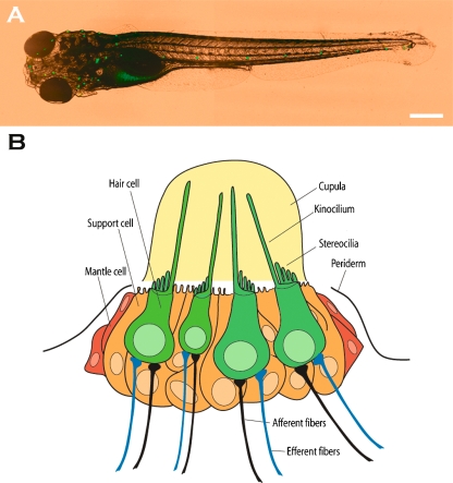
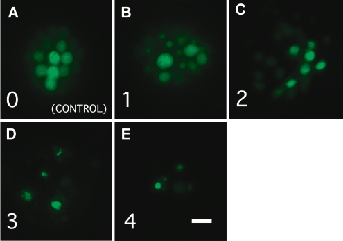
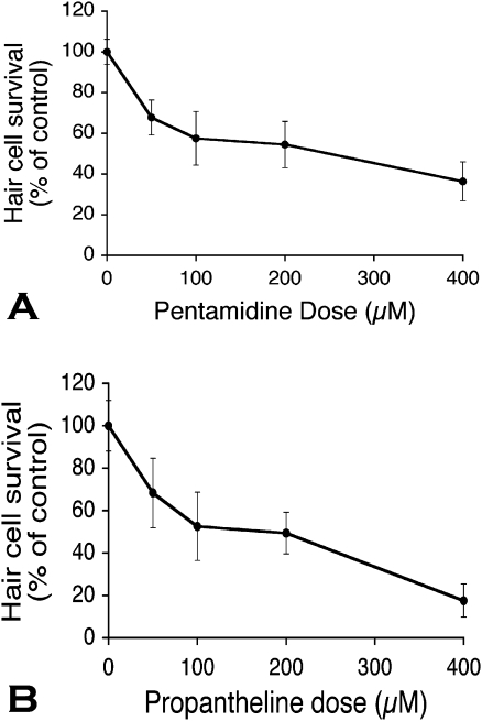
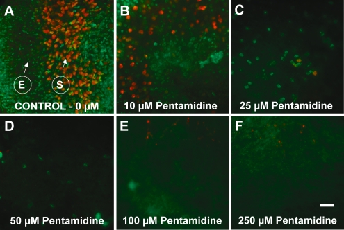
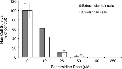
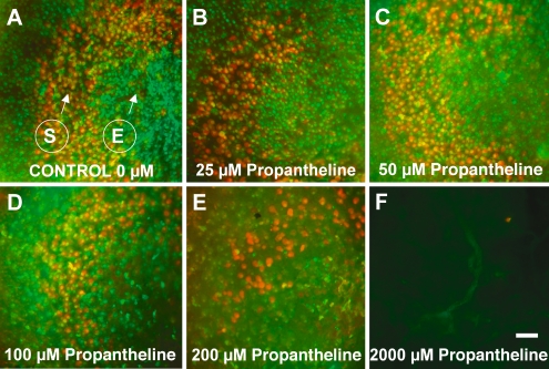
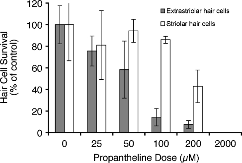
The zebrafish is a valuable model for studying hair cell development, structure, genetics, and behavior. Zebrafish and other aquatic vertebrates have hair cells on their body surface organized into a sensory system called the lateral line. These hair cells are highly accessible and easily visualized using fluorescent dyes. Morphological and functional similarities to mammalian hair cells of the inner ear make the zebrafish a powerful preparation for studying hair cell toxicity. The ototoxic potential of drugs has historically been uncovered by anecdotal reports that have led to more formal investigation. Currently, no standard screen for ototoxicity exists in drug development. Thus, for the vast majority of Food and Drug Association (FDA)-approved drugs, the ototoxic potential remains unknown. In this study, we used 5-day-old zebrafish larvae to screen a library of 1,040 FDA-approved drugs and bioactives (NINDS Custom Collection II) for ototoxic effects in hair cells of the lateral line. Hair cell nuclei were selectively labeled using a fluorescent vital dye. For the initial screen, fish were exposed to drugs from the library at a 100-muM concentration for 1 h in 96-well tissue culture plates. Hair cell viability was assessed in vivo using fluorescence microscopy. One thousand forty drugs were rapidly screened for ototoxic effects. Seven known ototoxic drugs included in the library, including neomycin and cisplatin, were positively identified using these methods, as proof of concept. Fourteen compounds without previously known ototoxicity were discovered to be selectively toxic to hair cells. Dose-response curves for all 21 ototoxic compounds were determined by quantifying hair cell survival as a function of drug concentration. Dose-response relationships in the mammalian inner ear for two of the compounds without known ototoxicity, pentamidine isethionate and propantheline bromide, were then examined using in vitro preparations of the adult mouse utricle. Significant dose-dependent hair cell loss in the mouse utricle was demonstrated for both compounds. This study represents an important step in validating the use of the zebrafish lateral line as a screening tool for the identification of potentially ototoxic drugs.


Figures

FIG. 1.
FIG. 1.
A Live preparation of fluorescently labeled zebrafish larva 5 dpf (Harris et al. 2003). Neuromasts of the lateral line are stained with YO-PRO1. Scale bar = 0.5 mm. B Schematic illustration of a neuromast. Hair cells are depicted in green with long kinocilia and shorter stereocilia projecting from the apical end of the cells and afferent and efferent nerve fibers at the basal end. Support cells (orange cells) intercalate between the hair cells.
FIG. 2.
FIG. 2.
AE Examples of hair cell damage along with the grading system used for the initial screen. Each panel shows a single neuromast stained with YO-PRO1. Neuromasts range from undamaged control (A, grade 0), to subtle disorganization of hair cells and nuclear condensation (B, grade 1), to near complete hair cell loss (E, grade 4). Scale bar in E = 10 μm and applies to all five panels.
FIG. 3.
FIG. 3.
Dose–response relationships of pentamidine and propantheline-exposed zebrafish. Zebrafish larvae at 5 dpf were exposed to various concentrations of pentamidine isethionate (A) or propantheline bromide (B) and were evaluated 1 h after exposure for surviving hair cells in neuromasts SO1, SO2, O1, and OC1 (n = 10–15 larvae per condition). Data points represent the percentage of hair cell survival compared to the control (no drug) condition (±1 SD). Hair cell survival decreased with increasing concentrations of pentamidine (p < 0.0001, one-way ANOVA) and propantheline (p < 0.0001, one-way ANOVA).
FIG. 4.
FIG. 4.
Cultured mouse utricles treated with pentamidine and labeled for calmodulin and calbindin. Utricles were cultured for 24 h without pentamidine (control; A), or with pentamidine at 10 (B), 25 (C), 50 (D), 100 (E), and 250 μM (F). Following culture, utricles were fixed and double-labeled with antibodies directed against calmodulin (green) that label all hair cells of the utricle and calbindin (red) that label hair cells of the striolar region only. S indicates striolar hair cells, while E indicates extrastriolar hair cells. Treatment with 10 μM pentamidine resulted in a significant reduction in hair cell density as well as a swollen, damaged appearance of the hair cells. Treatment with increasing doses results in progressive injury up to 100- to 250-μM levels when all hair cells are absent. Scale bar in F = 30 μm and applies to all panels.
FIG. 5.
FIG. 5.
Dose–response relationship of utricles treated with pentamidine. The density of hair cells of each utricle were quantified and averaged from four striolar and four extrastriolar representative regions of 900 μm2 each. Bars represent the mean hair cell survival (% control) ± 1 SD. Hair cell survival decreases significantly as pentamidine doses increases (p < 0.001, one-way ANOVA). Both striolar and extrastriolar regions are sensitive to pentamidine-induced injury (N = 6–8 per condition).
FIG. 6.
FIG. 6.
Cultured mouse utricles treated with propantheline and labeled for calmodulin and calbindin. Utricles were cultured for 24 h without propantheline (control; A) or with propantheline at 25 (B), 50 (C), 100 (D), 200 (E), and 2,000 μM (F). S indicates striolar hair cells, while E indicates extrastriolar hair cells. Treatment with increasing doses of propantheline resulted in progressively higher degrees of hair cell loss. Scale bar in F = 30 μm and applies to all panels.
FIG. 7.
FIG. 7.
Dose–response relationship of utricles treated with propantheline. Hair cells of each utricle were quantified and averaged from four striolar and four extrastriolar representative regions of 900 μm2 each. Bars represent the mean hair cell survival (% control) ± 1 SD (N = 3–8 per condition). Both extrastriolar and striolar hair cell survival decreased in a dose-dependent manner (p < 0.0001, one-way ANOVA). The striolar region exhibited less hair cell loss than the extrastriolar region.

Similar articles

Cited by

References

    1. {'text': '', 'ref_index': 1, 'ids': [{'type': 'PubMed', 'value': '16135282', 'is_inner': True, 'url': 'https://pubmed.ncbi.nlm.nih.gov/16135282/'}]}
    2. Bellamy R. Pneumocystis pneumonia in people with HIV. Clin. Evid. 13:854–868, 2006. - PubMed
    1. {'text': '', 'ref_index': 1, 'ids': [{'type': 'DOI', 'value': '10.1054/mehy.2001.1535', 'is_inner': False, 'url': 'https://doi.org/10.1054/mehy.2001.1535'}, {'type': 'PubMed', 'value': '12056877', 'is_inner': True, 'url': 'https://pubmed.ncbi.nlm.nih.gov/12056877/'}]}
    2. Borghi C, Modugno G, Pirodda A. Possible role of HMG-CoA reductase inhibitors for the treatment of sudden sensorineural hearing loss (SSHL). Med. Hypotheses 58:399–402, 2002. - PubMed
    1. {'text': '', 'ref_index': 1, 'ids': [{'type': 'PubMed', 'value': '11806628', 'is_inner': True, 'url': 'https://pubmed.ncbi.nlm.nih.gov/11806628/'}]}
    2. Calza L, Marinacci G, Manfredi R, Colangeli V, Fortunat L, Chiodo F. Pentamidine isethionate as treatment and secondary prophylaxis for disseminated cutaneous leishmaniasis during HIV infection: case report. J. Chemother. 13:653–657, 2001. - PubMed
    1. {'text': '', 'ref_index': 1, 'ids': [{'type': 'DOI', 'value': '10.1016/j.brainres.2006.01.128', 'is_inner': False, 'url': 'https://doi.org/10.1016/j.brainres.2006.01.128'}, {'type': 'PMC', 'value': 'PMC2668867', 'is_inner': False, 'url': 'https://pmc.ncbi.nlm.nih.gov/articles/PMC2668867/'}, {'type': 'PubMed', 'value': '16569399', 'is_inner': True, 'url': 'https://pubmed.ncbi.nlm.nih.gov/16569399/'}]}
    2. Cunningham L. The adult mouse utricle as an in vitro preparation for studies of ototoxic-drug-induced sensory hair cell death. Brain Res. 1091:277–281, 2006. - PMC - PubMed
    1. {'text': '', 'ref_index': 1, 'ids': [{'type': 'PMC', 'value': 'PMC6757801', 'is_inner': False, 'url': 'https://pmc.ncbi.nlm.nih.gov/articles/PMC6757801/'}, {'type': 'PubMed', 'value': '12351727', 'is_inner': True, 'url': 'https://pubmed.ncbi.nlm.nih.gov/12351727/'}]}
    2. Cunningham L, Cheng A, Rubel E. Caspase activation in hair cells of the mouse utricle exposed to neomycin. J. Neurosci. 22:8532–8540, 2002. - PMC - PubMed

Publication types

MeSH terms

LinkOut - more resources