. 2008 May 6;47(18):5111-26.
doi: 10.1021/bi702537s. Epub 2008 Apr 15.

Structure of Ca2+-bound S100A4 and its interaction with peptides derived from nonmuscle myosin-IIA

Affiliations

Structure of Ca2+-bound S100A4 and its interaction with peptides derived from nonmuscle myosin-IIA

Vladimir N Malashkevich et al. Biochemistry. .

Abstract

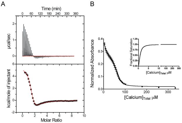
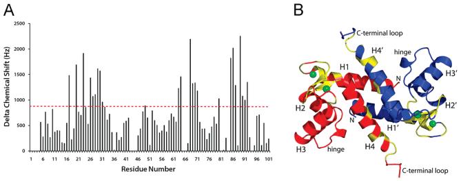
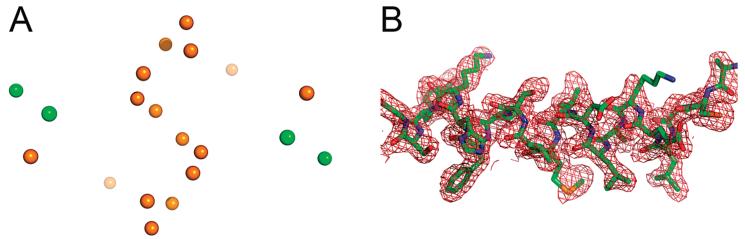
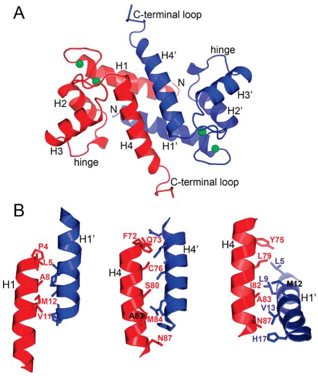
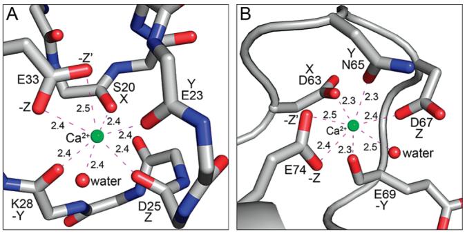
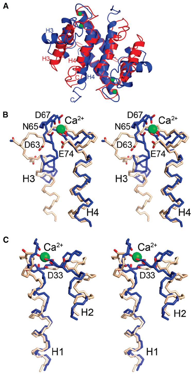
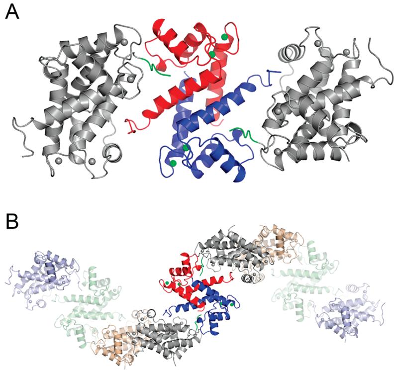
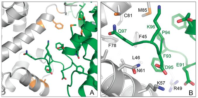
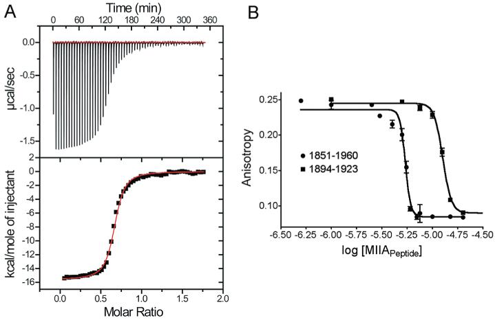
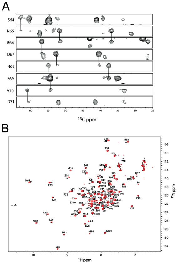
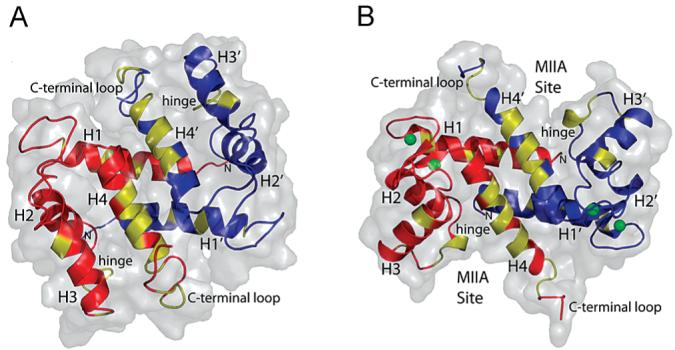
S100A4, also known as mts1, is a member of the S100 family of Ca2+-binding proteins that is directly involved in tumor invasion and metastasis via interactions with specific protein targets, including nonmuscle myosin-IIA (MIIA). Human S100A4 binds two Ca2+ ions with the typical EF-hand exhibiting an affinity that is nearly 1 order of magnitude tighter than that of the pseudo-EF-hand. To examine how Ca2+ modifies the overall organization and structure of the protein, we determined the 1.7 A crystal structure of the human Ca2+-S100A4. Ca2+ binding induces a large reorientation of helix 3 in the typical EF-hand. This reorganization exposes a hydrophobic cleft that is comprised of residues from the hinge region,helix 3, and helix 4, which afford specific target recognition and binding. The Ca2+-dependent conformational change is required for S100A4 to bind peptide sequences derived from the C-terminal portion of the MIIA rod with submicromolar affinity. In addition, the level of binding of Ca2+ to both EF-hands increases by 1 order of magnitude in the presence of MIIA. NMR spectroscopy studies demonstrate that following titration with a MIIA peptide, the largest chemical shift perturbations and exchange broadening effects occur for residues in the hydrophobic pocket of Ca2+-S100A4. Most of these residues are not exposed in apo-S100A4 and explain the Ca2+ dependence of formation of theS100A4-MIIA complex. These studies provide the foundation for understanding S100A4 target recognition and may support the development of reagents that interfere with S100A4 function.


Figures

Figure 1
Figure 1
Thermodynamic data showing Ca2+ binding to S100A4. (A) Isothermal titration calorimetry (ITC) data demonstrating the interaction of S100A4 with Ca2+ (n = 2). (B) Competition assay with the chelator 5,5′Br2-BAPTA to examine the affinity of S100A4 for Ca2+ in the presence of MIIA1851–1960. The decrease in absorbance was monitored at 263 nm for a mixture containing 25 μM 5,5′Br2-BAPTA, 12.5 μM S100A4 dimer, and 125 μM MIIA1851–1960. The inset shows the saturation curve representation for the best fit in Caligator.
Figure 2
Figure 2
Chemical shift perturbations following the addition of Ca2+ to apo-S100A4. (A) Bar graph of the cumulative 1H, 15N, and 13C chemical shift perturbations observed per residue upon the addition of Ca2+ to apo-S100A4. (B) Ribbon diagram of Ca2+-S100A4 showing the subunits in red and blue. Residues highlighted in yellow exhibited the largest perturbations (as per the dashed red line at 900 Hz in panel A).
Figure 3
Figure 3
(A) Heavy atom positions detected by SHELXD based on the SAD signal at 1.7 Å wavelength. Green and orange spheres denote Ca2+ ions and sulfur atoms, respectively. (B) Refined model of S100A4 (residues Ala2-Gly21) superimposed onto the 2FoFc electron density map, contoured at 1σ.
Figure 4
Figure 4
Molecular architecture of the Ca2+-S100A4 dimer. (A) Ribbon diagram of the dimer, where the two subunits are colored blue and red. The key structural elements common for the S100 family are indicated. Green spheres denote Ca2+ ions. (B) Details of the dimer interface of S100A4 involving helix 1–1′, 4–4′, and 1–4′ (1′–4) contact areas.
Figure 5
Figure 5
Structures of the Ca2+-loaded S100A4 EF-hands. (A) N-Terminal pseudo-EF-hand. The Ca2+ ion is coordinated by the main chain carbonyl oxygens of Ser20, Glu23, Asp25, and Lys28; the side chain carboxylate of Glu33 (both oxygens), and a water molecule. (B) C-Terminal typical EF-hand. The Ca2+ ion is coordinated by the side chains of Asp63, Asn65, Asp67, and Glu74 (both oxygens); the carbonyl oxygen of Glu69; and a water molecule. Green and red spheres denote Ca2+ and water atoms, respectively.
Figure 6
Figure 6
Conformational rearrangements in S100A4 caused by Ca2+ binding. (A) Ribbon diagrams comparing the three-dimensional structures of apo-S100A4 (red) and Ca2+-S100A4 (blue). Green spheres denote Ca2+ ions. Stereoviews of the (B) pseudo-EF-hand and (C) typical EF-hand of apo-S100A4 (beige) and Ca2+-S100A4 (blue). The Ca2+-coordinating oxygen atoms are colored red in both the apo and Ca2+-bound states.
Figure 7
Figure 7
Self-association of S100A4 molecules in the crystal lattice. (A) The S100A4 dimer is shown with one red and one blue subunit. The symmetry-related molecules (gray) in the S100A4 crystals are positioned such that their C-terminal tails (green) bind to the hydrophobic cleft of the central molecule. (B) The interacting S100A4 dimers form infinite superhelical structures in the crystal.
Figure 8
Figure 8
Interaction of the C-terminal tail of S100A4 (green) with the ligand binding site of the symmetry-related S100A4 molecule (gray). (A) Overall view of the ligand binding pocket. Residues showing chemical shift perturbations upon binding a peptide derived from the C-terminus of S100A4 (residues Glu88–Lys101) are colored orange. (B) Zoomed in view. Many of the interactions are nonpolar; however, Glu91 and Asp95 of the peptide may form ionic interactions with Arg49, Asn61, and Lys57 from the symmetry-related molecule.
Figure 9
Figure 9
Chemical shift perturbations of 1H–15N correlations in a 2D HSQC spectrum of Ca2+-S100A4 upon the addition of a peptide derived from the C-terminus of S100A4. (A) HSQC spectra of Ca2+-S100A4 in the absence (black contours) and presence of 0.6 mM (red contours) and 3.6 mM (blue contours) C-terminal S100A4 peptide. The inset shows an expanded region of the HSQC spectrum illustrating the perturbations to the 1H–15N correlation for F55. (B) Chemical shift perturbations for residues in Ca2+-S100A4 upon addition of 3.6 mM C-terminal S100A4 peptide.
Figure 10
Figure 10
Sedimentation equilibrium measurements taken at 25 °C. The solid lines represent the best fit from a global nonlinear least-squares analysis of data obtained at 15000 and 22000 rpm: (□) 38 μM wild-type S100A4 and CaCl2 (subunit concentration), (○) 54 μM wild-type S100A4 and EDTA/EGTA (subunit concentration), and (◇) 61 μM Δ13C S100A4 and CaCl2 (subunit concentration).
Figure 11
Figure 11
Thermodynamic data showing MIIA peptide binding to S100A4. (A) Isothermal titration calorimetry (ITC) of MIIA1893–1923 peptide (low salt; n = 1). (B) Competition fluorescence anisotropy of MIIA1893–1923 and MIIA1851–1960 at a physiological salt concentration.
Figure 12
Figure 12
Nuclear magnetic resonance (NMR) data together with the resonance assignments for Ca2+-S100A4 in the presence and absence of the MIIA1908–1923 peptide. (A) Strips from 15N planes of a three-dimensional HNCACB data set for 13C- and 15N-labeled S100A4 bound to the MIIA1908–1923 peptide in the presence of Ca2+, which illustrates the quality of the data used for making the resonance assignments. (B) Overlay of 1H–15N HSQC data for Ca2+-S100A4 in the MIIA1908–1923 peptide-bound (red contours) and free (black contours) states.
Figure 13
Figure 13
Ribbon and surface diagrams of S100A4 in the (A) apo and (B) Ca2+-bound states. Residues colored yellow show significant chemical shift perturbations and exchange broadening effects upon binding of the MIIA1908–1923 peptide.

References

    1. Donato R. S100: A multigenic family of calcium-modulated proteins of the EF-hand type with intracellular and extracellular functional roles. Int. J. Biochem. Cell Biol. 2001;33:637–668. - PubMed
    1. Marenholz I, Heizmann CW, Fritz G. S100 proteins in mouse and man: From evolution to function and pathology (including an update of the nomenclature) Biochem. Biophys. Res. Commun. 2004;322:1111–1122. - PubMed
    1. Zimmer DB, Cornwall EH, Landar A, Song W. The S100 protein family: History, function, and expression. Brain Res. Bull. 1995;37:417–429. - PubMed
    1. Santamaria-Kisiel L, Rintala-Dempsey AC, Shaw GS. Calcium-dependent and -independent interactions of the S100 protein family. Biochem. J. 2006;396:201–214. - PMC - PubMed
    1. Grigorian MS, Tulchinsky EM, Zain S, Ebralidze AK, Kramerov DA, Kriajevska MV, Georgiev GP, Lukanidin EM. The mts1 gene and control of tumor metastasis. Gene. 1993;135:229–238. - PubMed

Publication types

MeSH terms