. 2008 May 13;47(19):5368-77.
doi: 10.1021/bi800228w. Epub 2008 Apr 15.

The binding and release of oxygen and hydrogen peroxide are directed by a hydrophobic tunnel in cholesterol oxidase

Affiliations

The binding and release of oxygen and hydrogen peroxide are directed by a hydrophobic tunnel in cholesterol oxidase

Lin Chen et al. Biochemistry. .

Abstract

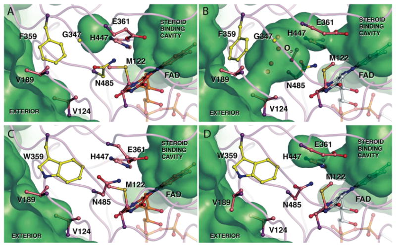
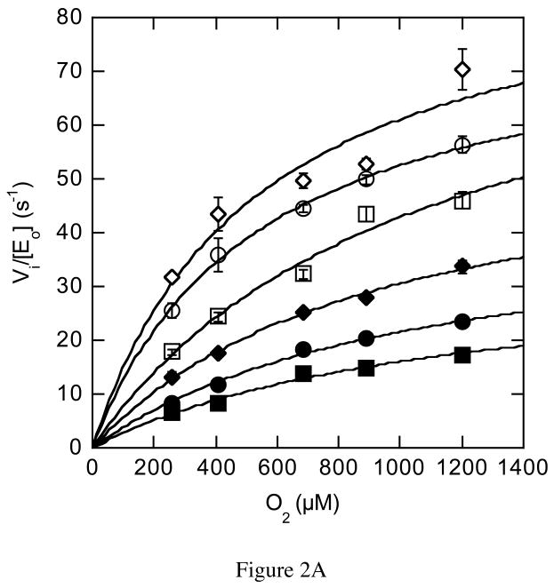
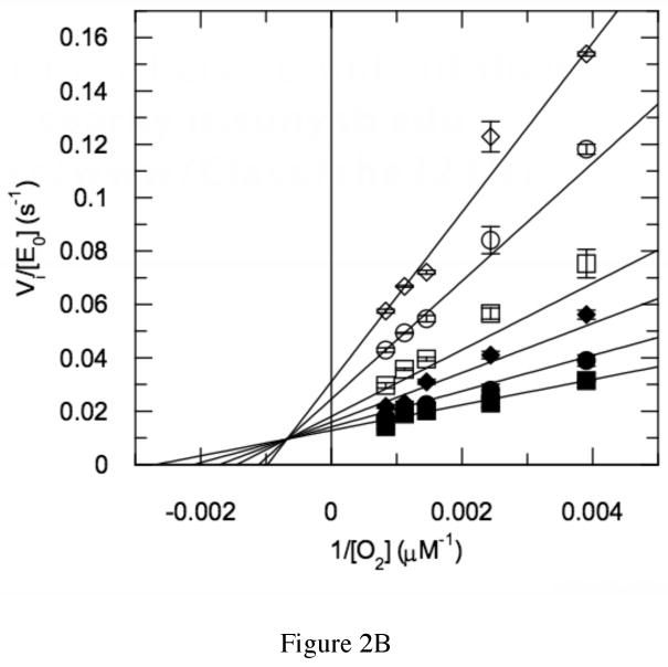
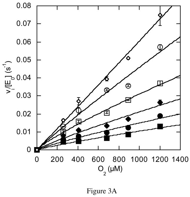
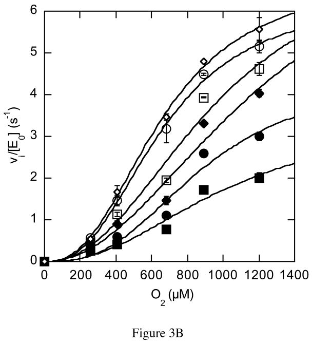
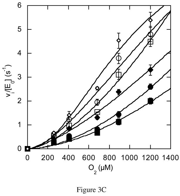
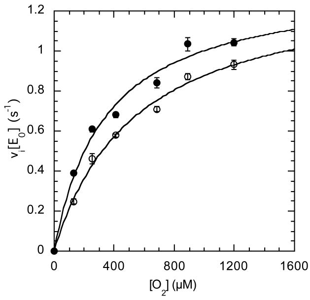
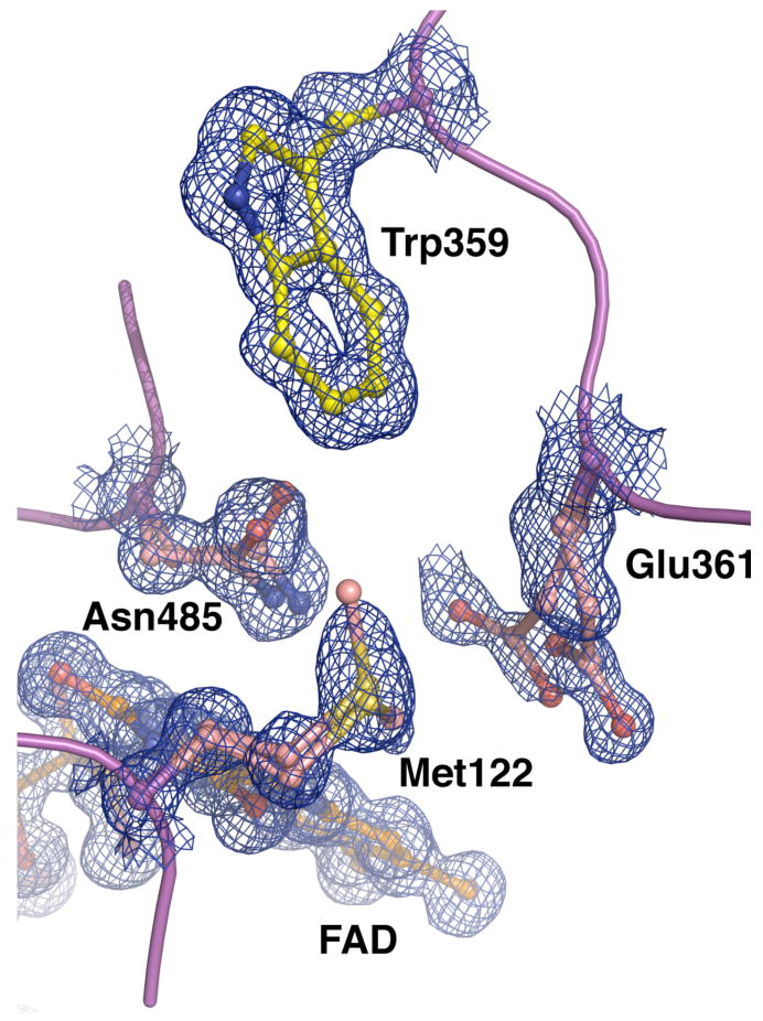
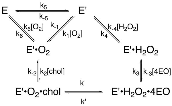
The usage by enzymes of specific binding pathways for gaseous substrates or products is debated. The crystal structure of the redox enzyme cholesterol oxidase, determined at sub-angstrom resolution, revealed a hydrophobic tunnel that may serve as a binding pathway for oxygen and hydrogen peroxide. This tunnel is formed by a cascade of conformational rearrangements and connects the active site with the exterior surface of the protein. To elucidate the relationship between this tunnel and gas binding and release, three mutant enzymes were constructed to block the tunnel or its putative gate. Mutation of the proposed gating residue Asn485 to Asp or tunnel residue Phe359 or Gly347 to Trp or Asn reduces the catalytic efficiency of oxidation. The K mO 2 increases from 300 +/- 35 microM for the wild-type enzyme to 617 +/- 15 microM for the F359W mutant. The k cat for the F359W mutant-catalyzed reaction decreases 13-fold relative to that of the wild-type-catalyzed reaction. The N485D and G347N mutants could not be saturated with oxygen. Transfer of hydride from the sterol to the flavin prosthetic group is no longer rate-limiting for these tunnel mutants. The steady-state kinetics of both wild-type and tunnel mutant enzymes are consistent with formation of a ternary complex of steroid and oxygen during catalysis. Furthermore, kinetic cooperativity with respect to molecular oxygen is observed with the tunnel mutants, but not with the wild-type enzyme. A rate-limiting conformational change for binding and release of oxygen and hydrogen peroxide, respectively, is consistent with the cooperative kinetics. In the atomic-resolution structure of F359W, the indole ring of the tryptophan completely fills the tunnel and is observed in only a single conformation. The size of the indole is proposed to limit conformational rearrangement of residue 359 that leads to tunnel opening in the wild-type enzyme. Overall, these results substantiate the functional importance of the tunnel for substrate binding and product release.


Figures

Figure 1
Figure 1
Atomic resolution structure of (A, B) wild-type (1MXT) and (C, D) F359W (####) cholesterol oxidases with key residues in the proposed oxygen tunnel shown in their closed (A, C) and open (B, D) conformations. The solvent accessible surface calculated using the probe radius of 1.4 Å is shown in green. The hydrophobic tunnel between the flavin and the exterior is present when the wild-type tunnel residues are in their open conformation (B) but blocked in the wild-type closed conformation (A). The open conformation includes a dioxygen species which was modeled in the tunnel with a fixed occupancy of 0.25 (37). However, the density may also represent two alternate water conformations. Inclusion in the model of either a dioxygen species or two alternate water molecules requires the adjacent active site and tunnel residues to be present in conformer B. Tryptophan 359 obstructs the tunnel in both conformations (C, D). However, the other tunnel residues, e.g., Glu361, Met122 or Val124, can adopt the closed (C) or open (D) conformation. Images were created using Pymol (41).
Figure 2
Figure 2
Steady-state kinetic profile of wild-type cholesterol oxidase. Initial velocities for wild-type cholesterol oxidase were measured over a range of O2 concentrations with varied cholesterol concentrations. The data were globally fit to equation (3) for a sequential ternary mechanism or they were fit to equation (4) for a cooperative mechanism at each individual cholesterol concentration. For the fit to equation (4), the Hill coefficient, h, was 1.0 at all cholesterol concentrations and the fit shown is for equation (3). The data shown are the average of two independent experiments, and the errors are the standard deviation of measurement. (A) Michaelis-Menten plots for νi/[Eo] versus [O2] at varied cholesterol concentrations: ◇, 30 μM; ○, 15 μM; □, 9 μM; ◆, 6 μM; ●, 3 μM; and ■, 2 μM cholesterol. (B) Double-reciprocal plot of the data in panel (A). The intersecting line pattern is consistent with the formation of a ternary complex between the enzyme, oxygen and steroid.
Figure 2
Figure 2
Steady-state kinetic profile of wild-type cholesterol oxidase. Initial velocities for wild-type cholesterol oxidase were measured over a range of O2 concentrations with varied cholesterol concentrations. The data were globally fit to equation (3) for a sequential ternary mechanism or they were fit to equation (4) for a cooperative mechanism at each individual cholesterol concentration. For the fit to equation (4), the Hill coefficient, h, was 1.0 at all cholesterol concentrations and the fit shown is for equation (3). The data shown are the average of two independent experiments, and the errors are the standard deviation of measurement. (A) Michaelis-Menten plots for νi/[Eo] versus [O2] at varied cholesterol concentrations: ◇, 30 μM; ○, 15 μM; □, 9 μM; ◆, 6 μM; ●, 3 μM; and ■, 2 μM cholesterol. (B) Double-reciprocal plot of the data in panel (A). The intersecting line pattern is consistent with the formation of a ternary complex between the enzyme, oxygen and steroid.
Figure 3
Figure 3
Steady-state kinetic profiles of mutant cholesterol oxidases. Initial velocities for mutant cholesterol oxidases were measured over a range of O2 concentrations with varied cholesterol concentrations and the data fit to equation (4) for a cooperative mechanism at each individual cholesterol concentration: ◇, 30 μM; ○, 15 μM; □, 9 μM; ◆, 6 μM; ●, 3 μM; and ■, 2 μM cholesterol. (A) N485D; (B) F359W; (C) G347N. The data shown are the average of two independent experiments, and the errors are the standard deviation of measurement.
Figure 3
Figure 3
Steady-state kinetic profiles of mutant cholesterol oxidases. Initial velocities for mutant cholesterol oxidases were measured over a range of O2 concentrations with varied cholesterol concentrations and the data fit to equation (4) for a cooperative mechanism at each individual cholesterol concentration: ◇, 30 μM; ○, 15 μM; □, 9 μM; ◆, 6 μM; ●, 3 μM; and ■, 2 μM cholesterol. (A) N485D; (B) F359W; (C) G347N. The data shown are the average of two independent experiments, and the errors are the standard deviation of measurement.
Figure 4
Figure 4
Initial velocities for wild-type and F359W cholesterol oxidases were measured over a range of O2 concentrations with a fixed dehydroepiandrosterone concentration of 140 μM and fit to equation (1). ○, WT; ●, F359W. The data shown are the average of two independent experiments, and the errors are the standard deviation of measurement.
Figure 5
Figure 5
Electron density for a portion of the F359W active site, including Trp359. The density (blue mesh) was calculated using the coefficients 2Fo-Fc and contoured at 1.0 σ. Alternate conformations for Glu361, Met122 and Asn485 could clearly be resolved, while no alternate conformations for Trp359 were observed.
Scheme 1
Scheme 1
Mnemonic model for kinetically cooperative oxygen reaction in the mutant-catalyzed reactions. Adapted from the work of Cornish-Bowden and Ricard (44, 45).

Similar articles

Cited by

References

    1. Tilton RF, Jr, Kuntz ID, Jr, Petsko GA. Cavities in proteins: structure of a metmyoglobin-xenon complex solved to 1.9 A. Biochemistry. 1984;23:2849–2857. - PubMed
    1. Whittington DA, Rosenzweig AC, Frederick CA, Lippard SJ. Xenon and halogenated alkanes track putative substrate binding cavities in the soluble methane monooxygenase hydroxylase. Biochemistry. 2001;40:3476–3482. - PubMed
    1. Duff AP, Trambaiolo DM, Cohen AE, Ellis PJ, Juda GA, Shepard EM, Langley DB, Dooley DM, Freeman HC, Guss JM. Using xenon as a probe for dioxygen-binding sites in copper amine oxidases. J Mol Biol. 2004;344:599–607. - PubMed
    1. de Sanctis D, Dewilde S, Pesce A, Moens L, Ascenzi P, Hankeln T, Burmester T, Bolognesi M. Mapping protein matrix cavities in human cytoglobin through Xe atom binding. Biochem Biophys Res Commun. 2004;316:1217–1221. - PubMed
    1. Raushel FM, Thoden JB, Holden HM. Enzymes with molecular tunnels. Acc Chem Res. 2003;36:539–548. - PubMed

Publication types