. 2008 Apr;14(4):507-22.
doi: 10.1016/j.devcel.2008.02.001.

Inactivation of a human kinetochore by specific targeting of chromatin modifiers

Affiliations

Inactivation of a human kinetochore by specific targeting of chromatin modifiers

Megumi Nakano et al. Dev Cell. 2008 Apr.

Abstract

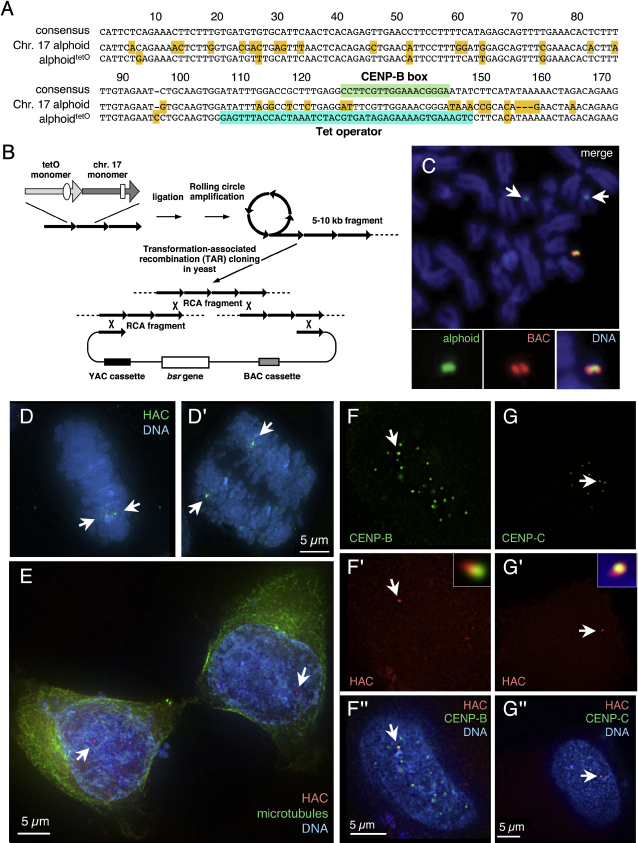
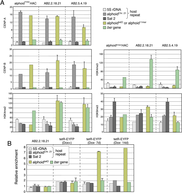
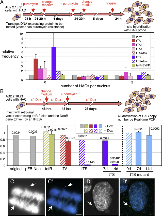
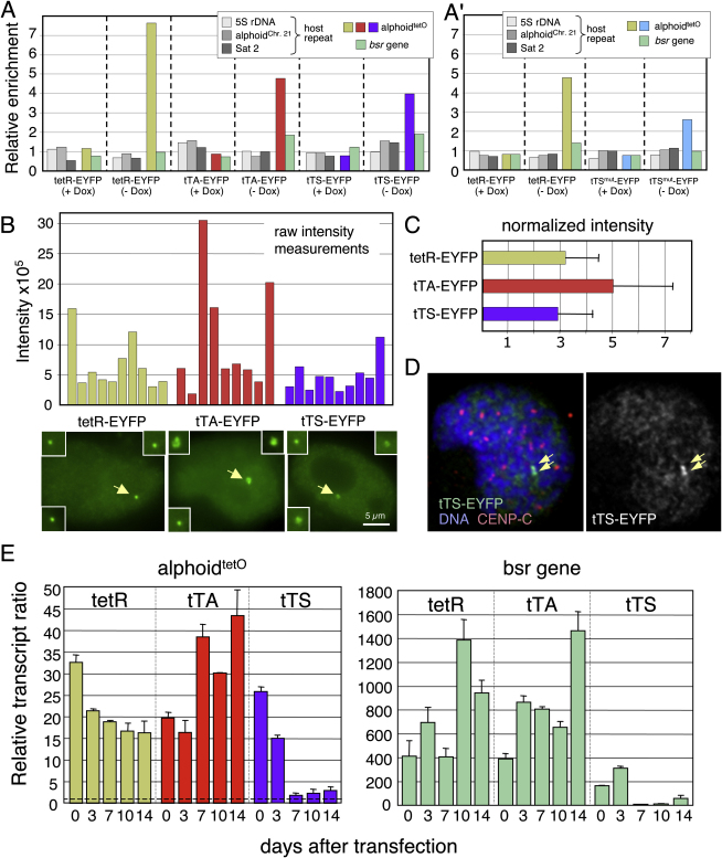
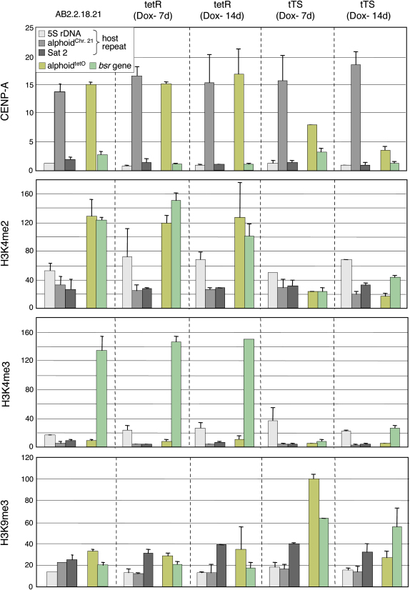
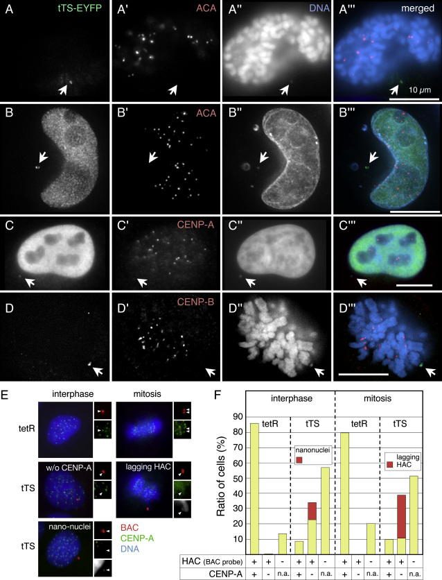
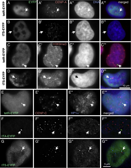
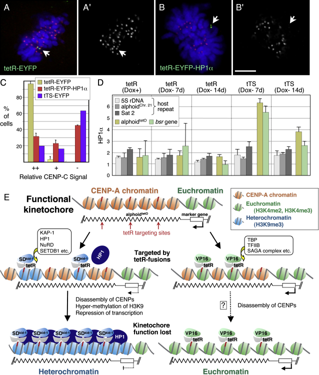
We have used a human artificial chromosome (HAC) to manipulate the epigenetic state of chromatin within an active kinetochore. The HAC has a dimeric alpha-satellite repeat containing one natural monomer with a CENP-B binding site, and one completely artificial synthetic monomer with the CENP-B box replaced by a tetracycline operator (tetO). This HAC exhibits normal kinetochore protein composition and mitotic stability. Targeting of several tet-repressor (tetR) fusions into the centromere had no effect on kinetochore function. However, altering the chromatin state to a more open configuration with the tTA transcriptional activator or to a more closed state with the tTS transcription silencer caused missegregation and loss of the HAC. tTS binding caused the loss of CENP-A, CENP-B, CENP-C, and H3K4me2 from the centromere accompanied by an accumulation of histone H3K9me3. Our results reveal that a dynamic balance between centromeric chromatin and heterochromatin is essential for vertebrate kinetochore activity.


Figures

Figure 1
Figure 1
Isolation of the AlphoidtetO HAC (A) Sequence comparison between the alphoid monomers used for the alphoidtetO DNA array and the alphoid consensus. One monomer of the alphoidtetO dimer is derived from a chromosome 17 alphoid type I 16-mer unit and contains a CENP-B box (shaded green). The second monomer is a wholly synthetic sequence derived from the Choo consensus (Choo et al., 1991), with sequences corresponding to the CENP-B box replaced by a 42 bp tetO motif (shaded blue). Other bases that differ from the consensus are shaded in yellow. (B) Diagram showing the construction of the alphoidtetO BAC by rolling circle amplification (RCA) in vitro and transformation-associated recombination (TAR) cloning in yeast cells. This yielded BAC clone, BAC32-2-mer(tetO), containing 50 kb of the alphoidtetO dimer DNA. (C) FISH analysis of a metaphase cell chromosome spread containing the alphoidtetO HAC (AB2.2.18.21) with alphoidtetO (green) and BAC vector probes (red). Chromosomes were counterstained with DAPI (blue in merged panels). Endogenous chromosome 17 centromeres were also detected by the alphoidtetO probe (arrows in upper panel). (D and D′) FISH on AB 2.2.18.21 cells in metaphase (D) and anaphase (D′). Arrows indicate the HAC undergoing normal segregation. (E) Cell in cytokinesis transfected with mRFP-TetR (red) and stained with anti-tubulin antibody (green) and DAPI (blue). mRFP-TetR binds to the HAC (arrows), in the two daughter cells. (F and G) mRFP-TetR binds to the alphoidtetO HAC in vivo where it colocalizes (arrows) with CENP-B (F), CENP-C (G) (both green), and DNA ([F″ and G″], blue). Size bars in (D–G) = 5 μm.
Figure 2
Figure 2
ChIP Analysis of CENP Assembly and Modified Histone H3 on Two Independent AlphoidtetO HACs (A) PCR analysis of immunoprecipitates using antibodies against CENP-A, CENP-B, H3K4me2, H3K4me3, or H3K9me3. Chromatin was precipitated from three HAC-containing cell lines: a control HAC with a 60 kb BAC array of chromosome 21 type I 11-mer alphoid DNA (alphoid11-mer) (left), or the independent AB2.2.18.21 and AB2.5.4.19 sublines bearing the alphoidtetO HAC (middle and right, respectively). The bars show the relative rate of recovery of various target DNA loci by immunoprecipitation with each antibody, calculated by dividing the percentage recovery of each DNA locus (as indicated) by that recovered in the control IP with mouse normal IgG. Error bars indicate SD (n = 3). CENP-A and CENP-B associate preferentially with the alphoidtetO DNA, alphoid11-mer from the wild-type control HAC, or endogenous alphoidchr. 21 relative to that seen with the marker genes, 5S ribosomal DNA and Sat2 (p < 0.05). The average recoveries of alphoidtetO sequences by anti-H3K4me2 antibody were significantly higher than the recovery of the alphoid11-mer from the control HAC and alphoidchr. 21 (p < 0.05, Student's t test). (B) AB2.2.18.21 cells bearing the alphoidtetO HAC and expressing tetR-EYFP were cultured in doxycycline-free medium for 7 or 14 d and analyzed by ChIP assay with anti-GFP antibody. The bars show the relative enrichment of various target DNA loci by immunoprecipitation with anti-GFP antibody (binds the EYFP moiety of tetR-EYFP). Error bars indicate SD (n = 2).
Figure 3
Figure 3
Targeting a Transcriptional Activator or Silencer into the HAC Kinetochore Induces HAC Loss (A) The HAC cell-by-cell stability assay. Coding regions of proteins to be tested were cloned into a vector that also expresses a puromycin resistance marker. Nontransfected cells were killed by puromycin and the remaining population was analyzed by FISH to quantify the HAC retention after 11–12 d of culture. To correct for variability in transfection and killing efficiency, all values were normalized to the results of transfection with empty vector bearing puromycin resistance (gray bars in lower panel). Lower panel: expression of transcriptional activators (tTA, tTA3, tTA4) and silencer tTS causes significant destabilization of the HAC with efficiency tTS > tTA > tTA3,tTA4 (n = 2–5). Constructs yielding results indistinguishable from the control have a value on the ordinate of 1.0. Addition of doxycycline, which prevents TetR from binding TetO, blocked the inactivation of HAC kinetochore by tTA and tTS (shaded bars). (B) Quantitative HAC stability assay using real-time PCR. The proteins to be tested were expressed using retroviral vectors. Virus-infected cells were maintained in medium containing neomycin ± doxycycline. After 30 d postinfection (left panel) (or additionally, 7 or 14 d for tTS or tTS mutant expressing cells, middle and right panels), the relative copy number of the alphoidtetO array was quantitated by real-time PCR. Numbers above the bars indicate the HAC loss rate (R) calculated using the formula: N30 = N0 × (1−R)30. Asterisk indicates HAC loss rate between 7 and 14 d after tTS binding. Cells expressing the tetR protein (tetR) or infected with empty vector (pFB-Neo) showed no HAC instability after 30 d of culture. The KAP-1 interaction deficient mutant (tTS mutant, right blue bars) failed to induce HAC instability over 14 d. Error bars indicate SD (n = 3). (C and C′) The HAC (detected by FISH, green in [C′]) fails to segregate with the bulk chromosomes (stained with DAPI, grayscale in [C], blue in [C′]) in anaphase cells expressing the tTA transactivator. Size bar = 5 μm. (D and D′) Nanonucleus revealed by DAPI staining (D) contains the HAC (D′), as revealed by FISH with the BAC probe (colors as in [C′]). HACs are indicated by arrows.
Figure 4
Figure 4
TetR-Fusion Proteins Specifically Associate with the AlphoidtetO Array and Change Its Transcriptional Activity (A) AB2.5.30 cells containing a stably integrated alphoidtetO dimer array and expressing tetR-EYFP, tTA-EYFP, or tTS-EYFP were cultured in doxycycline-free medium for 14 d and analyzed by ChIP using anti-GFP (binds EYFP). Bars show the relative enrichment of various target DNA loci. (A′) AB2.2.18.21 cells bearing the alphoidtetO HAC and expressing tetR-EYFP and tTS mutant (tTSmut)-EYFP were cultured in doxycycline-free medium for 14 d and analyzed by ChIP with anti-GFP. The bars show the relative enrichment of various target DNA loci by ChIP. All tetR-fusion proteins specifically bound to the alphoidtetO DNA in a doxycycline-dependent manner. (B and C) Quantitative analysis of the fluorescence intensity of various tetR-EYFP constructs bound to the HAC. Transfected cells were analyzed using a DeltaVision microscope under identical acquisition conditions. (B) The raw fluorescence intensity of the tetR-EYFP constructs is shown (n = 9–10), together with representative images. Size bar = 5 μm. (C) The fraction “enriched” on the HAC was calculated by normalizing for the unbound nucleoplasmic fraction. In some cells, the amount of tTA bound was relatively higher. In these cells, the HACs also appeared larger (insets in [B]), possibly because they contain more decondensed chromatin. (D) Mitotic cells with tTS-EYFP bound to the HAC. The presence of two sister chromatids confirms that the HAC had replicated in the previous S phase. Arrowheads indicate the HAC. (E) Levels of transcripts derived from the alphoidtetO HAC were analyzed by quantitative RT-PCR using total RNA purified from AB2.2.18.21 cells expressing tetR, tTA, or tTS. Cells were cultured in doxycycline-free medium for 3, 7, 10, and 14 d. The bars show the transcription levels of alphoidtetO dimer (left panel) or bsr gene (right panel) relative to that of the negative control, which contains no reverse transcriptase in the reaction mixture (dashed line on left panel). Levels of each transcript were corrected for the copy number of alphoidtetO HAC as determined by quantitative PCR using the genomic DNA of each cell line and normalized by that of endogenous alphoidchr.21 (for alphoidtetO) or human β-actin (for bsr gene). The levels of transcripts from alphoid DNA were extremely low (∼2.9-fold above the reverse transcriptase free control reaction for alphoidchr.21) compared to those from the bsr gene (∼1,500 fold) and β-actin (∼19,000 fold). Error bars indicate SD (n = 2).
Figure 5
Figure 5
tTS-Enhanced Heterochromatin Assembly and CENP-A Chromatin Disassembly on the AlphoidtetO HAC AB2.2.18.21 cells expressing tetR or tTS were cultured in doxycycline-free medium for 7 or 14 d and analyzed by ChIP with antibodies against CENP-A and modified histone H3. CENP-A assembly on alphoidtetO dimer was gradually decreased following induction of tTS binding for 7–14 d. Levels of H3K4me2 and H3K4me3 on the alphoidtetO HAC decreased rapidly upon tTS binding. In contrast, H3K9me3 levels on the alphoidtetO HAC increased transiently following tTS binding, but then declined again (possibly due to loss of the HAC). Error bars indicate SD (n = 2–3).
Figure 6
Figure 6
tTS Targeting to the AlphoidtetO HAC Induces Lagging Chromosomes and Formation of Nanonuclei AB2.2.18.21 cells were either transiently transfected with constructs expressing the tTS ([A], [B], and [D]) or AB2.2.18.21 cells stably expressing the tTS (C) were incubated in the absence of doxycycline to allow TetR binding to the tetO sequences in the alphoidtetO HAC. EYFP (green) was detected either via its own fluorescence ([A], [B], and [D]) or using an anti-GFP antibody (C). Cells were also stained for CENP antigens with ACA (red in [A′ and B′]), anti-CENP-A antibody (red, [C′]), or anti-CENP-B antibody (red, [D′]). Chromosomes were stained with DAPI (blue, [A″–D″]). Nonaligned alphoidtetO HACs targeted by tTS-EYFP were observed in mitotic cells (A and D). These HACs lacked detectible CENP-A, -B, and -C. In interphase cells, nanonuclei with associated tTS-EYFP were observed ([B], [C], and [E]). Nanonuclei also lacked detectible CENP-A and ACA antigens. Arrows indicate the HACs. Bar = 10 μm. (E) AB2.2.18.21 cells with alphoidtetO HAC expressing tetR (upper panels) or tTS (middle and lower panels) were cultured in doxycycline-free medium for 7 d and analyzed by immuno-FISH using anti-CENP-A antibody (green) and BAC vector probe (red). DNA was stained by DAPI (blue). Arrowheads indicate the alphoidtetO HAC. In tTS expressing cells, lagging HACs (middle right) and nanonuclei (lower left) were observed in mitosis and interphase, respectively. (F) Nanonuclei or lagging HAC were frequently observed in tTS-expressing cells (red bars, n > 50).
Figure 7
Figure 7
tTS Binding Induces Heterochromatin Formation and Kinetochore Disassembly on the AlphoidtetO HAC (A–D) AlphoidtetO HAC cell lines expressing tetR-EYFP (A and C) or tTS-EYFP (B and D) were cultured in doxycycline-free medium for 8 d and analyzed by indirect immunofluorescence using anti-GFP (green, [A–D]) and anti-CENP-A (red, [A′ and B′]) or anti-H3K9me3 (red, [C′ and D′]). DNA was stained by DAPI (blue, [A″–D″]). The HAC is indicated by arrows. Bar in (D″′) = 10 μm for (A–D). (E) tetR-EYFP has no effect on kinetochore structure and does not recruit HP1α. (F) tTA-EYFP inactivates the kinetochore, as shown by loss of CENP-C, but does not recruit HP1α. (G) The tTS inactivates the kinetochore, accompanied by a robust recruitment of HP1α. Bar in (G″′) = 5 μm for (E–G).
Figure 8
Figure 8
HP1α Recruitment Inactivates the AlphoidtetO Kinetochore (A) A duplicated HAC at metaphase has bound tetR-EYFP and CENP-C. (B) A duplicated HAC at metaphase has bound tetR-EYFP-HP1α, but lacks CENP-C. Bar in (B′) = 5 μm for (A and B). (C) Quantitation of the effects of expression of tetR-EYFP, tetR-EYFP-HP1α, and tTS-EYFP on the levels of CENP-C at kinetochores. (D) AB2.2.18.21 cells expressing tetR or tTS were cultured in doxycycline-free medium for 7 or 14 d and analyzed by ChIP with anti-HP1α antibody. The tTS induces targeting of HP1α to both the alphoidtetO array and the adjacent marker gene. The decrease seen at 14 d likely reflects loss of the HAC. Error bars indicate SD (n = 2–3). (E) Inactivation of the kinetochore by modulating the epigenetic status of the underlying chromatin. Centromere chromatin containing CENP-A (red) and H3K4me2 (not shown here) assembles on the alphoidtetO dimer array of the HAC, forming an active kinetochore structure. The chromatin of the marker gene contains H3K4me3 (green). Binding of the tTS (tetR-SDkid-1) induces H3K9 trimethylation (blue) at its target sites (red rectangle and arrow) on the alphoidtetO HAC. The resulting remodeling and compaction of the chromatin is incompatible with the structure of CENP-A chromatin and CENP-A quickly disappears from the heterochromatic alphoidtetO array. The centromere/kinetochore is inactivated. tTA (tetR-VP16) binding induces formation of open chromatin at the target site. In some cases, the open chromatin structure (euchromatin) somehow disrupts the CENP-A chromatin. However, in the case of tTA, the degree of HAC inactivation was less than that seen with the tTS. This suggests that the core of CENP-A chromatin is less sensitive to chromatin opening induced by VP16 and that in many cases HACs with open chromatin can still maintain a functional kinetochore. The data presented suggest an epigenetic mechanism to regulate the kinetochore activity based in part upon antagonism between centromere chromatin rich in CENP-A and H3K4me2 and heterochromatin rich in H3K9me3.

References

    1. Agata Y., Matsuda E., Shimizu A. Two novel Kruppel-associated box-containing zinc-finger proteins, KRAZ1 and KRAZ2, repress transcription through functional interaction with the corepressor KAP-1 (TIF1β/KRIP-1) J. Biol. Chem. 1999;274:16412–16422. - PubMed
    1. Allshire R.C., Javerzat J.-P., Redhead N.J., Cranston G. Position effect variegation at fission yeast centromeres. Cell. 1994;76:157–169. - PubMed
    1. Alonso A., Mahmood R., Li S., Cheung F., Yoda K., Warburton P.E. Genomic microarray analysis reveals distinct locations for the CENP-A binding domains in three human chromosome 13q32 neocentromeres. Hum. Mol. Genet. 2003;12:2711–2721. - PubMed
    1. Black B.E., Foltz D.R., Chakravarthy S., Luger K., Woods V.L., Jr., Cleveland D.W. Structural determinants for generating centromeric chromatin. Nature. 2004;430:578–582. - PubMed
    1. Blower M.D., Sullivan B.A., Karpen G.H. Conserved organization of centromeric chromatin in flies and humans. Dev. Cell. 2002;2:319–330. - PMC - PubMed

Publication types

MeSH terms