. 2008 May 19:1210:39-47.
doi: 10.1016/j.brainres.2008.02.059. Epub 2008 Mar 4.

Neuroprotective effects of neuregulin-1 on B35 neuronal cells following ischemia

Affiliations

Neuroprotective effects of neuregulin-1 on B35 neuronal cells following ischemia

DaJoie R Croslan et al. Brain Res. .

Abstract

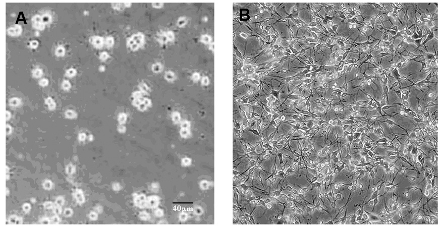
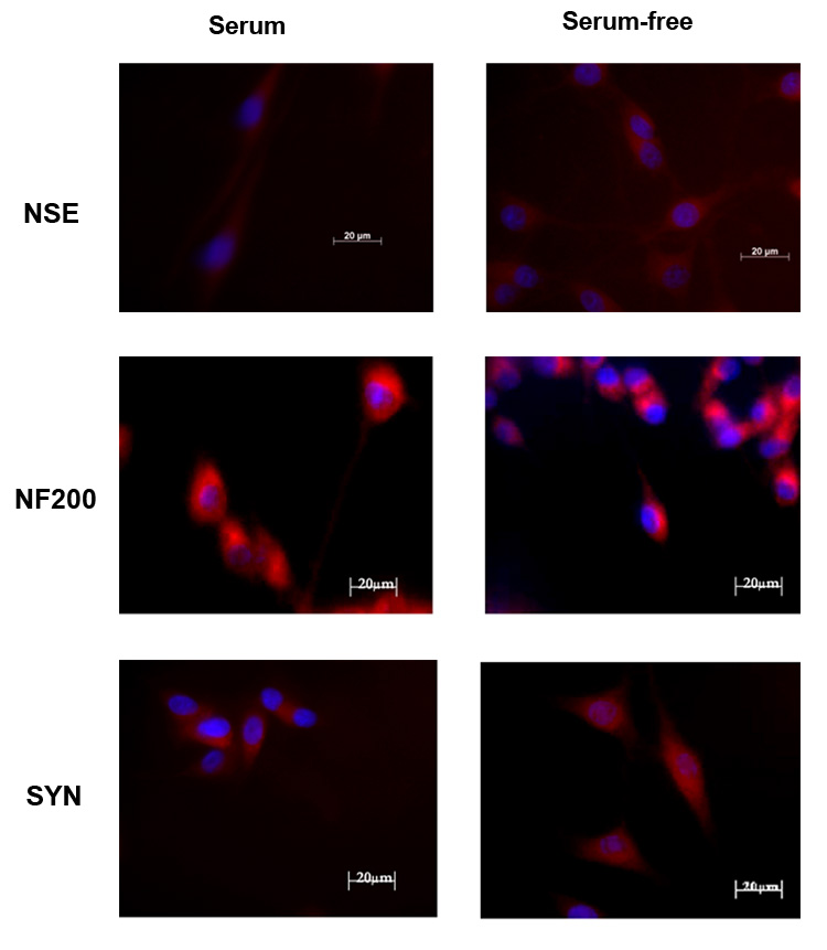
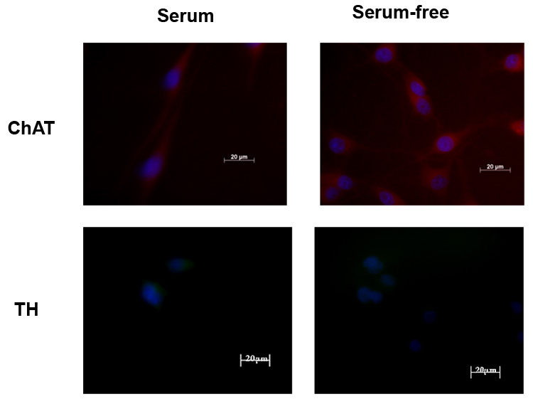
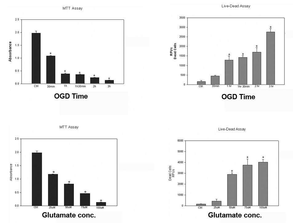
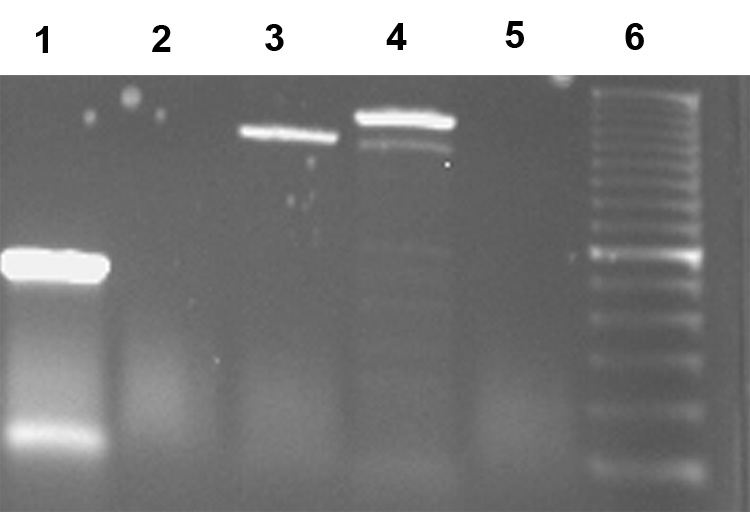
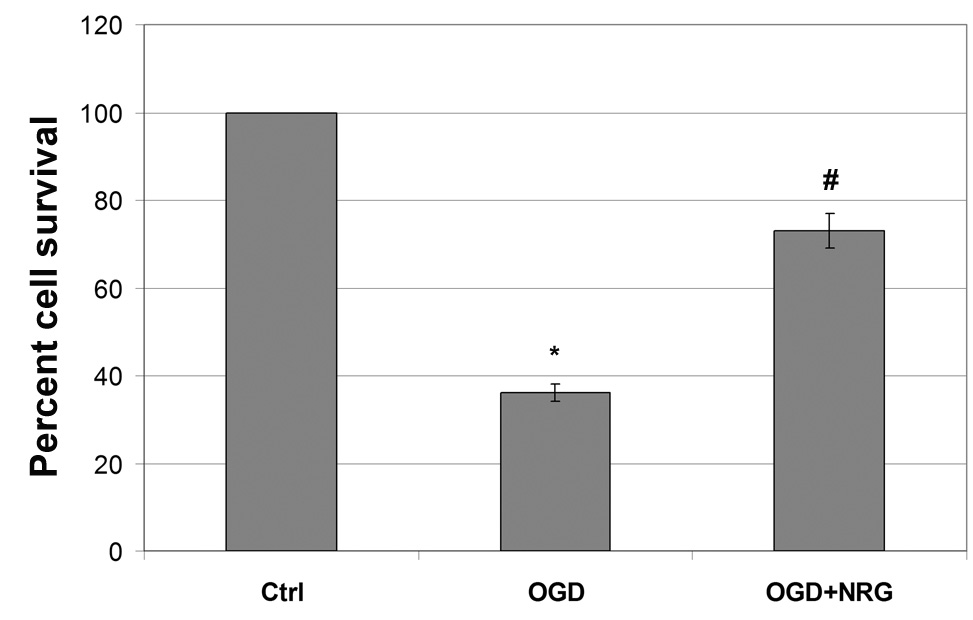
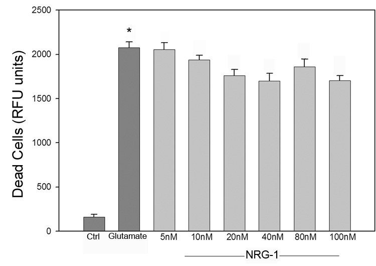
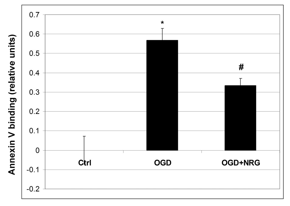
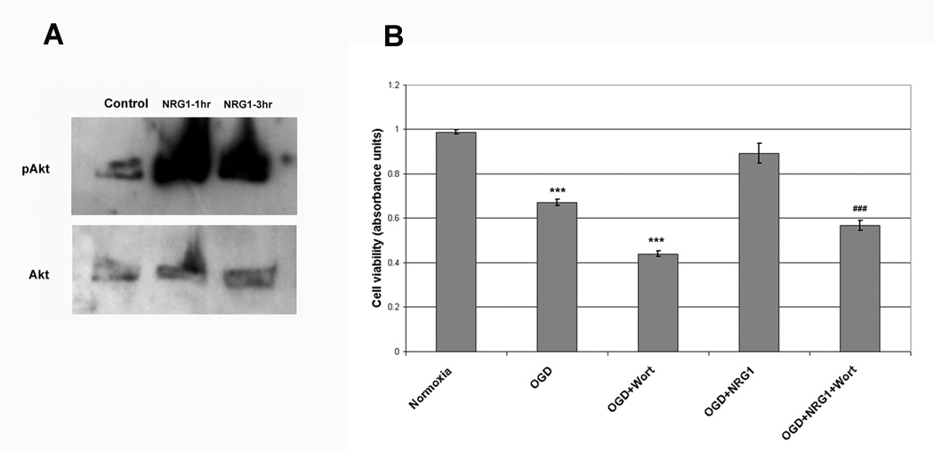
We previously showed that neuregulin-1 (NRG-1) protected neurons from death in vivo following focal ischemia. The goal of this study was to develop an in vitro rat ischemia model to examine the cellular and molecular mechanisms involved in the neuroprotective effects of NRG-1 on ischemia-induced neuronal death. Rat B-35 neuroblastoma cells differentiated by serum withdrawal, developed enhanced neuronal characteristics including, neurite extension and upregulation of neuronal markers of differentiation. When B35 neurons were subjected to oxygen glucose deprivation (OGD)/reoxygenation or glutamate, widespread neuronal death was seen after both treatments. Treatment with NRG-1 immediately after OGD significantly increased neuronal survival. NRG-1 administration also resulted in a significant decrease in annexin V, an early marker of apoptosis. However, the neurotoxic actions of glutamate were unaffected by NRG-1. The neuroprotective effects of NRG-1 were prevented by an inhibitor of the phosphatidylinositol-3-kinase/Akt pathway. These results provide a new model to gain insight into the mechanisms employed by NRG-1 to protect neurons from ischemic brain injury.


Figures

Figure 1
Figure 1
Differentiation of rat B-35 neuroblastoma cells. Rat B-35 neuroblastoma cells were grown in serum (A; undifferentiated) or serum-free medium (B; differentiated) for 3 days. Representative bright field images are shown. Upon serum withdrawal, cells begin to elaborate long processes within 2 days of culture which continues for several days in vitro. The scale bar is 40 µm.
Figure 2
Figure 2
Rat B-35 neuroblastoma cells were grown in serum (left column) or serum-free medium (right column) for 3 days. Representative images are shown (n = 3). B35 cell express neuron specific enolase (NSE) as well as neurofilament 200 (NF200) and synaptophysin (SYN) when grown in the absence or presence of serum. These levels of these neuronal markers appeared virtually unchanged in differentiated cells after serum withdrawal. The scale bar is 20 µm.
Figure 3
Figure 3
Rat B-35 neuroblastoma cells were grown in serum (left column) or serum-free medium (right column) for 3 days. Representative images are shown (n = 3). Consistent with previous studies [32], undifferentiated B-35 cells (left column) express choline acetyltransferase (ChAT). However, serum withdrawal (right column) resulted in an increase in the expression of ChAT. B-35 cells did not synthesize tyrosine hydroxylase (TH). The scale bar is 20 µm.
Figure 4
Figure 4
Effect of OGD and glutamate exposure time on B35 cell survival. B-35 cells were subjected to OGD at the indicated time followed by reoxygenation for a total of 24 hours. Results revealed a time-dependent decrease in cell death with increase in duration of OGD when using an MTT assay (upper left) or the Live-Dead cytotoxicity assay (upper right). B-35 cells were subjected to glutamate treatment for 1hr followed by 24 hours recovery and analyzed by the MTT (lower left) or the Live-Dead assay (lower right). Experiments were performed 3 or more times in triplicate. Values are represented as means ± SEM; * denotes significant difference from control.
Figure 5
Figure 5
B35 cells express erbB receptor mRNA. RNA was isolated from B35 cells and erbB expression was examined by RT-PCR. GAPDH (lane 1), erbB1 (lane 2), erbB2 (lane 3), erbB3 (lane 4) and erbB4 (lane 5) PCR products along with DNA ladder (lane 6) were separated by agarose gel electrophoresis.
Figure 6
Figure 6
NRG-1 increases survival after OGD but not glutamate exposure. B-35 cells were subjected to 90 minutes of OGD 22.5 hours of reoxygenation. NRG-1 treatment resulted in a two-fold increase in cell viability (A). When B-35 cells were incubated with 50 µM glutamate, NRG-1 did not protect neurons from glutamate excitotoxicity. Values are represented as means ± SEM; * denotes difference from control, # denotes difference from OGD (n = 5; p<0.05).
Figure 6
Figure 6
NRG-1 increases survival after OGD but not glutamate exposure. B-35 cells were subjected to 90 minutes of OGD 22.5 hours of reoxygenation. NRG-1 treatment resulted in a two-fold increase in cell viability (A). When B-35 cells were incubated with 50 µM glutamate, NRG-1 did not protect neurons from glutamate excitotoxicity. Values are represented as means ± SEM; * denotes difference from control, # denotes difference from OGD (n = 5; p<0.05).
Figure 7
Figure 7
NRG-1 decreases annexin V labeling after OGD. OGD results in an increase in annexin V labeling of cells that is reduced by NRG-1. Values are represented as means ± SEM;* denotes difference from control, # denotes difference from OGD (n = 5; p<0.05)
Figure 8
Figure 8
Neuroprotection by NRG-1 involved the PI3K/Akt pathway. Stimulation with NRG-1 resulted in a dramatic increase in pAkt levels while total Akt levels remained unchanged (A). Wortmannin, an inhibitor of PI3K, prevented NRG-1 from protecting neurons from OGD (B). Values are represented as means ± SEM; *** denotes difference from control, ### denotes difference from OGD (n = 3; p<0.001).

References

    1. Bermingham-McDonogh O, McCabe K, Reh T. Effects of GGF/neuregulins on neuronal survival and neurite outgrowth correlate with erbB2/neu expression in developing rat retina. Dev Suppl. 1996;122:1427–1438. - PubMed
    1. Bruno VM, Goldberg MP, Dugan LL, Giffard RG, Choi DW. Neuroprotective effect of hypothermia in cortical cultures exposed to oxygen-glucose deprivation or excitatory amino acids. J Neurochem. 1994;63:1398–1406. - PubMed
    1. Buonanno A, Fischbach GD. Neuregulin and ErbB receptor signaling pathways in the nervous system. Curr Opin Neurobiol. 2001;11:287–296. - PubMed
    1. Burden S, Yarden Y. Neuregulins and their receptors: a versatile signaling module in organogenesis and oncogenesis. Neuron. 1997;18:847–855. - PubMed
    1. Chan PH. Mitochondria and neuronal death/survival signaling pathways in cerebral ischemia. Neurochem Res. 2004;29:1943–1949. - PubMed

Publication types

MeSH terms