. 2008 May;36(9):3139-49.
doi: 10.1093/nar/gkn143. Epub 2008 Apr 13.

Structure and function of the regulatory C-terminal HRDC domain from Deinococcus radiodurans RecQ

Affiliations

Structure and function of the regulatory C-terminal HRDC domain from Deinococcus radiodurans RecQ

Michael P Killoran et al. Nucleic Acids Res. 2008 May.

Abstract

RecQ helicases are critical for maintaining genome integrity in organisms ranging from bacteria to humans by participating in a complex network of DNA metabolic pathways. Their diverse cellular functions require specialization and coordination of multiple protein domains that integrate catalytic functions with DNA-protein and protein-protein interactions. The RecQ helicase from Deinococcus radiodurans (DrRecQ) is unusual among RecQ family members in that it has evolved to utilize three 'Helicase and RNaseD C-terminal' (HRDC) domains to regulate its activity. In this report, we describe the high-resolution structure of the C-terminal-most HRDC domain of DrRecQ. The structure reveals unusual electrostatic surface features that distinguish it from other HRDC domains. Mutation of individual residues in these regions affects the DNA binding affinity of DrRecQ and its ability to unwind a partial duplex DNA substrate. Taken together, the results suggest the unusual electrostatic surface features of the DrRecQ HRDC domain may be important for inter-domain interactions that regulate structure-specific DNA binding and help direct DrRecQ to specific recombination/repair sites.


Figures

Figure 1.
Figure 1.
Structural features of D. radiodurans RecQ HRDC #3. (A) Schematic comparison of the conserved domains of DrRecQ and EcRecQ. Each protein is comprised of a Helicase, RecQ-Ct and HRDC domain. DrRecQ features two additional HRDC domains at its C-terminus numbered HRDC #2 and #3, respectively. (B) Stereodiagram of the 2Fo−Fc electron density map surrounding P790 in the α3 to α4 loop contoured at 2.0 σ. All structural figures were created using PyMol (39). (C) The α-helical fold of HRDC #3 shown as ribbons. Helix numbers are included in the representation on the right. (D) Sequence alignment of DrRecQ HRDC domains and HRDC #3's closest structural homologs. The secondary structure and corresponding residue numbers in DrRecQ are indicated at the top of the alignment. Structural homologs were identified using the DALI search engine (34) and resulting Z-scores are shown following each sequence. Basic residues contacting phosphate in the crystal structure are shown in blue and acidic residues comprising the acidic patch are in red. Residues conserved in HRDC #3's structural homologs for the HRDC #3 phosphate binding or acidic patch residues are also colored blue and red, respectively. (E) Phosphate ion present in the HRDC #3 crystal structure. The position of the phosphate ion and basic residues K805 and R806 are shown on the left side. To the right, a closer view of the surface of HRDC #3 and the phosphate ion surrounded by an omit map contoured at 5.0 σ. (F) Electrostatic surface diagram of HRDC #3 in orthogonal views [for positive (blue) and negative (red) surfaces].
Figure 2.
Figure 2.
DrRecQ variants have reduced affinity for ssDNA and dup-3′ DNA. (A) The positions of alanine-substitution mutations are indicated on ribbons (left) and surface (right) representations of the HRDC #3 structure. Residues in close proximity to a phosphate ion in the crystal structure are shown in blue, with those comprising the acidic patch in red. (BE) Fraction of DNA bound with either ssDNA (B and D) or dup-3′ (C and E) by DrRecQ variants with mutations in the phosphate binding site (B and C) or D816 in the acidic patch (D and E). DrRecQ (closed diamonds), K805A (open squares), R805A (closed circles) and D816A (open circles). A trend line for DrRecQ is shown in each plot for reference.
Figure 3.
Figure 3.
The D816A and K805A mutants have defects in synthetic HJ binding. Increasing concentrations (1.6 nM, 8 nM, 40 nM, 200 nM, 1 μM) of DrRecQ, K805A and D816A added to a synthetic HJ substrate. The position of the wells and the unbound substrate is indicated on the left side.
Figure 4.
Figure 4.
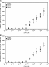
Variants in DrRecQ HRDC #3 do not affect DNA-dependent ATP hydrolysis with short ssDNA. The rate of ATP hydrolysis for DrRecQ and (A) phosphate-binding site variants K805A and R806A and (B) acidic patch variant D816A are plotted as a function of ssDNA concentration (in nucleotides). DrRecQ (closed diamonds), K805A (open squares), R806A (closed circles) and D816A (open circles).
Figure 5.
Figure 5.
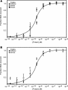
DrRecQ variants have differing helicase activities. The fraction of dup-3′ DNA unwound by DrRecQ, (A) phosphate binding site variants K805A and R806A, and (B) acidic patch variant D816A are plotted as a function of protein concentration (0.3 pM to 300 nM). DrRecQ (closed diamonds), K805A (open squares), R806A (closed circles) and D816A (open circles). A trend line for DrRecQ is shown for reference.

References

    1. Bachrati CZ, Hickson ID. RecQ helicases: suppressors of tumorigenesis and premature aging. Biochem. J. 2003;374:577–606. - PMC - PubMed
    1. Ellis NA, Groden J, Ye TZ, Straughen J, Lennon DJ, Ciocci S, Proytcheva M, German J. The Bloom's syndrome gene product is homologous to RecQ helicases. Cell. 1995;83:655–666. - PubMed
    1. Kitao S, Shimamoto A, Goto M, Miller RW, Smithson WA, Lindor NM, Furuichi Y. Mutations in RECQL4 cause a subset of cases of Rothmund–Thomson syndrome. Nat. Genet. 1999;22:82–84. - PubMed
    1. Yu CE, Oshima J, Fu YH, Wijsman EM, Hisama F, Alisch R, Matthews S, Nakura J, Miki T, Ouais S, et al. Positional cloning of the Werner's syndrome gene. Science. 1996;272:258–262. - PubMed
    1. Bennett RJ, Keck JL. Structure and function of RecQ DNA helicases. Crit. Rev. Biochem. Mol. Biol. 2004;39:79–97. - PubMed

Publication types

MeSH terms