. 2008 May 1;121(Pt 9):1559-68.
doi: 10.1242/jcs.021089. Epub 2008 Apr 14.

Organellar dynamics during the cell cycle of Toxoplasma gondii

Affiliations

Organellar dynamics during the cell cycle of Toxoplasma gondii

Manami Nishi et al. J Cell Sci. .

Abstract

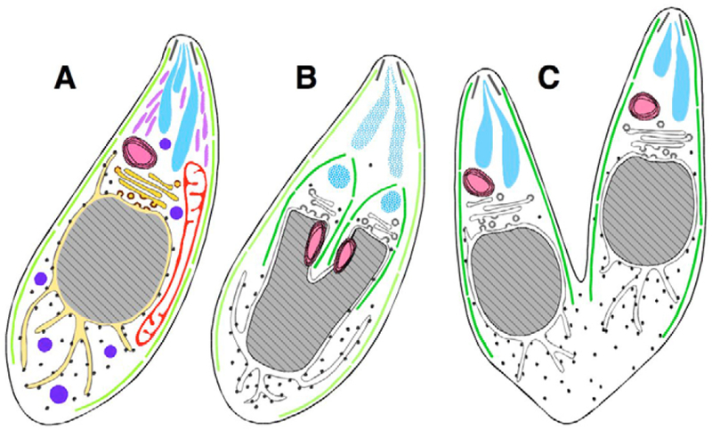
The protozoan phylum Apicomplexa encompasses approximately 5000 species of obligate intracellular parasites, including those responsible for malaria and toxoplasmosis. Rather than dividing by binary fission, apicomplexans use a remarkable mechanism for replication, assembling daughters de novo within the cytoplasm. Here, we exploit time-lapse microscopy of fluorescent markers targeted to various subcellular structures in Toxoplasma gondii tachyzoites to determine how these unicellular eukaryotes efficiently package a complete set of organelles, maintaining the highly polarized organization necessary for host cell invasion and pathogenesis. Golgi division and elongation of the apicoplast are among the first morphologically observable events, associated with an unusual pattern of centriolar migration. Daughter parasites are assembled on cytoskeletal scaffolding, whose growth proceeds from the apical end, first encapsulating the divided Golgi. Further extension of the cytoskeletal scaffold results in partitioning of the apicoplast, nucleus, endoplasmic reticulum, and finally the mitochondrion, which enters the developing daughters rapidly, but only very late during the division cycle. The specialized secretory organelles (micronemes and rhoptries) form de novo. This distinctive pattern of replication -- in which organellar segregation spans approximately 75% of the cell cycle, completely encompassing S phase -- suggests an unusual mechanism of cell cycle regulation.


Figures

Fig. 1.
Fig. 1.
Endodyogeny of Toxoplasma gondii tachyzoites. Diagrams of longitudinal sections of T. gondii parasites at various stages during replication. Subcellular structures (A, interphase) include, proceeding from the apical to the basal end: the conoid (black lines), inner-membrane complex (light green lines), rhoptries (turquoise), micronemes (lavender), dense granules (blue), apicoplast (pink), mitochondrion (red), Golgi (gold) and nucleus (grey), bordered by endoplasmic reticulum (yellow). Mid-way through daughter cell formation (B), the developing daughter IMC scaffolds (dark green) encompass the Golgi and apicoplast (which have already divided), and the nucleus begins to bifurcate. Hatched turquoise indicates breakdown of the maternal rhoptries, and de novo synthesis of daughter rhoptries, which have not yet acquired their mature club-like shape (other organelles not colored and mitochondrion not shown, for clarity). (C) Daughter IMC complexes then grow to establish two complete daughter parasites, which are about to emerge, acquiring their plasma membrane from the mother.
Fig. 2.
Fig. 2.
Coordination of Golgi, apicoplast, centriolar and nuclear replication. Left column: Time-lapse images of living parasites, labeled for simultaneous visualization of the inner membrane complex of mother and daughter parasites (IMC1-YFP, green) and the Golgi complex (GRASP-mRFP, red). Golgi elongation (D) and fission occurs before the initiation of daughter scaffold formation (J). Daughter Golgi are then encapsulated into the developing daughter parasites (M). Middle column, fixed parasites labeled to image the inner membrane complex (IMC1-YFP, green), apicoplast (anti-ACP, red) and nucleus (DAPI, blue). During interphase, the apicoplast is oval in shape and lies just apical to the nucleus (B). The apicoplast elongates (E,H) before daughter scaffolds are initiated (K), and finishes dividing near the mid point of scaffold formation, shortly before karyokinesis (N). The same sequence of events was observed in living cells using IMC1-YFP parasites transiently transfected with ACPL/FNRL-mRFP/DsRed to label the apicoplast. Right column, fixed parasites labeled to image the centrioles (EGFP-TgCentrin, green), apicoplast (anti-ACP, red) and nucleus (DAPI, blue). The apicoplast (open arrowhead) lies close to the centriole (filled arrowhead) during interphase (C), and begins to elongate (F) before centriole duplication (I). During apicoplast elongation, the centrioles migrate to the basal end of the nucleus (F), where they replicate (I, paired arrowheads) before returning to the apical end (L) and reassociating with the apicoplast, which remains apical throughout. The apicoplast and centrioles remain associated through plastid division (O), nuclear division and cytokinesis. Scale bar: 5 μm.
Fig. 3.
Fig. 3.
Dynamics of ER replication and segregation. Parasites expressing IMC1-YFP (green) and P30L-mRFP-HDEL (red) were examined through the cell cycle using time-lapse microscopy. During interphase (top row), the nucleus is surrounded by the endoplasmic reticulum (ER), from which branches extend both apically and basally. The perinuclear ER expands with the nucleus, and ramifies to form extensive branches coincident with daughter scaffold formation (t=0′). Emanations from the ER associate with the developing IMC (arrowheads; +40′), enter into the developing daughters along with the nucleus (+70′, +90′), and continue to segregate (+130′) until the emergence of daughter parasites from the mother, leaving a small amount of material behind in the residual body (+170′). Scale bar: 5 μm.
Fig. 4.
Fig. 4.
Dynamics of microneme and rhoptry biogenesis in T. gondii. Parasites expressing MIC3-GFP (left two columns, green) or ROP1-CAT-YFP (right two columns, green) were transfected with IMC1-mRFP (red) in order to track microneme or rhoptry biogenesis. Both organelles are located at the apical end of parasites during interphase (top row). As daughter cells begin to elongate (+40′), new micronemes and rhoptries, distinct from their maternal counterparts, associate with the pellicles of each daughter (white arrowheads). As daughter cells grow, the maternal micronemes and rhoptries (open arrowheads) disappear (+95′ for micronemes, +80′ for rhoptries) and residual material is packaged into residual bodies when daughters emerge (+140′ for micronemes, +130′ for rhoptries). Scale bars: 5 μm.
Fig. 5.
Fig. 5.
Dynamics of mitochondrial replication in T. gondii. (A) Time-lapse microscopy of parasites labeled with IMC1-YFP (green) and HSP60-RFP (red). During interphase, the single mitochondrion typically forms an elongated S or lasso shape. The mitochondrion branches at multiple locations during the early stage of daughter IMC formation (arrowheads, t=0′). These branches elongate and often surround the growing daughter IMCs (+60′), but rarely enter into the developing daughters until they begin to emerge from the mother (+110′). Once initiated, however, mitochondrial entry into the daughter cells is very rapid (+120′). A portion of the mitochondrion often remains within the residual material left behind when daughter parasites emerge after the completion of endodyogeny (+160′). Scale bar: 5 μm. (B) HSP60L-YFP transgenics were fixed and labeled with anti-ACP antibody to reveal transient association of the apicoplast (red) and mitochondrion (green) during the G1 phase and the early stages of apicoplast elongation (before the initiation of daughter scaffold formation). Scale bar: 5 μm. (C) Apicoplast or mitochondrial association is also observed by transmission EM (image kindly provided by G. Warren, University of Vienna, Austria). Scale bar: 0.5 μm.
Fig. 6.
Fig. 6.
Effects of oryzalin on the dynamics of organellar replication. In contrast to control cultures (left), T. gondii tachyzoites grown for 24 hours in 1 μM oryzalin (right) fail to organize daughter parasites (green), owing to the absence of subpellicular microtubules (Stokkermans et al., 1996; Morrissette and Sibley, 2002). Early organellar division events, including Golgi division (filled arrowheads), apicoplast elongation (open arrowheads), and ER and mitochondrial ramification still occur in the presence of oryzalin, but later events, such as nuclear, apicoplast, ER and mitochondrial division, do not. In all panels, DNA is labeled with DAPI (blue) and the inner-membrane complex scaffolding is labeled using IMC1-YFP (green). Red markers: row 1, GRASP-mRFP (Golgi); row 2, anti-ACP (apicoplast); row 3, P30L-mRFP-HDEL (ER); row 4, HSP60L-RFP (mitochondrion). Note the ER and mitochondrial material left behind in the central residual body after the emergence of daughter parasites (bottom two control panels on left). Scale bar: 5 μm.
Fig. 7.
Fig. 7.
Organellar replication: the T. gondii cell cycle. The timeline shows the coordination of major events during T. gondii tachyzoite cell division (see also Table 1). Morphologically visible events span ~5 hours, representing ~75% of the entire cell cycle [different strains and culture conditions may produce slightly different timing, usually because of differences in the duration of G1 (Fichera et al., 1995; Radke and White, 1998; Radke et al., 2001; Hu et al., 2002; Hu et al., 2004)]. Images show the typical appearance of various organelles during the major morphological transitions associated with their replication (colored circles indicate which organelles are labeled with which color). Data compiled from current studies and previous reports (see Table 1).

References

    1. Aikawa M (1971). Parasitological review. Plasmodium: the fine structure of malarial parasites. Exp. Parasitol 30, 284–320. - PubMed
    1. Aurrecoechea C, Heiges M, Wang H, Wang Z, Fischer S, Rhodes P, Miller J,Kraemer E, Stoeckert CJ Jr, Roos DS et al. (2007). ApiDB: integrated resources for the apicomplexan bioinformatics resource center. Nucleic Acids Res 35, D427–D430. - PMC - PubMed
    1. Bannister LH, Hopkins JM, Fowler RE, Krishna S and Mitchell GH (2000a). A brief illustrated guide to the ultrastructure of Plasmodium falciparum asexual blood stages. Parasitol. Today 16, 427–433. - PubMed
    1. Bannister LH, Hopkins JM, Fowler RE, Krishna S and Mitchell GH (2000b).Ultrastructure of rhoptry development in Plasmodium falciparum erythrocytic schizonts. Parasitology 121, 273–287. - PubMed
    1. Bereiter-Hahn J and Voth M (1994). Dynamics of mitochondria in living cells: shape changes, dislocations, fusion, and fission of mitochondria. Microsc. Res. Tech 27, 198–219. - PubMed

Publication types

MeSH terms