. 2008 Jul;76(7):3116-23.
doi: 10.1128/IAI.00173-08. Epub 2008 Apr 14.

The surface-exposed carboxyl region of Mycoplasma pneumoniae elongation factor Tu interacts with fibronectin

Affiliations

The surface-exposed carboxyl region of Mycoplasma pneumoniae elongation factor Tu interacts with fibronectin

Sowmya Balasubramanian et al. Infect Immun. 2008 Jul.

Abstract

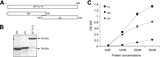
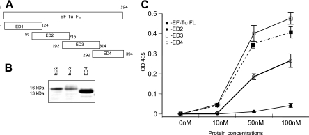
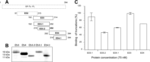
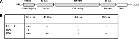
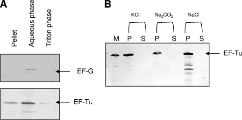
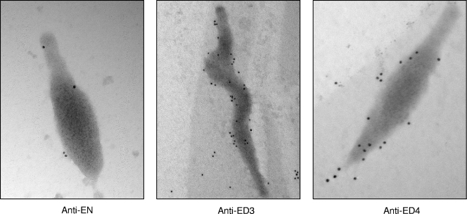
Mycoplasma pneumoniae is a bacterial pathogen of the human respiratory tract that causes a wide range of airway diseases as well as extrapulmonary symptoms. It possesses a distinct, differentiated terminal structure, termed the attachment organelle, that mediates adherence to the host respiratory epithelium. Previously, we reported that surface-associated M. pneumoniae elongation factor Tu (EF-Tu, also called MPN665) serves as a fibronectin (Fn)-binding protein, facilitating interactions between mycoplasmas and extracellular matrix. In the present study, we determined that binding of M. pneumoniae EF-Tu to Fn is primarily mediated by the EF-Tu carboxyl region. A 179-amino-acid region spanning the carboxyl terminus (designated EC; amino acids 192 to 394) binds Fn in a dose-dependent manner. Further analysis of carboxyl constructs (ED3 and ED4) and their deletion truncations (ED3.1, ED3.2, and ED4.1) revealed that the carboxyl region possessed two distinct sites with different Fn-binding efficiencies. Immunogold electron microscopy using antibodies raised against recombinant ED3 and ED4 demonstrated the surface accessibility of the EF-Tu carboxyl region. Competitive binding assays using intact radiolabeled mycoplasmas and purified recombinant ED3 and ED4 proteins, along with antibody blocking assays, reinforced the role of the surface-exposed EF-Tu carboxyl region in Fn binding. Alkali and high-salt treatment of mycoplasma membranes and Triton X-114-partitioned mycoplasma fractions confirmed the stable association of EF-Tu within the mycoplasma membrane. These observations highlight the unique, multifaceted, and unpredictable role of the classically defined cytoplasmic protein EF-Tu relative to cellular function, compartmentalization, and topography.


Figures

FIG. 1.
FIG. 1.
Identification of Fn-binding region of M. pneumoniae EF-Tu. (A) Schematic representation of EF-Tu amino and carboxyl termini. EF-Tu FL (amino acids 1 to 394) was divided into two overlapping amino-terminal (EN, residues 1 to 215) and carboxyl-terminal (EC, residues 192 to 394) constructs. (B) Purification of EF-Tu FL, EN, and EC proteins. Regions encoding EF-Tu FL, EN, and EC were cloned, expressed, and purified as described in Materials and Methods, separated on 4 to 12% Nu-PAGE gels, and stained with Coomassie blue. (C) Binding of recombinant EF-Tu FL, EN, and EC to immobilized Fn. Microtiter plates were coated with 0.1 μg of human Fn. Increasing concentrations of recombinant proteins were incubated in individual wells for 2 h at RT. Bound proteins were detected with mouse anti-His MAb and goat anti-mouse AP-conjugated antibodies, followed by color development with p-nitrophenyl phosphate substrate. Results are expressed as means ± standard deviations. Each sample point is based upon triplicate values. OD 405, optical density at 405 nm.
FIG. 2.
FIG. 2.
Characterization of minimal Fn-binding regions of M. pneumoniae EF-Tu. (A) Schematic representation of deletion constructs of amino and carboxyl regions of EF-Tu. EN was further divided into ED1 (residues 1 to 124) and ED2 (residues 91 to 215) with 23 overlapping amino acids while EC was divided into ED3 (residues 192 to 314) and ED4 (residues 292 to 394) with 22 overlapping amino acids. (B) Purification of ED2, ED3, and ED4. All recombinant proteins were purified as described in Materials and Methods, separated by electrophoresis with 4 to 12% Nu-PAGE gels, and stained with Coomassie blue. ED1 could not be expressed. (C) Dose-dependent binding of recombinant EF-Tu-related proteins to immobilized Fn. ELISAs were performed with recombinant ED2, ED3, and ED4 as described in the legend of Fig. 1C. OD 405, optical density at 405 nm.
FIG. 3.
FIG. 3.
Construction and Fn binding of overlapping carboxyl regions of M. pneumoniae EF-Tu. (A) Schematic representation of deletion constructs ED3.1, ED3.2, and ED4.1. ED3.1 and ED3.2 were constructed by deleting the overlapping regions of ED3 and ED4. ED3.2 additionally had a deletion of the overlapping region of ED3 and ED2. ED4.1 was constructed by deleting the ED3-overlapping region. (B) Purification of ED3.1, ED3.2, and ED4.1. ED3.1, ED3.2, and ED4.1 were purified under denatured conditions and then refolded in 50 mM Tris buffer. Purified proteins were separated by electrophoresis with 4 to 12% Nu-PAGE gels and stained with Coomassie blue. (C) Interaction of ED3.1, ED3.2, and ED4.1 with Fn. ELISAs were performed using 75 nM concentrations of recombinant proteins in triplicate as described in Materials and Methods. Values were determined, and binding percentages were calculated using ED3 binding values at 75 nM as 100%. Results are expressed as means ± standard deviations. Each sample point is based upon triplicate values.
FIG. 4.
FIG. 4.
Interaction of M. pneumoniae EF-Tu FL and its truncations with different Fn domains. (A) Schematic representation of different domains of Fn. The figure was adapted from Virkola et al. (53). Fn domains used for the ligand blot analyses are indicated in cross-hatched boxes. (B) Interaction of EF-Tu FL, ED3, and ED4 with domains of Fn. Equal amounts of available Fn fragments were separated individually on 4 to 12% Nu-PAGE gels and transferred onto nitrocellulose membranes, which were incubated with 10 μg/ml recombinant EF-Tu proteins in 3% Blotto in 1× TBS for 2 h at RT (see Materials and Methods). Membranes were probed with mouse anti-His MAb (1:5,000) and subsequently with goat anti-mouse AP (1:2,000) reagent. Based on the mean intensities of His tag antibody recognition of recombinant EF-Tu proteins, interactions were classified as demonstrating weak (+), intermediate (++), strong (+++) or no (−) binding.
FIG. 5.
FIG. 5.
M. pneumoniae EF-Tu association with mycoplasma membrane. (A) Triton X-114 phase partitioning of intact M. pneumoniae cells. Equal amounts of sample were separated on Nu-PAGE gels under reducing conditions and transferred onto nitrocellulose membranes. Immunoblotting was performed with mouse anti-EF-Tu (1:3,000) or rabbit anti-EF-G (1:3,000) antiserum in 3% Blotto for 1 h at RT. (B) Alkali and high-salt treatment of M. pneumoniae membranes. M. pneumoniae S1 membranes (M) were treated with 3 M KCl, 0.1 M Na2CO3, and 1 M NaCl. Supernatant (S) and pellet (P) fractions were subjected to Nu-PAGE under reducing conditions, transferred onto nitrocellulose membranes, and immunoblotted with anti-EF-Tu antiserum as described in Materials and Methods.
FIG. 6.
FIG. 6.
Immunogold labeling of EF-Tu on intact M. pneumoniae cells. Mycoplasmas were incubated with antisera (1:1,000) generated against EN, ED3, or ED4 truncations followed by anti-mouse IgG gold complex (10 nm). Mycoplasma membrane-associated gold labeling of EF-Tu is readily observed with ED3 and ED4 antisera and much less so with EN antiserum. EN magnification, ×71,000; ED3 magnification, ×44,000; and ED4 magnification, ×71,000.

References

    1. Alvarez, R. A., M. W. Blaylock, and J. B. Baseman. 2003. Surface localized glyceraldehyde-3-phosphate dehydrogenase of Mycoplasma genitalium binds mucin. Mol. Microbiol. 481417-1425. - PubMed
    1. Balish, M. F., T. W. Hahn, P. L. Popham, and D. C. Krause. 2001. Stability of Mycoplasma pneumoniae cytadherence-accessory protein HMW1 correlates with its association with the triton shell. J. Bacteriol. 1833680-3688. - PMC - PubMed
    1. Baseman, J. B., R. M. Cole, D. C. Krause, and D. K. Leith. 1982. Molecular basis for cytadsorption of Mycoplasma pneumoniae. J. Bacteriol. 1511514-1522. - PMC - PubMed
    1. Bergmann, S., M. Rohde, G. S. Chhatwal, and S. Hammerschmidt. 2001. Alpha-Enolase of Streptococcus pneumoniae is a plasmin(ogen)-binding protein displayed on the bacterial cell surface. Mol. Microbiol. 401273-1287. - PubMed
    1. Berrier, C., A. Garrigues, G. Richarme, and A. Ghazi. 2000. Elongation factor Tu and DnaK are transferred from the cytoplasm to the periplasm of Escherichia coli during osmotic downshock presumably via the mechanosensitive channel MscL. J. Bacteriol. 182248-251. - PMC - PubMed

Publication types

MeSH terms

LinkOut - more resources