. 2008 May 12;205(5):1037-48.
doi: 10.1084/jem.20072528. Epub 2008 Apr 14.

Essential role of TNF receptor superfamily 25 (TNFRSF25) in the development of allergic lung inflammation

Affiliations

Essential role of TNF receptor superfamily 25 (TNFRSF25) in the development of allergic lung inflammation

Lei Fang et al. J Exp Med. .

Abstract

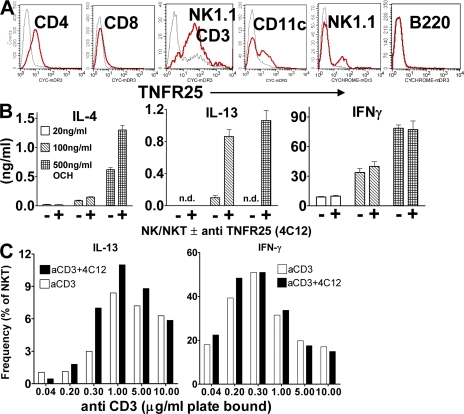
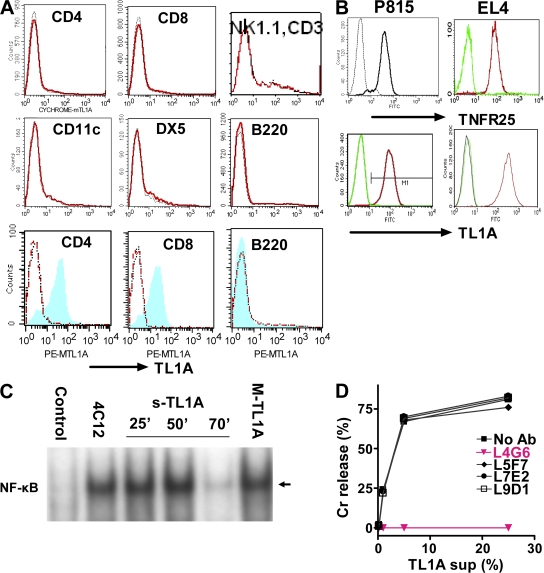
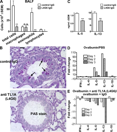
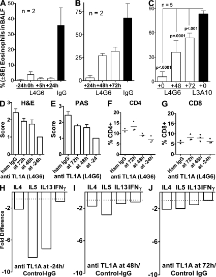
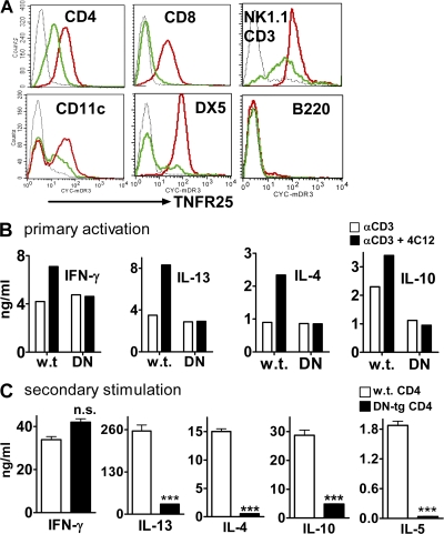
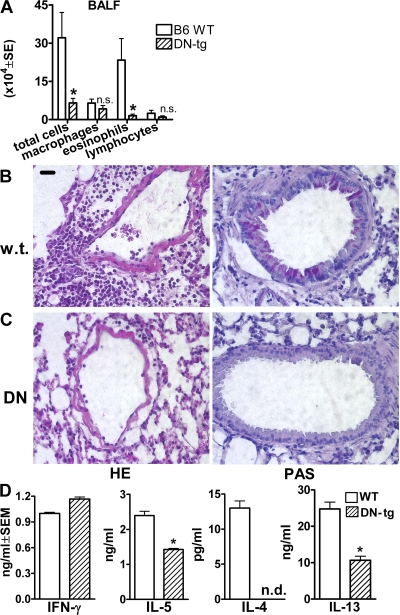
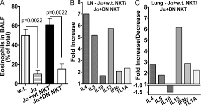
We identify the tumor necrosis factor receptor superfamily 25 (TNFRSF25)/TNFSF15 pair as critical trigger for allergic lung inflammation, which is a cardinal feature of asthma. TNFRSF25 (TNFR25) signals are required to exert T helper cell 2 (Th2) effector function in Th2-polarized CD4 cells and co-stimulate interleukin (IL)-13 production by glycosphingolipid-activated NKT cells. In vivo, antibody blockade of TNFSF15 (TL1A), which is the ligand for TNFR25, inhibits lung inflammation and production of Th2 cytokines such as IL-13, even when administered days after airway antigen exposure. Similarly, blockade of TNFR25 by a dominant-negative (DN) transgene, DN TNFR25, confers resistance to lung inflammation in mice. Allergic lung inflammation-resistant, NKT-deficient mice become susceptible upon adoptive transfer of wild-type NKT cells, but not after transfer of DN TNFR25 transgenic NKT cells. The TNFR25/TL1A pair appears to provide an early signal for Th2 cytokine production in the lung, and therefore may be a drug target in attempts to attenuate lung inflammation in asthmatics.


Figures

Figure 1.
Figure 1.
TNFR25 is constitutively expressed on resting NKT cells and co-stimulates IL-4 and -13 cytokine production. (A) Expression of TNFR25 in lymph node cells. Cells were gated for CD4, CD8, B220, CD11c, or NK1.1-positive and CD3-negative cells or NK1.1/CD3 double-positive cells and TNFR25 fluorescence displayed in the histogram. Red curve, anti-TNFR25; black curve, isotype control. (B) TNFR25 co-stimulation of IL-4 and -13 production by glycosphingolipid-activated NKT cells. NKT cells were enriched from pooled spleen cells from 10 mice by positive selection using the EasySep mouse Pan NK Positive Selection kit. The ratio of NK to NKT cells in the enriched population was 65:35 × 105 cells were incubated for 48 h in flat bottom 96-microtiter plates in triplicate in the presence of 10 ng/ml IL-15 and stimulated with 20, 100, or 500 ng/ml OCH-glycosphingolipid. Co-stimulation was achieved with 5 μg/ml agonistic anti-TNFR25 antibody 4C12 as indicated; cytokines were analyzed in supernatants by ELISA. n.d., not detectable. Representative of three independent experiments. (C) TNFR25 co-stimulation increases the frequency of IL-13–producing NKT cells. Enriched NK/NKT cells purified as described above were incubated with increasing concentrations of plate-bound anti-CD3, as indicated in the presence and absence of 5 μg/ml agonistic anti-TNFR25 (4C12) for 72 h. Details of the method for intracellular cytokine staining are given in Materials and methods. In negative controls for unspecific staining, anti–IL-13 was blocked with recombinant IL-13 before use in intracellular staining (see Materials and methods) and were given values of <1%. Data for intracellular staining of IL-13 and IFN-γ in NKT cells were obtained by gating on NK1.1 and CD3 double-positive cells. Data are taken from two experiments. Error bars represent the mean ± the SEM.
Figure 2.
Figure 2.
TL1A-triggered TNFR25 signals are blocked by antagonistic anti-TL1A L4G6. (A) TL1A is not expressed on resting lymphocytes and up-regulated on activated T cells (top 6 graphs). Resting splenocyte cell suspensions were gated using the respective labeled antibody as a population marker and the TL1A histogram displayed. Red curve, anti-TL1A; black curve, isotype control (bottom 3 graphs). Splenocytes were activated for 24 h with plate-bound anti-CD3 or with LPS and stained with anti-TL1A and with the population marker, as indicated. After gating on the population marker, TL1A expression on activated cells is shown as blue/shaded histogram. Red curve, resting cells; black curve, isotype control. Representative of more than three experiments. (B) TNFR25 and TL1A expression on cDNA transfected P815 and EL4. Transfected (right curve in each histogram) and untransfected cells were stained with the appropriate antibody and isotype controls and analyzed by flow cytometry. (C) TNFR25 activates NF-κB when triggered by agonistic antibody 4C12, by soluble TL1A or by membrane-bound TL1A. NF-κB activation was measured in EL4 cells transfected with TNFR25 in response to TNFR25 triggering. Cells were treated with the agonistic anti-TNFR25 antibody 4C12 (5 μg/ml) for 50 min; soluble TL1A was given for 25, 50, or 70 min, as indicated in the form of 25% supernatants from TL1A-transfected EL4 cells; membrane-bound TL1A (MTL1A) was given for 50 min by adding TL1A-transfected EL4-cells directly to TNFR25-transfected EL4. Controls received EL4 (untransfected) supernatants for 50 min. Nuclear extracts were prepared and analyzed by EMSA; the arrow indicates activated NF-κB. (D) Anti-TL1A antibody L4G6 blocks TL1A induced cell death of TNFR25-transfected cells. Soluble TL1A harvested from supernatants of P815-TL1A–transfected cells were mixed with 51Cr-labeled P815-TNFR25 target cells. Different anti-murine TL1A monoclonal antibodies were added into the assay, and 51Cr release was analyzed 5 h later. L4G6 antibody completely blocked the ability of TL1A to induce apoptosis in TNFR25-transfected P815 cells.
Figure 3.
Figure 3.
Antagonistic anti-TL1A antibody (L4G6) blocks lung inflammation. (A) Diminished cellular exudation in BALF in anti-TL1A (L4G6)–treated mice. Mice were primed i.p. on day 0 and 5 with 66 μg ovalbumin absorbed to alum. On day 12, mice were aerosol challenged with 0.5% ovalbumin in PBS for 1 h using an ultrasonic nebulizer. Mice received 4 daily doses of 50 μg purified L4G6-IgG i.p. (anti-TL1A), beginning 1 d before aerosol. Controls received the same amount and schedule of purified hamster IgG. All mice were analyzed 3 d after aerosol antigen exposure (n = 4; representative of >10 experiments). *, P < 0.05; **, P < 0.01. (B) TL1A-blocking antibody L4G6 suppresses mucus production and lung inflammation. Lung histology after PAS staining after treatment of mice with control IgG (top) or L4G6-IgG (anti-TL1A; bottom). Notice the lack of mucus production and cell infiltration in L4G6-treated animals (arrows point to mucus in mice treated with control IgG). Experiments were repeated three times. (C) Diminished IL-5 and -13 production by ovalbumin restimulated bronchial lymph node cells after TL1A blockade with L4G6. Bronchial lymph node cells were harvested 3 d after aerosol and restimulated in vitro with 100 μg/ml ovalbumin for 4 d. IL-4 was not detectable (not depicted), even in the absence of anti-TL1A. n = 4; **, P < 0.01; ***, P < 0.001. Experiments were repeated more than three times. (D) Cytokine expression in lung parenchyma after ovalbumin aerosol exposure. Lungs were harvested 1, 2, or 3 d after ovalbumin aerosol treatment. RNA was extracted, and after reverse transcription it was analyzed by Taqman PCR. Values are normalized to GAPDH cDNA and expressed as the fold increase of ovalbumin aerosol–treated over untreated mice. (E) Blocking anti-TL1A antibody L4G6 suppresses ovalbumin-induced cytokine expression in lung parenchyma. Mice were immunized twice with ovalbumin/alum, as described. 1 d before ovalbumin aerosol and for the next 3 d, mice received 50 μg blocking TL1A antibody L4G6 or control IgG i.p. Lungs were analyzed for expression of cytokine mRNA on day 1–3 after aerosol administration by Taqman PCR, as above. Data are presented as anti-TL1A–induced suppression of cytokine mRNA over control IgG.
Figure 4.
Figure 4.
Kinetics of inhibition of lung inflammation by administration of blocking and nonblocking anti-TL1A. Mice were immunized and subjected to ovalbumin aerosol according to our standard protocol. Administration of 50 μg blocking (L4G6) or nonblocking (L3A10) anti-Tl1A, or control hamster IgG i.p. was started at the time indicated relative to aerosol exposure and continued daily until analysis, which was at 76 h after aerosol. In the 72-h time points, anti-TL1A was administered 4 h before analysis. A, B, and C are separate experiments testing different schedules and controls. Note that nonblocking TL1A (L3A10) does not affect eosinophil exudation, similar to hamster IgG. (A and B) Data from three experiments with two mice. (C) Data from two experiments with five mice. (D and E) Histopathology of lung sections stained with HE and PAS. Five sections from each of three mice in each group were evaluated in a blinded fashion according to the scoring system described in Materials and methods. (F and G) Relative frequency of CD4 and CD8 cells in lung parenchyma after aerosol exposure and blockade of TL1A for different periods of time. Single-cell suspensions were analyzed by flow cytometry gating on the lymphocyte gate. (H–J) RNA was isolated from whole lungs and analyzed by real-time PCR as in Fig. 3. Error bars represent the mean ± the SEM.
Figure 5.
Figure 5.
DN TNFR25 transgene blocks TNFR25 signaling and interferes with Th2 cytokine production upon secondary stimulation. (A) Expression of DN-TNFR25-tg on lymph node cells. Lymph node cells were gated on CD4, CD8, CD11c, B220, DX5, or NK1.1/CD3; TNFR25 expression is displayed in the histogram. Black curves, isotype controls; red curves, DN TNFR25-tg lymph node cells; green curves, nontransgenic lymph node cells from littermates; all cells are in resting, nonactivated state. (B) DN TNFR25-tg blocks cytokine co-stimulation by agonistic anti-TNFR25 (4C12). Purified WT and DN TNFR25-tg (DN) CD4 cells were stimulated for 3 d with anti-CD3 with or without the agonistic anti-TNFR25 antibody 4C12 (5 μg/ml). The supernatants were analyzed for cytokines by ELISA. (C) DN TNFR25-tg CD4 T cells do not exert Th2 cytokine production in secondary activation. WT and DN TNFR25-tg CD4 T cells were purified by negative selection and activated with 2 μg/ml immobilized anti-CD3 and 1 μg/ml soluble anti-CD28 for 3 d under Th cell neutral conditions (no cytokines were added). Cells were harvested, washed, replated, and restimulated with 1 μg/ml immobilized anti-CD3 for 2 d. The supernatants were collected for cytokine ELISA assay. All experiments were performed more than three times with reproducible results. Error bars represent the mean ± the SEM. ***, P < 0.001.
Figure 6.
Figure 6.
Genetic blockade of TNFR25 abolishes lung inflammation. (A) Diminished cellular exudation in BALF in DN TNFR25-tg mice. Mice were primed and aerosol challenged with ovalbumin according to our standard protocol. n = 5. *, P < 0.05. (B and C) Suppression of lung inflammation in DN TNFR25-tg mice after immunization and airway challenge. Lung inflammation was induced by ovalbumin immunization, and subsequent aerosol exposure as described. (B) Wild-type B6. (C) DN TNFR25-tg B6 mice. Note absence of perivascular infiltrates in DN TNFR25-tg mice after antigen aerosol exposure and absence of mucus over production and goblet cell hyperplasia in DN TNFR25-tg mice. (D) Suppression of Th2, but not Th1, cytokine production in DN TNFR25-tg bronchial lymph nodes from aerosol-challenged mice. Bronchial lymph nodes were harvested 3 d after aerosol challenge, and lymph node cells were prepared and restimulated with 100 μg/ml ovalbumin for 4 d. Supernatants were then analyzed for cytokine production by ELISA. This figure is the representative of two independent experiments (n = 4). n.d., not detected. *, P < 0.05. Error bars represent the mean ± the SEM.
Figure 7.
Figure 7.
TNFR25 signals on NKT cells are required for induction of lung inflammation. NKT-deficient Jα18 KO mice (28) were primed with ovalbumin and alum as in our standard protocol in Materials and methods. On day 11, the mice received 3.1 million purified NK/NKT cells containing 1 million WT NKT cells or DN TNFR25-tg NKT cells (DN NKT) or PBS by i.v. adoptive transfer, as indicated. The mice were exposed to ovalbumin aerosol on day 12 and analyzed on day 15. WT mice and Jα18 KO mice receiving WT NKT cells by adoptive transfer served as positive controls for induction of lung inflammation. Jα18 KO mice, immunized and ovalbumin aerosolized without adoptive cell transfer, served as negative controls. The data of three independent experiments and two mice in each group are shown. (A) Eosinophils in BALF. Error bars represent the mean ± the SEM. (B and C) Cytokine and TL1A mRNA expression in bronchial lymph nodes (LN; B) and lung parenchyma (C) determined by real time Taqman PCR. The fold increase or decrease of mRNA in Jα18 KO mice (Jα) adoptively transferred with WT NKT cells over mice adoptively transferred with DN TNFR25-tg NKT (DN NKT) cells is plotted.

References

    1. Chinnaiyan, A.M., K. O'Rourke, G.L. Yu, R.H. Lyons, M. Garg, D.R. Duan, L. Xing, R. Gentz, J. Ni, and V.M. Dixit. 1996. Signal transduction by DR3, a death domain-containing receptor related to TNFR-1 and CD95. Science. 274:990–992. - PubMed
    1. Kitson, J., T. Raven, Y.P. Jiang, D.V. Goeddel, K.M. Giles, K.T. Pun, C.J. Grinham, R. Brown, and S.N. Farrow. 1996. A death-domain-containing receptor that mediates apoptosis. Nature. 384:372–375. - PubMed
    1. Screaton, G.R., X.N. Xu, A.L. Olsen, A.E. Cowper, R. Tan, A.J. McMichael, and J.I. Bell. 1997. LARD: a new lymphoid-specific death domain containing receptor regulated by alternative pre-mRNA splicing. Proc. Natl. Acad. Sci. USA. 94:4615–4619. - PMC - PubMed
    1. Bodmer, J.L., K. Burns, P. Schneider, K. Hofmann, V. Steiner, M. Thome, T. Bornand, M. Hahne, M. Schroter, K. Becker, et al. 1997. TRAMP, a novel apoptosis-mediating receptor with sequence homology to tumor necrosis factor receptor 1 and Fas(Apo-1/CD95). Immunity. 6:79–88. - PubMed
    1. Marsters, S.A., J.P. Sheridan, C.J. Donahue, R.M. Pitti, C.L. Gray, A.D. Goddard, K.D. Bauer, and A. Ashkenazi. 1996. Apo-3, a new member of the tumor necrosis factor receptor family, contains a death domain and activates apoptosis and NF-kappa B. Curr. Biol. 6:1669–1676. - PubMed

Publication types

MeSH terms