. 2008 Aug 1;112(3):856-65.
doi: 10.1182/blood-2007-12-130567. Epub 2008 Apr 14.

Placenta growth factor augments endothelin-1 and endothelin-B receptor expression via hypoxia-inducible factor-1 alpha

Affiliations

Placenta growth factor augments endothelin-1 and endothelin-B receptor expression via hypoxia-inducible factor-1 alpha

Nitin Patel et al. Blood. .

Abstract

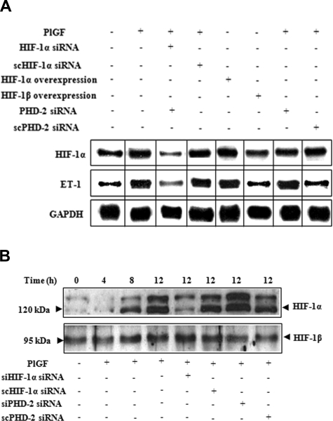
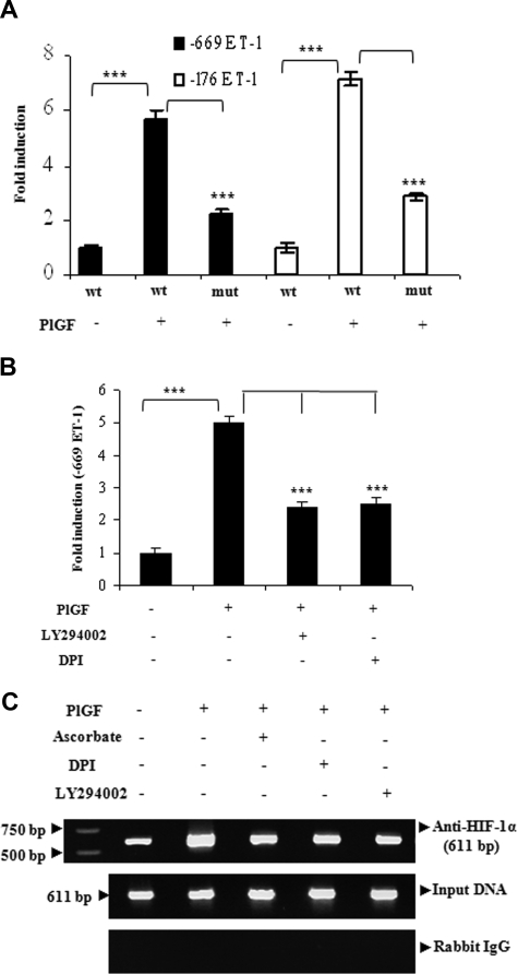
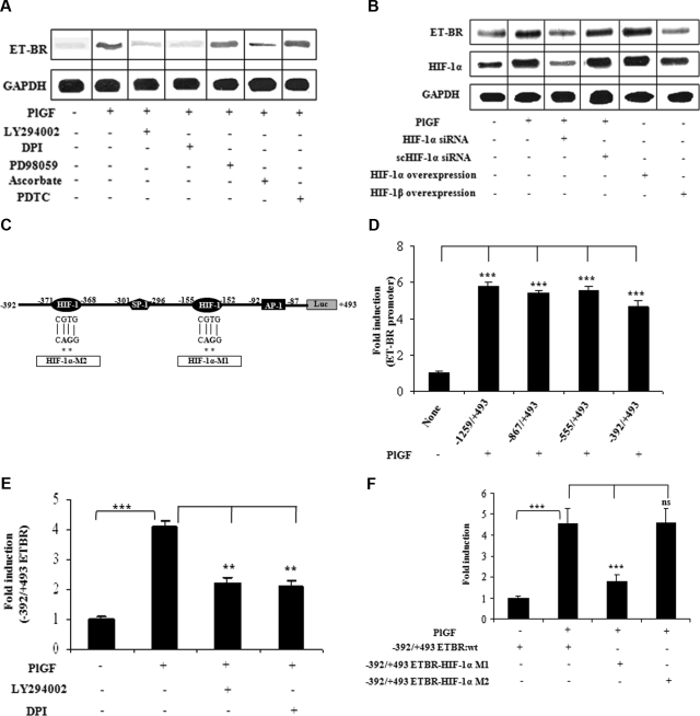
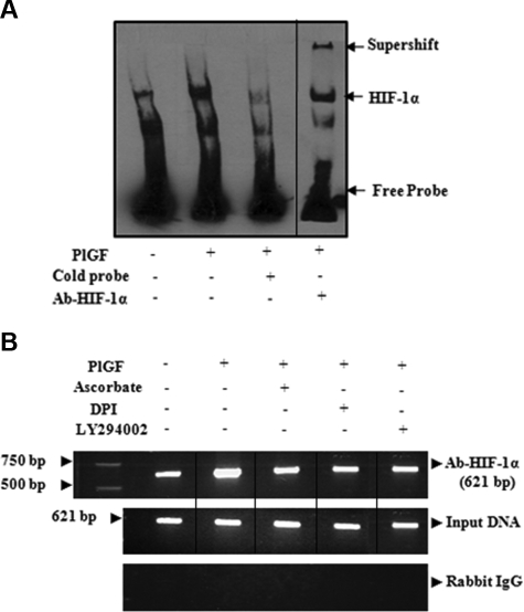
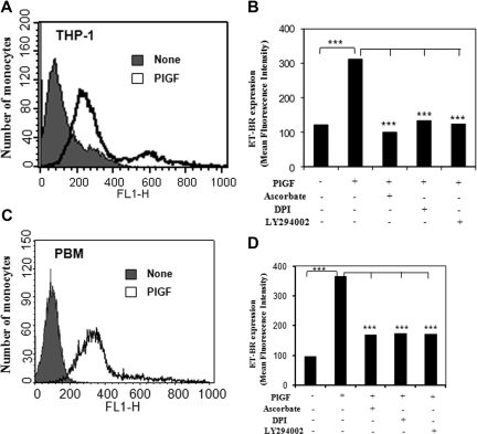
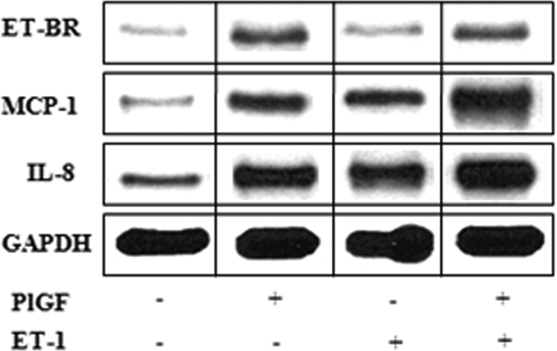
Pulmonary hypertension (PHT) develops in sickle cell disease (SCD) and is associated with high mortality. We previously showed that erythroid cells produce placenta growth factor (PlGF), which activates monocytes to induce proinflammatory cytochemokines, contributing to the baseline inflammation and severity in SCD. In this study, we observed that PlGF increased expression of endothelin-1 (ET-1) and endothelin-B receptor (ET-BR) from human pulmonary microvascular endothelial cells (HPMVECs) and monocytes, respectively. PlGF-mediated ET-1 and ET-BR expression occurred via activation of PI-3 kinase, reactive oxygen species and hypoxia inducible factor-1 alpha (HIF-1 alpha). PlGF increased binding of HIF-1 alpha to the ET-1 and ET-BR promoters; this effect was abrogated with mutation of hypoxia response elements in the promoter regions and HIF-1 alpha siRNA and confirmed by chromatin immunoprecipitation analysis. Furthermore, PlGF-mediated ET-1 release from HPMVECs and ET-BR expression in monocytes creates a PlGF-ET-1-ET-BR loop, leading to increased expression of MCP-1 and IL-8. Our studies show that PlGF-induced expression of the potent vasoconstrictor ET-1 and its cognate ET-BR receptor occur via activation of HIF-1 alpha, independent of hypoxia. PlGF levels are intrinsically elevated from the increased red cell turnover in SCD and in other chronic anemia (eg, thalassemia) and may contribute to inflammation and PHT seen in these diseases.


Figures

Figure 1
Figure 1
PlGF-mediated ET-1 expression involves PI-3 kinase, ROS, and HIF-1α. HPMVECs were treated with PlGF (250 ng/mL) for 6 hours, unless otherwise indicated, in the absence and presence of pharmacologic inhibitors or transfected with plasmids. (A-C) RNase protection assay. Data are representative of 3 different experiments. (D,E) ET-1 release by ELISA. Data are expressed as percentage change relative to untreated cells and represent means (± SE) of 3 independent experiments. ***P < .001; **P < .01; ns, P > .05. Where indicated, the vertical lines show the repositioned gel lanes.
Figure 2
Figure 2
The role of HIF-1α in PlGF-mediated ET-1 mRNA expression. HPMVECs were transfected with indicated plasmids followed by PlGF treatment for 6 hours. (A) RPA. (B) Western blot analysis of nuclear extracts (20 μg) using antibodies to HIF-1α and HIF-1β. Data are representative of 3 different experiments. Where indicated, the vertical lines show the repositioned gel lanes.
Figure 3
Figure 3
The regulation of ET-1 promoter by PlGF involves HIF-1α response elements. HPMVECs were transfected with indicated wild-type and mutant constructs followed by treatment with PlGF for 6 hours in the absence or presence of pharmacologic inhibitors. (A and B) relative luciferase activity. Data are expressed as means (± SE) of 3 independent experiments. ***P < .001. (C) HIF-1α binding to native chromatin was demonstrated by ChIP assay using HIF-1α or normal rabbit IgG antibodies. The ChIP products were amplified by PCR using primers flanking HIF-1α binding sites in the ET-1 promoter as shown in Table 1. Data are representative of 2 different experiments.
Figure 4
Figure 4
PlGF-mediated ET-BR expression involves PI-3kinase, ROS and HIF-1α. THP-1 monocytes were treated with PlGF for 24 hours in the absence and presence of inhibitors (A) and after transfection with siRNA constructs (B) and transfection with HIF expression plasmids (B). (A,B) RNase protection assay. (C) Schematics of ET-BR promoter showing the locations and sequence of HIF-1α binding sites and corresponding mutations as shown by asterisk. (D) PlGF-mediated ET-BR promoter luciferase activity of full-length and deletion constructs. (E) THP-1 cells were transfected with − 392/+493 ET-BR luciferase promoter construct, followed by treatment with PlGF in the absence and presence of LY294002 (15 μM) or DPI (10 μM). (F) PlGF-mediated − 392/+493 ET-BR luciferase promoter activity in THP-1 cells transfected with proximal HIF-1α binding site mutant (HIF-1α-MI) and distal HIF-1α binding site mutant (HIF-1α-M2) as shown in panel C. Data in panels D through F are expressed as means (± SE) of 3 independent experiments. ***P < .001; **P < .01; ns, P > .05. Where indicated, the vertical lines show the repositioned gel lanes.
Figure 5
Figure 5
PlGF promotes HIF-1α binding to DNA in vitro and in vivo. (A) EMSA demonstrating binding activity of HIF-1α in THP-1 nuclear extracts to its consensus DNA binding sequence. Where indicated, 50-fold excess of unlabeled probe or antibody to HIF-1α was added. Complex formation was visualized by autoradiography. (B) THP-1 cells were treated with PlGF for 4 hours in the absence and presence of pharmacologic inhibitors. The soluble chromatin was isolated and immunoprecipitated with either antibody to HIF-1α (top panel) or control rabbit IgG (bottom panel). The primers flanking HIF-1α binding sites in the ET-BR promoter as indicated in Table 1 were used to amplify the products by PCR. The bottom panel is amplification of input DNA before immunoprecipitation. Data are representative of 2 independent experiments. Where indicated, the vertical lines show the repositioned gel lanes.
Figure 6
Figure 6
FACS analysis of ET-BR surface expression in THP-1 and PBM. (A,C) THP-1 cells and PBM were treated with PlGF for 24 hours and surface expression of ET-BR was analyzed by flow cytometry. (B,D) The mean fluorescence intensity of ET-BR in PlGF treated THP-1 cells and PBM in the presence or absence of indicated pharmacologic inhibitors is shown as a histogram. Data are representative of 3 independent experiments in duplicates.
Figure 7
Figure 7
PlGF and ET-1 augment cytochemokine mRNA expression. THP-1 (106 cells/mL) were either treated with PlGF for 24 hours (250 ng/mL) or ET-1 (250 nmol/L) for 30 minutes. For priming, THP-1 cells were first treated with PlGF for 24 hours, washed with PBS and then treated with ET-1 for 30 minutes. Total mRNA was isolated and RPA was performed as described under “RNase protection assay.” The data are representative of 3 independent experiments. Where indicated, the vertical lines show the repositioned gel lanes.

Comment in

  • It really IS the red cell.
    Telen MJ. Telen MJ. Blood. 2008 Aug 1;112(3):459-60. doi: 10.1182/blood-2008-05-155754. Blood. 2008. PMID: 18650459 No abstract available.

References

    1. Francis RB, Jr., Johnson CS. Vascular occlusion in sickle cell disease: current concepts and unanswered questions. Blood. 1991;77:1405–1414. - PubMed
    1. Platt OS, Thorington BD, Brambilla DJ, et al. Pain in sickle cell disease. Rates and risk factors. N Engl J Med. 1991;325:11–16. - PubMed
    1. Buchanan G R. Infection. In: Embury SH, Hebbel RP, Mohandas N, Steinberg MH, editors. Sickle Cell Disease—Basic Principles and Clinical Practice. New York: Raven Press; 1994. pp. 567–587.
    1. Wong WY, Powars DR, Chan L, et al. Polysaccharide encapsulated bacterial infection in sickle cell anemia: a thirty year epidemiologic experience. Am J Hematol. 1992;39:176–182. - PubMed
    1. Embury S H, Hebbel R P, Steinberg M H, Mohandas N. Pathogenesis of vasoocclusion. In: Embury SH, Hebbel RP, Mohandas N, Steinberg MH, editors. Sickle Cell Disease—Basic Principles and Clinical Practice. New York: Raven Press; 1994. pp. 311–326.

Publication types

MeSH terms