. 2009 Apr;30(4):1051-9.
doi: 10.1002/hbm.20563.

White matter tract integrity in aging and Alzheimer's disease

Affiliations

White matter tract integrity in aging and Alzheimer's disease

Jessica S Damoiseaux et al. Hum Brain Mapp. 2009 Apr.

Abstract

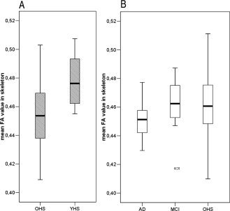
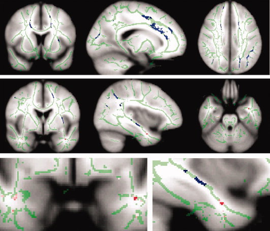
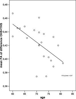
The pattern of degenerative changes in the brain white matter (WM) in aging, mild cognitive impairment (MCI), and Alzheimer's disease (AD) has been under debate. Methods of image analysis are an important factor affecting the outcomes of various studies. Here we used diffusion tensor imaging (DTI) to obtain fractional anisotropy (FA) measures of the WM in healthy young (n = 8), healthy elderly (n = 22), MCI (n = 8), and AD patients (n = 16). We then applied "tract-based spatial statistics" (TBSS) to study the effects of aging, MCI, and AD on WM integrity. Our results show that changes in WM integrity (that is, decreases in FA) are different between healthy aging and AD: in healthy older subjects compared with healthy young subjects decreased FA was primarily observed in frontal, parietal, and subcortical areas whereas in AD, compared with healthy older subjects, decreased FA was only observed in the left anterior temporal lobe. This different pattern of decreased anatomical connectivity in normal aging and AD suggests that AD is not merely accelerated aging.


Figures

Figure 1
Figure 1
Box plots depicting mean global FA values per group. The effect of aging and disease was computed separately, with a slightly (sub‐study specific) different mean FA‐skeleton. Therefore (A) aging and (B) disease are depicted separately in this figure. A significant decrease in global FA integrity (i.e. average FA across all skeleton voxels) is shown in figure (A) for normal aging (older healthy subjects (OHS) < younger healthy subjects (YHS); P = 0.008, one‐tailed) and in figure (B) for disease in AD < MCI (P < 0.047, one‐tailed). For AD < OHS a trend towards reduced global FA is shown (P < 0.065, one‐tailed); this is less significant than the AD < MCI difference because of the high spread in mean FA across the older healthy subjects. MCI and healthy older subjects do not differ in global FA.
Figure 2
Figure 2
Spatial maps of the results of between group voxel‐wise statistics. Differences in WM tract integrity are displayed in blue for older compared with younger healthy subjects and in red for AD compared with older healthy subjects. Images (coronal, sagittal, and axial view) are t‐statistics, P < 0.05 corrected. For visualization purposes the differences between AD and healthy older subjects is also displayed in pink at a more liberal threshold (P < 0.001, uncorrected). In green the “mean_FA_skeleton” is shown, only FA values projected on the skeleton were compared. These images are overlaid on the MNI152 standard brain. The left hemisphere of the brain corresponds to the right side of the image.
Figure 3
Figure 3
Correlations between FA integrity and age. Scatter plot of the correlation between mean FA within the WM areas affected by aging and age within the older healthy controls. A significant inverse correlation (ρ = −0.66, P = 0.0004) was observed, i.e. older age was related to lower mean FA.

References

    1. Ashburner J,Friston KJ ( 2000): Voxel‐based morphometry—The methods. Neuroimage 11: 805–821. - PubMed
    1. Behrens TE,Johansen‐Berg H,Woolrich MW,Smith SM,Wheeler‐Kingshott CA,Boulby PA,Barker GJ,Sillery EL,Sheehan K,Ciccarelli O,Thompson AJ,Brady JM,Matthews PM ( 2003): Non‐invasive mapping of connections between human thalamus and cortex using diffusion imaging. Nat Neurosci 6: 750–757. - PubMed
    1. Bookstein FL ( 2001): Voxel‐based morphometry should not be used with imperfectly registered images. Neuroimage 14: 1454–1462. - PubMed
    1. Bozzali M,Falini A,Franceschi M,Cercignani M,Zuffi M,Scotti G,Comi G,Filippi M ( 2002): White matter damage in Alzheimer's disease assessed in vivo using diffusion tensor magnetic resonance imaging. J Neurol Neurosurg Psychiatry 72: 742–746. - PMC - PubMed
    1. Conturo TE,Lori NF,Cull TS,Akbudak E,Snyder AZ,Shimony JS,McKinstry RC,Burton H,Raichle ME ( 1999): Tracking neuronal fiber pathways in the living human brain. Proc Natl Acad Sci USA 96: 10422–10427. - PMC - PubMed

Publication types