. 2008 Apr 15:9:15.
doi: 10.1186/1471-2172-9-15.

Dendritic cells are preferentially targeted among hematolymphocytes by Modified Vaccinia Virus Ankara and play a key role in the induction of virus-specific T cell responses in vivo

Affiliations

Dendritic cells are preferentially targeted among hematolymphocytes by Modified Vaccinia Virus Ankara and play a key role in the induction of virus-specific T cell responses in vivo

Luzheng Liu et al. BMC Immunol. .

Abstract

Background: Modified Vaccinia Ankara (MVA) is a highly attenuated strain of vaccinia virus (VV) that has lost approximately 15% of the VV genome, along with the ability to replicate in most mammalian cells. It has demonstrated impressive safety and immunogenicity profile in both preclinical and clinical studies, and is being actively explored as a promising vaccine vector for a number of infectious diseases and malignancies. However, little is known about how MVA interacts with the host immune system constituents, especially dendritic cells (DCs), to induce strong immune responses despite its inability to replicate in vivo. Using in vitro and in vivo murine models, we systematically investigated the susceptibility of murine DCs to MVA infection, and the immunological consequences of the infection.

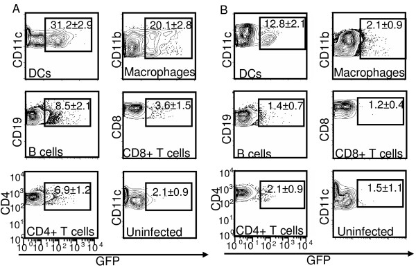
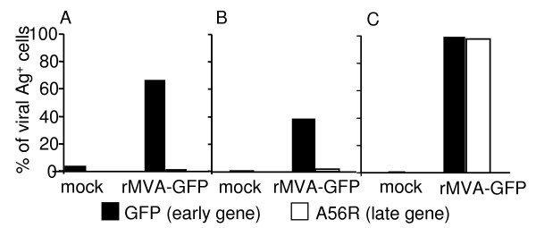
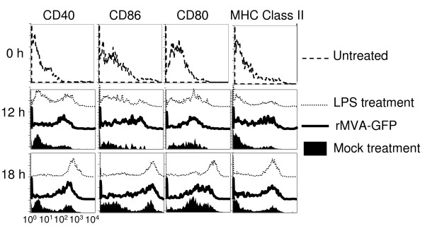
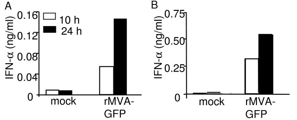
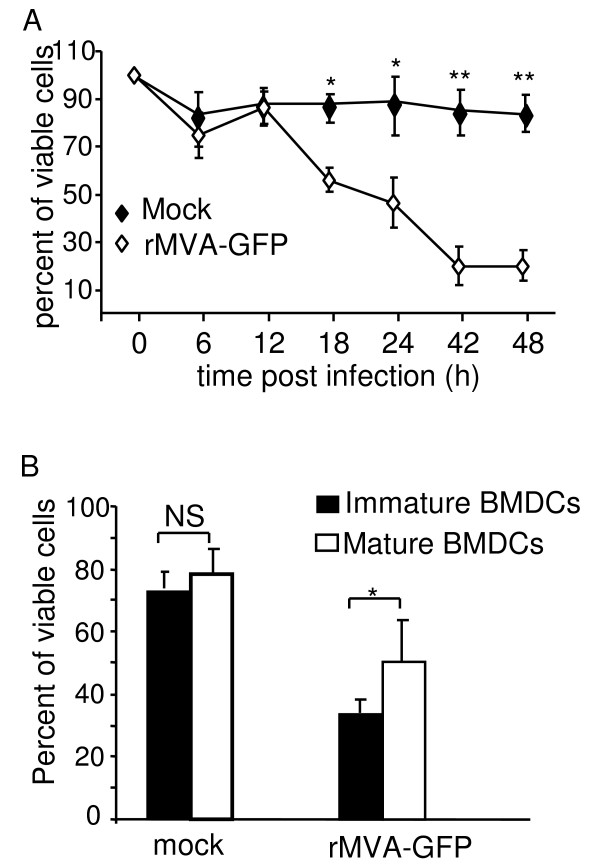
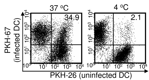
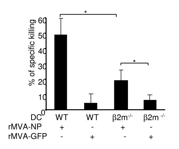
Results: Our data demonstrate that MVA preferentially infects professional antigen presenting cells, especially DCs, among all the subsets of hematolymphoid cells. In contrast to the reported blockage of DC maturation and function upon VV infection, DCs infected by MVA undergo phenotypic maturation and produce innate cytokine IFN-alpha within 18 h of infection. Substantial apoptosis of MVA-infected DCs occurs after 12 h following infection and the apoptotic DCs are readily phagocytosed by uninfected DCs. Using MHC class I - deficient mice, we showed that both direct and cross-presentation of viral Ags are likely to be involved in generating viral-specific CD8+ T cell responses. Finally, DC depletion abrogated the T cell activation in vivo.

Conclusion: We present the first in vivo evidence that among hematolymphoid cells, DCs are the most susceptible targets for MVA infection, and DC-mediated Ag presentation is required for the induction of MVA-specific immune responses. These results provide important information concerning the mechanisms by which strong immune responses are elicited to MVA-encoded antigens and may inform efforts to further improve the immunogenicity of this already promising vaccine vector.


Figures

Figure 1
Figure 1
MVA preferentially targeted professional APCs, especially DCs, both in vitro and in vivo. (A) Splenocytes from naïve BALB/c mice were infected with rMVA-GFP at a MOI of 10 in vitro for 8 h followed by cell surface marker staining and flow cytometry analysis. Uninfected splenocytes were used as negative control. (B) BALB/c mice were either uninfected or infected with rMVA-GFP at 3 × 10⁸ PFU/mouse by i.v. injection. Spleens were harvested at 9 h post infection and GFP expression in various subsets of splenocytes was monitored by flow cytometry. DCs: CD11c+; macrophages: CD1b+CD11c-; B cells: CD19+; CD8+ T cells: CD8+ CD11c-; CD4+ T cells: CD4+ CD11c-. Numbers shown are the percentages (average ± standard deviation (SD)) of GFP+ cells in the corresponding cell subsets. The data are representative of three independent experiments.
Figure 2
Figure 2
Only early but not late MVA genes were expressed from infected DCs. BMDCs (A), splenic CD11c+ DCs (B), and DF-1 cells (C) were infected with rMVA-GFP at a MOI of 10. Mock-treated cells were included as negative controls. MVA-encoded early gene (GFP) and late gene (A56R) expression was monitored 20 h post infection by flow cytometry. The data shown are representative of three independent experiments.
Figure 3
Figure 3
MVA infection induced rapid BMDC maturation. Immature BMDCs were mock treated, infected with rMVA-GFP at a MOI of 10, or stimulated with 10 μg/ml LPS as described in Methods. At 0, 12 and 18 h post treatment, cells were harvested and subjected to cell surface staining for the indicated maturation markers. Samples were analyzed by flow cytometry. Histograms were gated on GFP+ cells. These data are representative of five independent experiments.
Figure 4
Figure 4
MVA infection induced IFN-αproduction from DCs. Immature BMDCs (A) or splenic CD11c+ DCs (B) were either mock treated or infected with rMVA-GFP at a MOI of 10. Supernatant samples were collected at 10 h and 24 h post infection for IFN-α detection by ELISA. These data are representative of three independent experiments.
Figure 5
Figure 5
MVA infection significantly reduced the viability of BMDCs after 12 h of infection. (A) Kinetic analysis of DC viability following MVA infection. Immature BMDCs were either mock treated, or infected with rMVA-GFP at a MOI of 10. At various time points, cells were harvested and viable cells were counted with trypan blue exclusion method. These data represent the average ± SD viability of 6 replicates from 2 experiments. (B) Immature BMDCs and LPS-stimulated mature BMDCs were mock treated or infected with rMVA-GFP at a MOI of 10. Twenty four hours later, cell viability was examined by trypan blue exclusion. These data represent the average (± SD) viability of 15 replicates from 5 experiments. NS: statistically non-significant; *: P < 0.05; **: P < 0.01.
Figure 6
Figure 6
MVA-infected DCs were phagocytosed by uninfected DCs. Immature BMDCs were labeled with PKH-67 and infected with MVA at a MOI of 10 for 18 h. They were then extensively washed and mixed with PKH-26-labeled uninfected immature BMDCs for 4 h at either 37 C° or 4 C°. Phagocytosis of MVA-infected DCs by uninfected DCs were detected by flow cytometry. This data is representative of two independent experiments.
Figure 7
Figure 7
MVA-infected DCs induced Ag-specific CTL in vivo via both direct Ag presentation and cross-priming. BMDCs derived from either WT C57BL/6 (WT DCs) or MHC class I-deficient β2m-/- mice (β2m-/- DCs) were infected with rMVA-NP or control rMVA-GFP at a MOI of 10 for 6 h. The cells were washed and UV-irradiated to remove and inactivate any residual viruses. DCs were then intravenously injected into LCMV immune C57BL/6 mice 1 × (106). Five days later, NP396–404-specific CTL activity in spleens was assessed by fluorescent cellular cytotoxicity assay, as described in Methods. *: P < 0.05.
Figure 8
Figure 8
DCs were required for the generation of IFN-γ-producing effector T cells following MVA infection. CD11c-DTR mice were depleted of DC by i.p. injection of DT (12ng/g body weight). Six hours after DT treatment, mice were i.p. infected with 5 × 10⁶ PFU MVA. DT treatment was repeated on day 2 post infection. WT littermates were included as controls. At day 6 following the infection, spleens were harvested to prepare single cell suspension. (A) The efficiency of DC depletion was determined by surface cell lineage marker staining and flow cytometry. Numbers were the percentages of the gated population in total viable splenocytes. (B, C) Spleen single cell suspension (effector) was re-stimulated with MVA-infected C57BL/6 splenocytes (target) for 12 h in the presence of Brefeldin A. Following incubation, the frequencies of IFN-γ-producing T cells in the effector splenocytes were determined by intracellular cytokine staining, as described in Methods. Numbers were the percentages of IFN-γ+ cells in CD3+ T cell population. (C) The average ± SD percentages of IFN-γ+ cells for each group (n = 4). Data are representative of two independent experiments. **: P < 0.01.

References

    1. Willis NJ. Edward Jenner and the eradication of smallpox. Scott Med J. 1997;42:118–121. - PubMed
    1. Neff JM, Levine RH, Lane JM, Ager EA, Moore H, Rosenstein BJ, Millar JD, Henderson DA. Complications of smallpox vaccination United States 1963. II. Results obtained by four statewide surveys. Pediatrics. 1967;39:916–923. - PubMed
    1. Neff JM, Lane JM, Pert JH, Moore R, Millar JD, Henderson DA. Complications of smallpox vaccination. I. National survey in the United States, 1963. New England Journal of Medicine. 1967;276:125–132. - PubMed
    1. Mayr A, Stickl H, Muller HK, Danner K, Singer H. [The smallpox vaccination strain MVA: marker, genetic structure, experience gained with the parenteral vaccination and behavior in organisms with a debilitated defence mechanism (author's transl)] Zentralbl Bakteriol [B] 1978;167:375–390. - PubMed
    1. Meyer H, Sutter G, Mayr A. Mapping of deletions in the genome of the highly attenuated vaccinia virus MVA and their influence on virulence. J Gen Virol. 1991;72:1031–1038. - PubMed

Publication types

MeSH terms

Substances