

Parietal lobe deficits in frontotemporal lobar degeneration caused by a mutation in the progranulin gene
- PMID: 18413474
- PMCID: PMC2578869
- DOI: 10.1001/archneur.65.4.506
Parietal lobe deficits in frontotemporal lobar degeneration caused by a mutation in the progranulin gene
Abstract
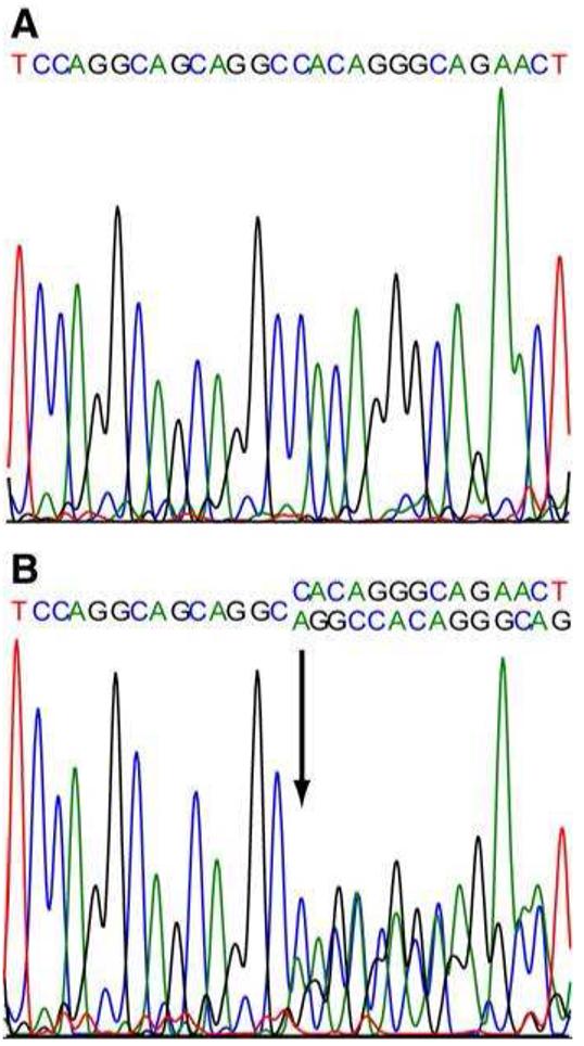
Objective: To describe the clinical, neuropsychologic, and radiologic features of a family with a C31LfsX35 mutation in the progranulin gene CCDS11483.1).
Design: Case series.
Patients: A large British kindred (DRC255) with a PGRN mutation was assessed. Affected individuals presented with a mean age of 57.8 years (range, 54-67 years) and a mean disease duration of 6.1 years (range, 2-11 years).
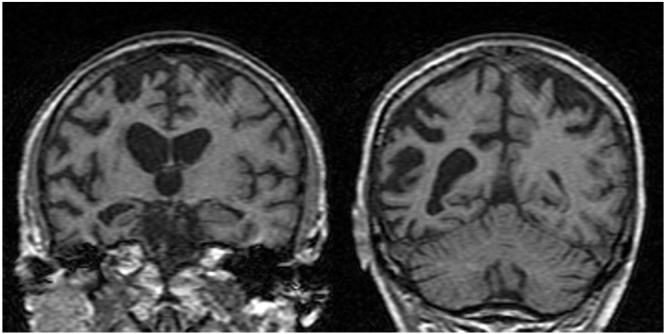
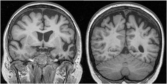
Results: All patients exhibited a clinical and radiologic phenotype compatible with frontotemporal lobar degeneration based on current consensus criteria. However, unlike sporadic frontotemporal lobar degeneration, parietal deficits, consisting of dyscalculia, visuoperceptual /visuospatial dysfunction, and/or limb apraxia, were a common feature, and brain imaging showed posterior extension of frontotemporal atrophy to involve the parietal lobes. Other common clinical features included language output impairment with either dynamic aphasia or nonfluent aphasia and a behavioral syndrome dominated by apathy.
Conclusion: We suggest that parietal deficits may be a prominent feature of PGRN mutations and that these deficits may be caused by disruption of frontoparietal functional pathways.
Figures

References
-
- Neary D, Snowden JS, Gustafson L, et al. Frontotemporal lobar degeneration: a consensus on clinical diagnostic criteria. Neurology. 1998 Dec;51(6):1546–54. - PubMed
-
- Kertesz A, Davidson W, Munoz DG. Clinical and pathological overlap between frontotemporal dementia, primary progressive aphasia and corticobasal degeneration: the Pick complex. Dement Geriatr Cogn Disord. 1999;10(Suppl 1):46–9. - PubMed
-
- Lomen-Hoerth C, Anderson T, Miller B. The overlap of amyotrophic lateral sclerosis and frontotemporal dementia. Neurology. 2002 Oct 8;59(7):1077–9. - PubMed
-
- Stevens M, van Duijn CM, Kamphorst W, et al. Familial aggregation in frontotemporal dementia. Neurology. 1998;50(6):1541–5. - PubMed
-
- Stanford PM, Brooks WS, Teber ET, et al. Frequency of tau mutations in familial and sporadic frontotemporal dementia and other tauopathies. J Neurol. 2004 Sep;251(9):1098–104. - PubMed
Publication types
MeSH terms
Grants and funding
LinkOut - more resources
Full Text Sources
Medical
Molecular Biology Databases
