. 2008 Apr 15;105(15):5705-9.
doi: 10.1073/pnas.0800691105. Epub 2008 Apr 14.

Activation of the lifespan regulator p66Shc through reversible disulfide bond formation

Affiliations

Activation of the lifespan regulator p66Shc through reversible disulfide bond formation

Melanie Gertz et al. Proc Natl Acad Sci U S A. .

Abstract

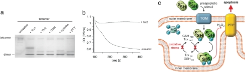
Cell fate and organismal lifespan are controlled by a complex signaling network whose dysfunction can cause a variety of aging-related diseases. An important protection against these failures is cellular apoptosis, which can be induced by p66(Shc) in response to cellular stress. The precise mechanisms of p66(Shc) action and regulation and the function of the p66(Shc)-specific N terminus remain to be identified. Here, we show that the p66(Shc) N terminus forms a redox module responsible for apoptosis initiation, and that this module can be activated through reversible tetramerization by forming two disulfide bonds. Glutathione and thioredoxins can reduce and inactivate p66(Shc), resulting in a thiol-based redox sensor system that initiates apoptosis once cellular protection systems cannot cope anymore with cellular stress.


Conflict of interest statement

The authors declare no conflict of interest.

Figures

Fig. 1.
Fig. 1.
Dimer–tetramer transition of p66CH2-CB regulating its apoptosis-inducing activity. (a) SEC elution profile of p66CH2-CB in the absence of reducing agent. (b) SEC elution profile of p66CH2-CB in the presence of 2.5 mM DTT, overlaid with the elution profile obtained with the mutant p66CH2-CB C59S in the absence of DTT (dashed line). (c) Stoichiometry of the two p66CH2-CB forms determined by BN-PAGE. (d) Standard experiment of the mitochondria swelling assay in the presence of 5 mM succinate. Rupture of mitochondria was observed after sequential addition of 7 μM CaCl2 and 20 μM p66CH2-CB instead of buffer (control). (e) Comparison of the apoptosis-inducing activity of dimeric and tetrameric p66CH2-CB and p66CH2-CB C59S. The initial sensitization with CaCl2 has been omitted for clarity. The decrease in OD620 observed for the dimeric p66CH2-CB and the p66CH2-CB C59S mutant beyond ≈400 s was also observed in buffer controls and is likely caused by sedimentation of the mitochondria during extended incubation.
Fig. 2.
Fig. 2.
Disulfide bridging through the conserved residue Cys-59 and accompanied structural rearrangements in p66CH2-CB. (a) Alignment of p66CH2-CB from various organisms around the conserved Cys-59 residue (▿). (b) Detection of free sulfhydryl groups in the dimer but not in the tetramer form by using Ellman's reagent. (c) Tetrameric p66CH2-CB subjected to reducing and nonreducing SDS/PAGE. (d) MALDI-TOF-MS spectrum of the chymotryptic digest of the p66CH2-CB tetramer including the disulfide-linked peptide Leu-68–Cys-59–S–S–Cys-59′–Leu-68′ (framed). (e) SEC elution profile of the tetramer after treatment with 2.5 mM DTT. (f and g) Time-dependent increase in p66CH2-CB-induced rupture of mitochondria (f) and its correlation with the protein's oxidation level determined by nonreducing SDS/PAGE (g). Dimeric p66CH2-CB was analyzed (t = 0), subsequently incubated on ice for 4 h, and then tested again (t = 4 h). (h) Limited proteolysis of p66CH2-CH dimer and tetramer with elastase. Even before the addition of elastase, fragmentation of the dimer but not the tetramer was observed, caused by proteases of the expression host. (i) Thermal denaturation of p66CH2-CH dimer and tetramer.
Fig. 3.
Fig. 3.
Copper-dependent ROS formation by p66CH2-CB and contribution of Cyt c to the apoptosis-inducing process. (a–c) Changes in fluorescence of H2DDFDA after sequential addition of the p66CH2-CB forms (20 μM) or buffer (control), 85 μM Na-dithionite (a–c) or 20 μM Cyt c (c), and 50 μM CuSO4 (a and c) or 50 μM of other metal ions (b). (d) Rupture of mitochondria after addition of 7 μM CaCl2 followed by buffer (control) or 0.3 μg/ml antimycin A, respectively, followed by addition of tetrameric p66CH2-CB. The experiment was done in the presence (control and + antimycin A) or absence (− succinate) of 5 mM succinate. (e) BN-PAGE of tetrameric p66CH2-CB treated with 85 μM Na-dithionite. (f) Rupture of mitochondria after addition of 7 μM CaCl2 followed by buffer (control), 280 units of catalase or 20 mM DMTU, and 20 μM tetrameric p66CH2-CB in the presence of 5 mM succinate. (g) Changes in fluorescence of H2DDFDA after sequential addition of mitochondria (100 μg total mitochondrial protein), succinate (5 mM), and dimeric or tetrameric p66CH2-CB (40 μM); as a control, buffer was added instead of protein.
Fig. 4.
Fig. 4.
Reduction of tetrameric p66CH2-CB by Trxs and GSH and model for the redox-mediated regulation of p66Shc. (a) Nonreducing SDS/PAGE of tetrameric p66CH2-CB incubated with a 2-fold molar excess of Trx1 and Trx2, 5 mM GSH, 5 mM cysteine, or 5 mM DTT. (b) Pretreatment of the p66CH2-CB tetramer with Trx2 reduces its apoptosis inducing activity. (c) Model of the redox-mediated regulation of p66Shc. After cellular stress dimeric p66Shc translocates to mitochondria. Under conditions of lower stress the Trx and GSH systems reduce tetrameric p66Shc and prevent apoptosis, whereas excessive oxidative stress leads to an overload of the redox systems and accumulation of active, tetrameric p66Shc. p66Shc-generated ROS then activate the PTP, which results in rupture of the mitochondria and release of apoptotic factors. MC, metal chaperone; TOM, translocase of the outer membrane.

References

    1. Balaban RS, Nemoto S, Finkel T. Mitochondria, oxidants, and aging. Cell. 2005;120:483–495. - PubMed
    1. Ranganathan AC, Adam AP, Aguirre-Ghiso JA. Opposing roles of mitogenic and stress signaling pathways in the induction of cancer dormancy. Cell Cycle. 2006;5:1799–1807. - PMC - PubMed
    1. Lee HC, Wei YH. Oxidative stress, mitochondrial DNA mutation, and apoptosis in aging. Exp Biol Med. 2007;232:592–606. - PubMed
    1. Hekimi S. How genetic analysis tests theories of animal aging. Nat Genet. 2006;38:985–991. - PubMed
    1. Migliaccio E, et al. The p66shc adaptor protein controls oxidative stress response and life span in mammals. Nature. 1999;402:309–313. - PubMed