. 2008 Apr 22;105(16):5955-62.
doi: 10.1073/pnas.0711220105. Epub 2008 Apr 14.

Global regulatory logic for specification of an embryonic cell lineage

Affiliations

Global regulatory logic for specification of an embryonic cell lineage

Paola Oliveri et al. Proc Natl Acad Sci U S A. .

Abstract

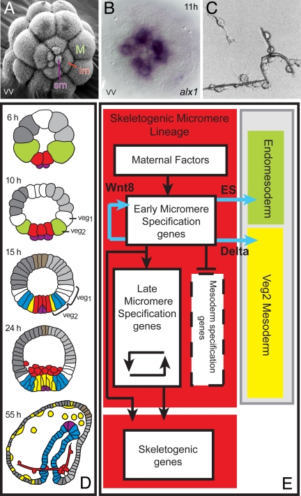
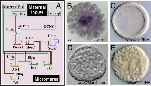
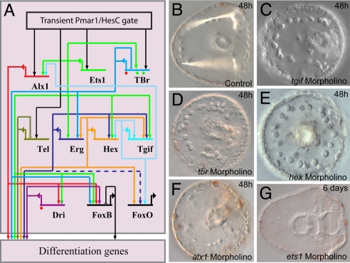
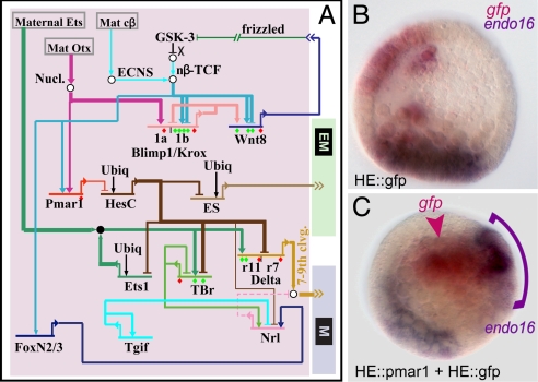
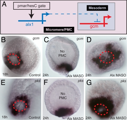
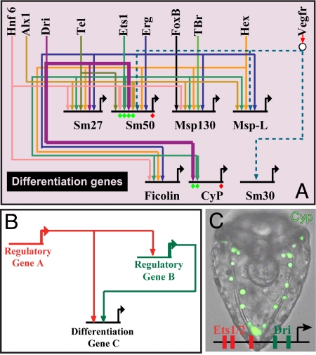
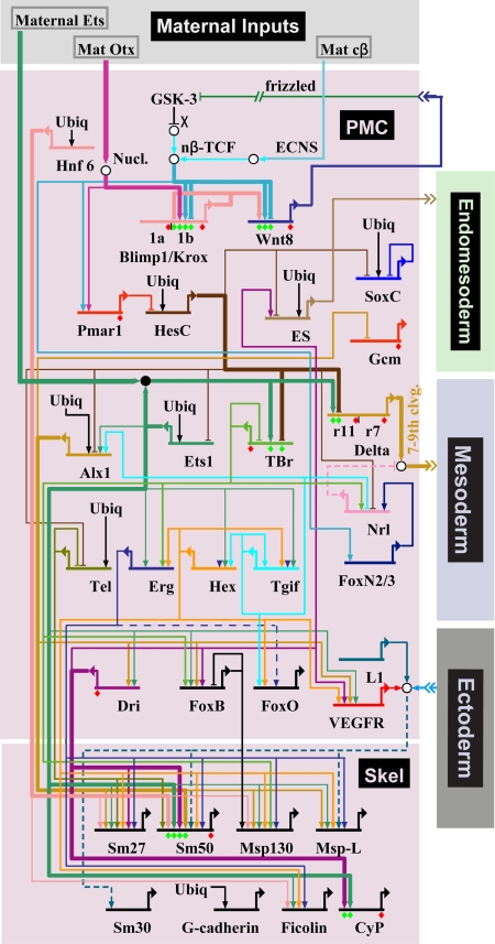
Explanation of a process of development must ultimately be couched in the terms of the genomic regulatory code. Specification of an embryonic cell lineage is driven by a network of interactions among genes encoding transcription factors. Here, we present the gene regulatory network (GRN) that directs the specification of the skeletogenic micromere lineage of the sea urchin embryo. The GRN now includes all regulatory genes expressed in this lineage up to late blastula stage, as identified in a genomewide survey. The architecture of the GRN was established by a large-scale perturbation analysis in which the expression of each gene in the GRN was cut off by use of morpholinos, and the effects on all other genes were measured quantitatively. Several cis-regulatory analyses provided additional evidence. The explanatory power of the GRN suffices to provide a causal explanation for all observable developmental functions of the micromere lineage during the specification period. These functions are: (i) initial acquisition of identity through transcriptional interpretation of localized maternal cues; (ii) activation of specific regulatory genes by use of a double negative gate; (iii) dynamic stabilization of the regulatory state by activation of a feedback subcircuit; (iv) exclusion of alternative regulatory states; (v) presentation of a signal required by the micromeres themselves and of two different signals required for development of adjacent endomesodermal lineages; and (vi) lineage-specific activation of batteries of skeletogenic genes. The GRN precisely predicts gene expression responses and provides a coherent explanation of the biology of specification.


Conflict of interest statement

The authors declare no conflict of interest.

Figures

Fig. 1.
Fig. 1.
Regulatory specificity and specification functions of the skeletogenic micromere lineage. (A) SEM image of fifth-cleavage embryo viewed from vegetal pole (vv), displaying small micromeres (sm), large micromeres (lm), and macromeres (M) (photograph by J. B. Morrill and L. Marcus, 2005). The embryos in this and all following figures are ≈70 μm in diameter. (B) Distinct regulatory state in large and small micromeres: expression of alx1 at midcleavage stage in large micromeres only. (C) Synthesis of calcite biomineral skeletal rods in vitro by descendants of isolated micromeres cultured in sea water with 2% horse serum. [Reproduced with permission from ref. (Copyright 1991, Japanese Society of Developmental Biologists).] (D) Territorial components of the sea urchin embryo in lateral view: green, macromeres; red, skeletogenic micromere lineage; purple, small micromeres; yellow, nonskeletogenic mesoderm; blue, gut endoderm; brown, apical neurogenic territory; dark gray, aboral ectoderm; light gray, oral ectoderm. Stages are: 6 h, fifth cleavage; 10 h, seventh cleavage; 15 h, early blastula; 24 h, mesenchyme blastula showing skeletogenic micromere lineage ingressed; 55 h, late gastrula with forming skeleton. (E) Process diagram (4), summarizing specification functions. Color coding of background is as in D; signal ligands produced by skeletogenic micromere lineage cells are in blue, transcriptional regulatory functions are in black.
Fig. 2.
Fig. 2.
Initial regulatory state and circuitry of the double negative gate. (A) Portion of GRN indicating initial inputs to the pmar1 gene, the double negative gate, and the target regulatory genes. The GRN in this and succeeding figures is represented in BioTapestry software (49). Thick lines indicate inputs validated by cis-regulatory analysis. ECNS, early cytoplasmic nuclear localization system. (B) WMISH display of pmar1 transcripts in micromeres, midcleavage, (vv, vegetal view). (C) Effect of blocking β-catenin nuclearization by injection of Δ-cadherin mRNA; all endomesodermal specification is blocked, including that of the micromere lineage, as shown earlier by others (16, 31). (D) Transformation of gastrula-stage embryo into solid ball of mesenchyme by ectopic expression of pmar1 mRNA. (E) Transformation of gastrula-stage embryo into solid ball of mesenchyme by use of anti-hesC morpholino.
Fig. 3.
Fig. 3.
State stabilization circuitry. (A) Portion of GRN displaying feedback circuitry and linkages among the regulatory genes that comprise the climax regulatory state of the skeletogenic micromere lineage during its specification period. Dashed line into foxo indicates the input from erg, which although supported by QPCR data, could be indirect, via other parallel linkages shown in the GRN. (B) Control late gastrula embryo displaying birefringent skeletal rods. (C–G) Failure of skeletogenesis when expression of the indicated regulatory genes is blocked [as also shown earlier for tbr (35) and alx1 (29)].
Fig. 4.
Fig. 4.
Control of signaling functions by the double negative gate. (A) Portion of the GRN displaying regulatory circuitry by which expression of signals (Wnt8, ES, Delta) is controlled in the micromere lineage. (B and C) Demonstration using double WMISH that the ES signal is a target of the double negative gate. (B) Control blastula stage embryo in which endogenous endo16 expression is displayed in purple in the vegetal plate and GFP mRNA produced by a construct under control of a hatching enzyme (HE) cis-regulatory module is shown in red, in the ectoderm. (C) Embryo bearing two cis-regulatory constructs under HE cis-regulatory control, one expressing pmar1, and the other expressing GFP, which marks the location of the ectopic pmar1 expression. The constructs are concatenated together in the egg and are incorporated together into the same cells. Ectopic endo16 transcript (purple arch) can be seen adjacent to the cells expressing gfp and pmar1 mRNA (red arrowhead), evidently in response to the ectopic production of the ES. [Reproduced with permission from ref. (Copyright 2003, Elsevier).]
Fig. 5.
Fig. 5.
Regulatory exclusion of mesodermal fate. (A) GRN subcircuit showing repression of gcm transcription by alx1 in micromere lineage (dashed line indicates this is not known to be a direct interaction). (B–D) WMISH demonstration of derepression of gcm in embryos in which alx1 expression is blocked: control (B); alx1 morpholino, lateral view (C); vegetal view comparable to B (D). (E–G) Same for the pks gene, a pigment cell differentiation gene downstream of gcm. Red dashed circle delimits the skeletogenic lineage territory in the vegetal plate.
Fig. 6.
Fig. 6.
Control of the differentiation gene batteries. (A) Portion of GRN displaying regulatory linkages to differentiation genes. sm27 and sm50 are biomineral matrix genes (27) expressed during the specification period. sm30 is a matrix gene expressed only after ingression, probably under control of signals from the ectoderm. Msp130, msp-L, ficolin, and cyclophilin (Cyp) are genes encoding cell biology functions of skeletogenesis (20). All genes shown on the top row encode transcription factors (compare Fig. 4) except for the receptor vegfR. (B) Canonical feed forward design of linkages to differentiation genes; compare Table S1. (C) GFP expression in skeletogenic mesenchyme, driven by a cis-regulatory construct from the cyclophilin differentiation gene. In the cases where there are multiple inputs shown of factors that might see the same target sites, such as Ets and Erg, and Tgif and Hex, and these genes are also interlocked in regulatory loops, the inputs shown could be redundant. [Reproduced with permission from ref. (copyright 2005, Elsevier).]
Fig. 7.
Fig. 7.
Overall current GRN for specification of the skeletogenic micromere lineage.

Comment in

Similar articles

Cited by

References

    1. Oliveri P, Davidson EH. Gene regulatory network controlling embryonic specification in the sea urchin. Curr Opin Genet Dev. 2004;14:351–360. - PubMed
    1. Davidson EH. The Regulatory Genome: Gene Regulatory Networks in Development and Evolution. San Diego: Academic; 2006.
    1. Davidson EH, et al. A genomic regulatory network for development. Science. 2002;295:1669–1678. - PubMed
    1. Sodergren E, et al. The genome of the sea urchin Strongylocentrotus purpuratus. Science. 2006;314:941–952. - PMC - PubMed
    1. Howard-Ashby M, et al. Identification and characterization of homeobox transcription factor genes in Strongylocentrotus purpuratus and their expression in embryonic development. Dev Biol. 2006;300:74–89. - PubMed

Publication types