. 2008 Apr 15;68(8):2764-72.
doi: 10.1158/0008-5472.CAN-07-6349.

Epigenetic inactivation of the canonical Wnt antagonist SRY-box containing gene 17 in colorectal cancer

Affiliations

Epigenetic inactivation of the canonical Wnt antagonist SRY-box containing gene 17 in colorectal cancer

Wei Zhang et al. Cancer Res. .

Erratum in

  • Cancer Res. 2008 Jul 15;68(14):6030

Abstract

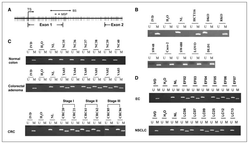
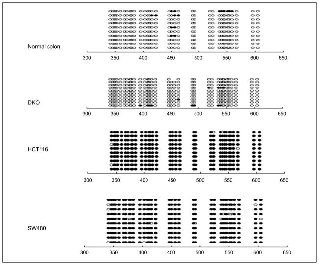
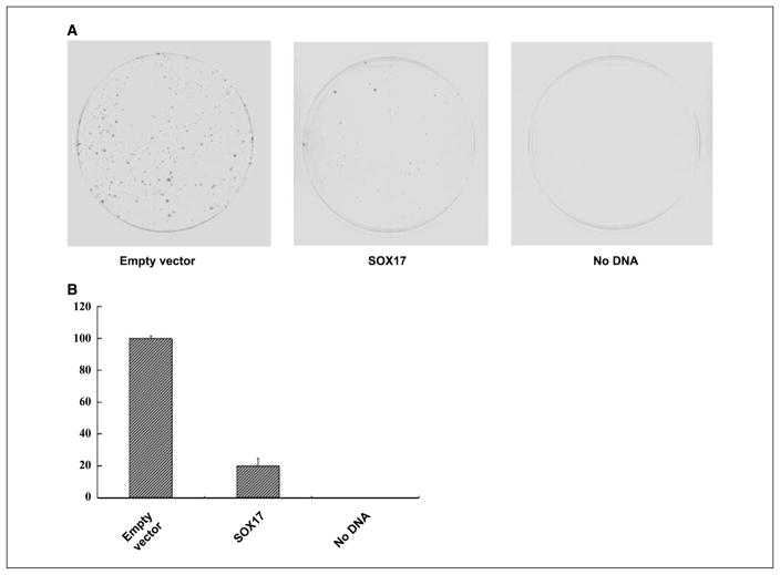
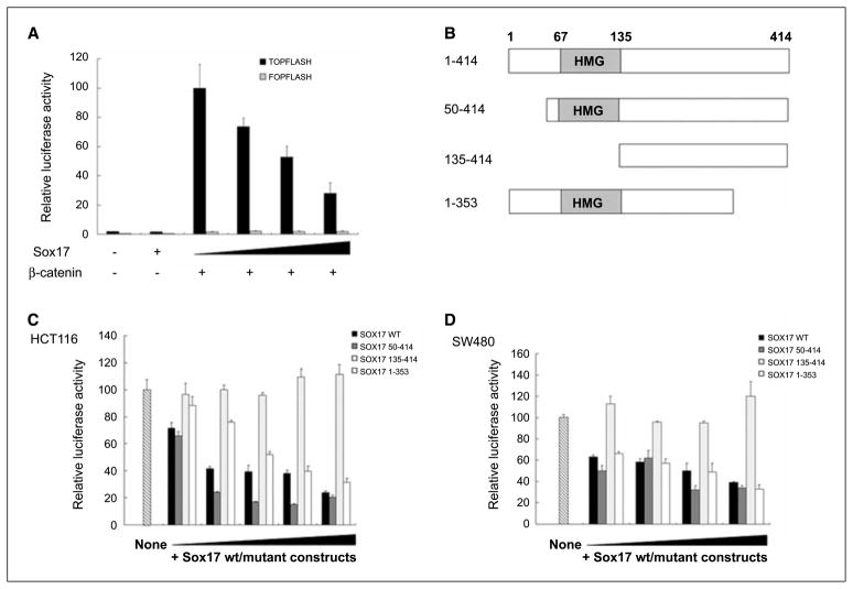
SRY-box containing gene 17 (Sox17) is a member of the high mobility group (HMG) transcription factor superfamily, which plays critical roles in the regulation of development and stem/precursor cell function, at least partly through repression of Wnt pathway activity. Modulators controlling aberrant Wnt signaling activation are frequently disrupted in human cancers through complementary effects of epigenetic and genetic changes. Our recent global analysis of CpG island hypermethylation and gene expression in colorectal cancer (CRC) cell lines revealed that SOX17 gene silencing is associated with DNA hypermethylation of a CpG island in the promoter region. Here, we report that CpG island methylation-dependent silencing of SOX17 occurs in 100% of CRC cell lines, 86% of colorectal adenomas, 100% of stage I and II CRC, 89% of stage III CRC, 89% of primary esophageal cancer, and 50% of non-small cell lung cancer. Overexpression of SOX17 in HCT116 CRC cells inhibits colony growth and beta-catenin/T-cell factor-dependent transcription. Structure-based deletion analysis further shows the presence of a Wnt signaling repression domain in the SOX17 HMG box. Together, our studies suggest that SOX17 is a negative modulator of canonical Wnt signaling, and that SOX17 silencing due to promoter hypermethylation is an early event during tumorigenesis and may contribute to aberrant activation of Wnt signaling in CRC.


Figures

Figure 1
Figure 1
Identification of SOX17 as a hypermethylated gene in CRC cells. A, the distribution of gene expression changes for HCT116 cells treated with trichostatin A (X axis) or DAC (Y axis) is analyzed and displayed (18). Black dots, individual genes; red dot, SOX17, which is a candidate DNA hypermethylated gene. SOX17 is identified by its position in a zone where gene expression did not respond to trichostatin A (<1.4-fold) but increased >2-fold with DAC treatment. B, RT-PCR analysis for expression of SOX genes in HCT116, DKO, and normal colonic mucosa. Gene symbols are indicated on the left; cell lines are indicated above the data; and NC indicates normal colon. β-Actin is used as a internal control. C, RT-PCR analysis for expression of SOX genes before and after treatment of cells with 1 μmol/L DAC (+) for 96 h. Restoration of SOX17 expression is observed in seven CRC cells. As a control for SOX17 expression, PCR was performed on DNase I–treated RNA without reverse transcription. D, Western blot analysis of SOX17 in different CRC cells. The positive control for SOX17 protein detection uses whole-cell lysate from HCT116 cells transfected with the pcDNA3.1-SOX17 expression vector. Left, of the five cell lines tested, endogenous SOX17 protein was only detected in DKO cells. β-Actin is shown as a gel loading control for the different cell lysates. Right, SOX17 protein is seen after treatment with 1 μmol/L DAC (+) for 96 h in HCT116 and Caco-2 cells.
Figure 2
Figure 2
Methylation of the SOX17 CpG island in cell lines and primary tumors. A, schematic of the SOX17 CpG island spanning the 5′ upstream region, exon1, intron 1, and exon 2. Bold vertical lines, individual CpG sites. TS, transcriptional start site in exon 1. Two small solid arrows, the location of primers used in the MSP assay. Double-headed arrow, the bisulfite genomic sequencing region (marked BS). B, aberrant methylation of SOX17 in human CRC cell lines. A visible PCR product in lanes marked U indicates the presence of unmethylated alleles; a visible PCR product in lanes marked M indicates the presence of methylated alleles. Note that in DKO cells, SOX17 is unmethylated, whereas in HCT116, RKO, SW48, Caco-2, SW480, LOVO, and DLD1 cells, the gene region queried is completely methylated. C, representative results of MSP analysis for SOX17 hypermethylation in human normal colon, adenomas, and stage I to III CRC. SOX17 is not methylated in normal colon but is frequently methylated in adenomas and in different stages of CRC. D, representative results of SOX17 methylation analysis in human esophageal squamous cancer (EC) and non–small cell lung cancer (NSCLC).
Figure 3
Figure 3
Bisulfite genomic DNA sequencing results of SOX17 in normal colon, DKO, HCT116, and SW480 cells. ○, unmethylated CpG sites; ●, methylated CpG sites. Locations of CpG sites are given relative to the transcription start site.
Figure 4
Figure 4
Suppression of cancer cell growth by SOX17. A, expression vectors encoding wild-type SOX17 or empty control vectors were transfected into HCT116 cells, which were then selected for G418 resistance. After 10 d, the cells were fixed with 10% formaldehyde and stained with Giemsa. B, quantitative analysis of surviving colonies after G418 selection. Each experiment was repeated three times and the average number of colonies is indicated with error bars on the histogram.
Figure 5
Figure 5
SOX17 inhibits Wnt stimulated transcription. A, SOX17 inhibits wild-type β-catenin–activated transcription. HEK293T cells were transfected with 70 ng of TOPFLASH or FOPFLASH plasmids, 7 ng of pRL-TK, and increasing amounts of pcDNA3.1-SOX17 or empty vector control (0, 30, 50, and 100 ng; black triangle, increasing dose), and stimulated for 48 h with cotransfection of 70 ng wild-type β-catenin expression vectors. The results are normalized to those for empty control vectors and are expressed as a relative ratio of firefly luciferase to Renilla luciferase. Bars, +1 SD. B, deletion analysis of SOX17. Individual SOX17 deletion mutants are depicted. The HMG box of SOX17 is shown in schematic form. C, SOX17 inhibits endogenous TCF/β-catenin–mediated transcription through the NH2-terminal HMG box. HCT116 cells were transfected with 70 ng of TOPFLASH, 7 ng of pRL-TK, and increasing amounts of pcDNA3.1-SOX17 or the different deletion mutants (SOX17 constructs 50–414, 135–414, and 1–353) or empty vectors (2, 10, 30, 50, and 100 ng; black triangle, increasing dose). Transfection of TOPFLASH reporter vectors reveals that HCT116 cells have high levels of endogenous β-catenin/TCF transcription activity (lane marked “None,” and set at 100% for normalization of data in all other lanes). Columns, mean of three independent experiments; bars, SD. D, a similar suppression effect mediated by the SOX17 HMG box is observed in SW480 cells. The SW480 cells were transfected with 10, 30, 50, or 100 ng SOX17 expression constructs, respectively. Data are expressed relative to the high basal activity in nontransfected cells as in C.

References

    1. Gubbay J, Collignon J, Koopman P, et al. A gene mapping to the sex-determining region of the mouse Y chromosome is a member of a novel family of embryonically expressed genes. Nature. 1990;346:245–50. - PubMed
    1. Wegner M. From head to toes: the multiple facets of Sox proteins. Nucleic Acids Res. 1999;27:1409–20. - PMC - PubMed
    1. Sohn J, Natale J, Chew LJ, et al. Identification of Sox17 as a transcription factor that regulates oligodendrocyte development. J Neurosci. 2006;26:9722–35. - PMC - PubMed
    1. Matsui T, Kanai-Azuma M, Hara K, et al. Redundant roles of Sox17 and Sox18 in postnatal angiogenesis in mice. J Cell Sci. 2006;119:3513–26. - PubMed
    1. Park KS, Wells JM, Zorn AM, Wert SE, Whitsett JA. Sox17 influences the differentiation of respiratory epithelial cells. Dev Biol. 2006;294:192–202. - PubMed

Publication types