. 2008 May;154(2):429-39.
doi: 10.1038/bjp.2008.95. Epub 2008 Apr 14.

Inhibition of the rapid component of the delayed rectifier potassium current in ventricular myocytes by angiotensin II via the AT1 receptor

Affiliations

Inhibition of the rapid component of the delayed rectifier potassium current in ventricular myocytes by angiotensin II via the AT1 receptor

Y H Wang et al. Br J Pharmacol. 2008 May.

Abstract

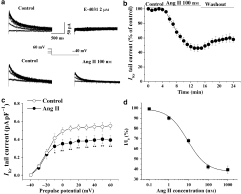
Background and purpose: There is increasing evidence that angiotensin II (Ang II) is associated with the occurrence of ventricular arrhythmias. However, little is known about the electrophysiological effects of Ang II on ventricular repolarization. The rapid component of the delayed rectifier K(+) current (I(Kr)) plays a critical role in cardiac repolarization. Hence, the aim of this study was to assess the effect of Ang II on I(Kr) in guinea-pig ventricular myocytes.

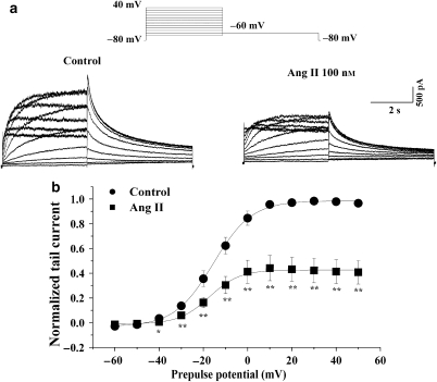
Experimental approach: The whole-cell patch-clamp technique was used to record I(Kr) in native cardiocytes and in human embryonic kidney (HEK) 293 cells, co-transfected with human ether-a-go-go-related gene (hERG) encoding the alpha-subunit of I(Kr) and the human Ang II type 1 (AT(1)) receptor gene.

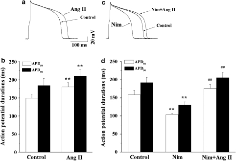
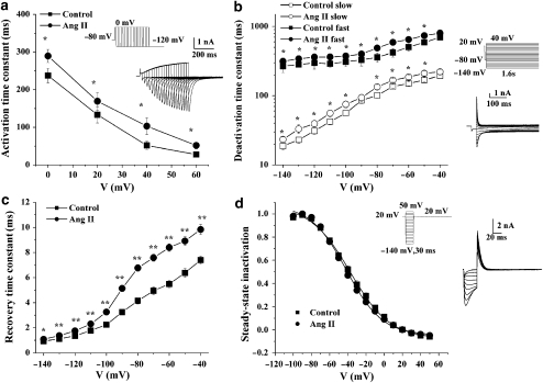
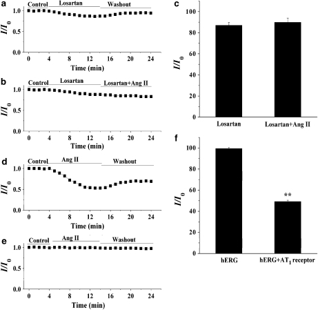
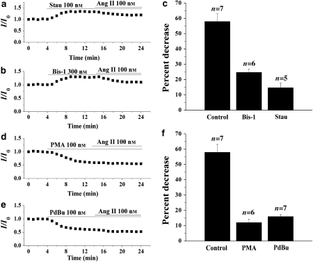
Key results: Ang II decreased the amplitude of I(Kr) in a concentration-dependent manner with an IC(50) of 8.9 nM. Action potential durations at 50% (APD(50)) and 90% (APD(90)) repolarization were prolonged 20% and 16%, respectively by Ang II (100 nM). Ang II-induced inhibition of the I(Kr) was abolished by the AT(1) receptor blocker, losartan (1 muM). Ang II decreased hERG current in HEK293 cells and significantly delayed channel activation, deactivation and recovery from inactivation. Moreover, PKC inhibitors, stausporine and Bis-1, significantly attenuated Ang II-induced inhibition of I(Kr).

Conclusions and implications: Ang II produces an inhibitory effect on I(Kr)/hERG currents via AT(1) receptors linked to the PKC pathway in ventricular myocytes. This is a potential mechanism by which elevated levels of Ang II are involved in the occurrence of arrhythmias in cardiac hypertrophy and failure.


Figures

Figure 1
Figure 1
Effects of angiotensin II (Ang II) on the rapid component of IK (IKr) in guinea-pig isolated ventricular cardiomyocytes. (a) Representative tail traces of IKr before (left), and after application of 2 μM E-4031 in one myocyte and 100 nM Ang II (right) to another myocyte. The ventricular myocyte was pulsed as shown. (b) The time course of the IKr current reduction by Ang II (100 nM). The effect of Ang II on IKr was quantitatively evaluated by measuring the amplitude of tail currents elicited on return to the potential of −40 mV after 2 s depolarizing pulse to 40 mV every minute. The percentage of decrease in the tail current was calculated in six cells. (c) Voltage-dependent activation of IKr was calculated from IKr tail current density before and 10 min after an application of 100 nM Ang II (n=6, *P<0.05, **P<0.01, versus before Ang II). (d) The concentration–response curve for the effect of Ang II on the IKr, IC50=8.9 nM (n=4–6 cells at each concentration). IK, the delayed rectifier K+ currents.
Figure 2
Figure 2
Effects of angiotensin II (Ang II) on action potentials. (a) Superimposed action potentials recorded before and 10 min after exposure to 100 nM Ang II. (b) A summary of the data for changes in APD at 50% of repolarization (APD50) and APD at 90% of repolarization (APD90; n=6, **P<0.01, Ang II versus control). (c) Superimposed action potentials recorded during exposure to Ang II (100 nM) in the presence of nimodipine (Nim, 1 μM). (d) A summary of the data for changes in APD50 and APD90 during exposure to Ang II in the presence of Nim (n=6, **P<0.01, Nim versus control, ##P<0.01, Nim plus Ang II versus Nim).
Figure 3
Figure 3
Effects of angiotensin II (Ang II) on ether-a-go-go-related gene (hERG) current. (a) Representative traces of hERG current recorded, using the pulse protocol shown, before (left) and after application of 100 nM Ang II (right) in a human embryonic kidney 293 (HEK293) cell. (b) Normalized IV relationships for tail currents in control conditions and in the presence of 100 nM Ang II (n=5, *P<0.05, **P<0.01, Ang II versus control). The solid lines represent fits to a Boltzmann function.
Figure 4
Figure 4
Effects of angiotensin II (Ang II; 100 nM) on ether-a-go-go-related gene (hERG) channel kinetics. (a) Activation time constants at test potential of 0, 20, 40 and 60 mV (n=5, *P<0.05, control versus Ang II). Inset: representative tracings for hERG current activation and pulse protocol. Activation time constants were obtained by fit with a single exponential function. (b) Time constants for fast and slow deactivation were plotted against the membrane potentials (n=6, *P<0.05, Ang II versus control). Inset: representative traces for hERG current deactivation and pulse protocol. Deactivation time constants were obtained by fit with double exponential function to the decay phase of the tail current. (c) The recovery time constants were plotted against the membrane potentials (n=5, *P<0.05, **P<0.01, control versus Ang II). Recovery from inactivation was determined by fitting a single exponential function to the initial ‘hook' preceding slower deactivation of tail currents shown in the inset of (d). (d) Normalized steady-state inactivation curves for control and after application of Ang II (n=5). Solid lines represent fits with Boltzmann function. Inset: representative current traces for steady-state inactivation and pulse protocol.
Figure 5
Figure 5
The angiotensin II (Ang II) type 1 (AT1) receptor mediates the inhibition of the rapid component of IK (IKr)/ether-a-go-go-related gene (hERG) current by Ang II. For the measurement of the amplitude of IKr tail currents, myocytes were stepped to 0 mV for 2 s from a holding potential of −40 mV and then repolarized to a test potential of −40 mV. In human embryonic kidney 293 (HEK293) cells, hERG tail currents were elicited on repolarization to a test potential of −60 mV after a 0-mV, 4-s long step from the holding potential of −80 mV. (a) The effect of losartan (1 μM) on IKr. (b) The effect of Ang II (100 nM) on IKr in the presence of losartan. (c) A summary of the percentage of decrease in the amplitude of IKr tail current (n=5). (d) Effect of Ang II (100 nM) on the hERG current recorded from HEK293 cells co-expressing hERG and AT1 receptors. (e) Effect of Ang II (100 nM) on the hERG current recorded from cells expressing the hERG channel without the AT1 receptor. (f) A summary of the results obtained from (e) (n=5, **P<0.01, hERG versus hERG plus AT1 receptor). IK, the delayed rectifier K+ currents.
Figure 6
Figure 6
Effects of buffering intracellular Ca2+ on the action of angiotensin II (Ang II; 100 nM). (a) The effects of an internal application of 20 mM BAPTA on the response of the rapid component of IK (IKr) to Ang II. (b) A summary of the data for the normalized IKr tail current in the presence of Ang II in myocytes dialysed with control pipette solution containing 5 mM EGTA and pipette solution containing 20 mM BAPTA (n=6). The amplitude of the IKr tail currents was measured using the same pulse protocol as that described in Figure 5. IK, the delayed rectifier K+ currents.
Figure 7
Figure 7
Effects of protein kinase C (PKC) inhibitors and activators on the response of the rapid component of IK (IKr) to angiotensin II (Ang II; 100 nM). (a and b) Effects of (a) the nonspecific PKC inhibitor, staurosporin (Stau; 100 nM) and (b) the specific PKC inhibitor, Bis-1 (300 nM), on the inhibition of IKr tail current by Ang II. (c) A summary of the data expressed as a percentage of decrease of IKr tail current by Ang II in the presence of Stau or Bis-1. (d and e) Effects of the PKC activators (d) PMA (100 nM) and (e) PdBu (100 nM), on the inhibition of IKr tail current by Ang II. (f) A summary of the data expressed as a percentage of decrease of IKr tail current by Ang II in the presence of PMA or PdBu. The amplitude of the IKr tail currents was measured using the same pulse protocol described in Figure 5. IK, the delayed rectifier K+ currents.
Figure 8
Figure 8
The effect of the protein kinase A (PKA) inhibitor, H-89, on the response of the rapid component of IK (IKr) to angiotensin II (Ang II). (a) Effects of H-89 (10 μM) on the inhibition of IKr by Ang II. (b) A summary of the data expressed as a percentage of decrease of IKr tail current by Ang II in the presence of H-89. The amplitude of the IKr tail currents was measured using the same pulse protocol described in Figure 5. IK, the delayed rectifier K+ currents.

Similar articles

Cited by

References

    1. Ahmmed GU, Dong PH, Song G, Ball NA, Xu Y, Walsh RA, et al. Changes in Ca(2+) cycling proteins underlie cardiac action potential prolongation in a pressure-overloaded guinea pig model with cardiac hypertrophy and failure. Circ Res. 2000;86:558–570. - PubMed
    1. Aiello EA, Cingolani HE. Angiotensin II stimulates cardiac L-type Ca(2+) current by a Ca(2+)- and protein kinase C-dependent mechanism. Am J Physiol Heart Circ Physiol. 2001;280:H1528–H1536. - PubMed
    1. Alberte C, Zipes DP. Use of nonantiarrhythmic drugs for prevention of sudden cardiac death. J Cardiovasc Electrophysiol. 2003;14:S87–S95. - PubMed
    1. Bragat AC, Blumenfeld J, Sealey JE. Effect of high-performance liquid chromatography on plasma angiotensin II measurements in treated and untreated normotensive and hypertensive patients. J Hypertens. 1997;15:459–465. - PubMed
    1. Brooksby P, Robinson PJ, Segal R, Klinger G, Pitt B, Cowley AJ. Effects of losartan and captopril on QT dispersion in elderly patients with heart failure. ELITE Study Group. Lancet. 1999;354:395–396. - PubMed

Publication types

MeSH terms