. 2008 Jun;154(3):675-87.
doi: 10.1038/bjp.2008.114. Epub 2008 Apr 14.

Aldosterone and the autocrine modulation of potassium currents and oxidative stress in the diabetic rat heart

Affiliations

Aldosterone and the autocrine modulation of potassium currents and oxidative stress in the diabetic rat heart

Y Shimoni et al. Br J Pharmacol. 2008 Jun.

Abstract

Background and purpose: Aldosterone plays a major role in cardiac pathology. This study was designed to investigate the role of cardiac aldosterone in modulating K(+) currents and oxidative stress in the streptozotocin-induced diabetic rat heart.

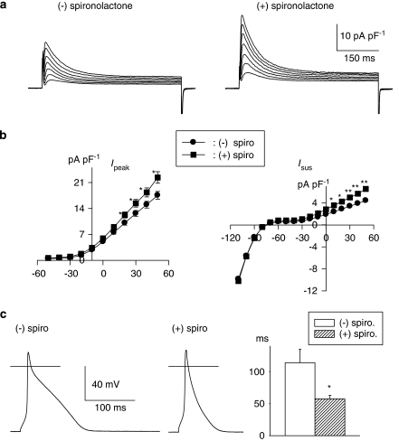
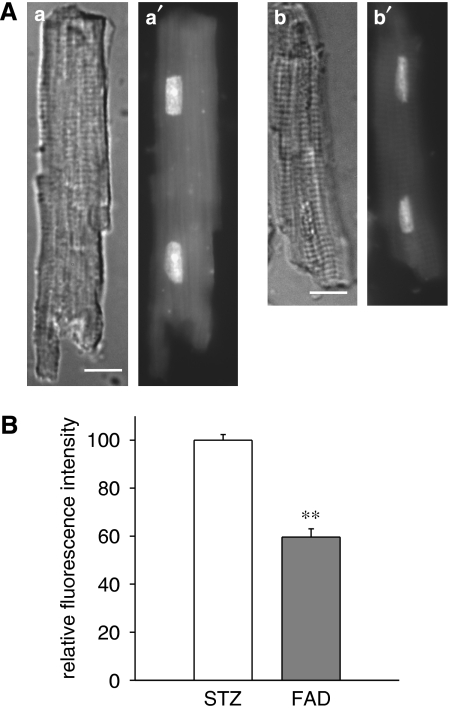
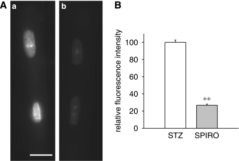
Experimental approach: Transient and sustained K(+) currents were measured in ventricular myocytes by voltage clamp. Plasma and cellular aldosterone were measured by ELISA. Fluorescent dihydroethidium (DHE) was used to assess superoxide ions as markers of oxidative stress.

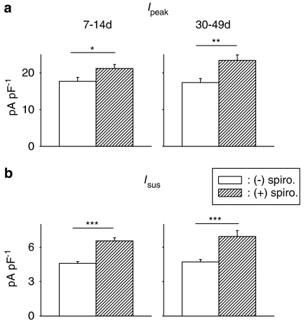
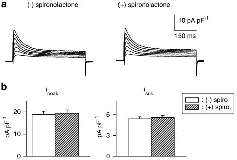
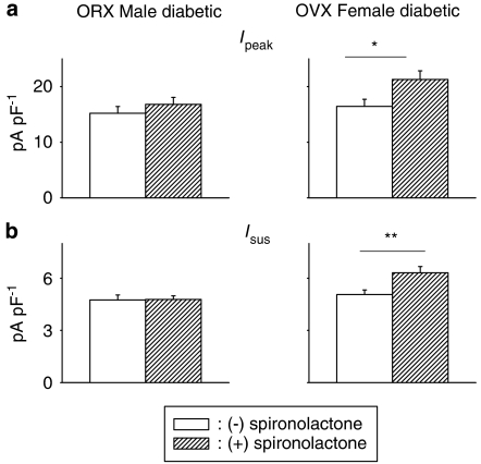
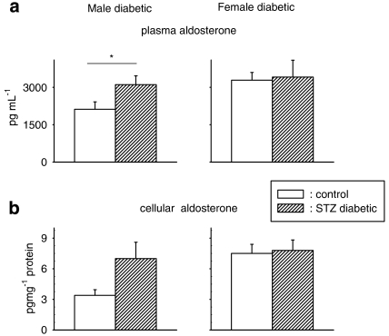
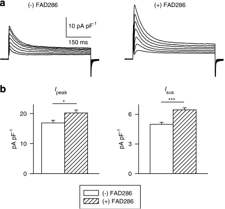
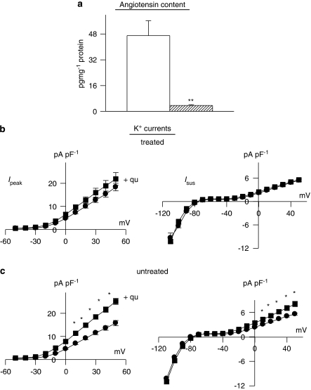
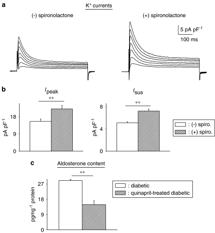
Key results: The mineralocorticoid antagonist spironolactone (1 microM, 5-9 h) significantly augmented both K(+) currents in diabetic males, with a concomitant shortening of the action potential but had no effect in myocytes from control males or from diabetic females. Effects of spironolactone were restored in ovariectomized diabetic females and abolished in orchidectomized diabetic males. The aldosterone synthase inhibitor FAD286 (1 microM, 5-9 h) significantly augmented K(+) currents in cells from diabetic males, but not females. Spironolactone and FAD286 significantly reduced oxidative stress in cells from diabetic males. Plasma aldosterone content was elevated in diabetic males (relative to control), but not in females. Cellular aldosterone was also elevated, but not significantly. The elevation in aldosterone was only partly dependent on a concomitant increase in cellular angiotensin II.

Conclusions and implications: A gender-related, sex-hormone-dependent elevation in plasma and cardiac cell aldosterone contributed to oxidative stress and to attenuation of K(+) currents in diabetic male rats. Aldosterone may thus contribute to diabetes-associated cardiac arrhythmias. Aldosterone elevation was partly related to levels of angiotensin II, but residual, angiotensin II-independent, aldosterone maintains functional relevance.


Figures

Figure 1
Figure 1
Effects of spironolactone on outward K+ currents and action potentials in ventricular cells from diabetic male rats. (a) Current traces in response to 500 ms pulses from a holding potential of −80 mV to potentials ranging from −10 to +50 mV in cells without (left) or following 8 h in 1 μM spironolactone (right). (b) Mean current densities (±s.e.mean) as a function of membrane potential (Ipeak on the left and Isus on the right) in the absence (n=33) or presence (n=40) of spironolactone (spiro; 1 μM, 5–9 h). Both currents were significantly (*P<0.05; **P<0.005) augmented by the aldosterone receptor antagonist at potentials at or above +10 mV. (c) Action potentials (at 1 Hz), recorded from two cells obtained from a diabetic rat heart. The left panel shows an action potential from an untreated cell, whereas the middle panel shows an action potential from a cell treated with 1 μM spironolactone for 6.5 h. The right panel shows the summary data for the action potential durations at −60 mV, in the absence (n=10) or presence (1 μM, >5 h, n=12) of spironolactone (spiro). *P<0.015.
Figure 2
Figure 2
Comparison of spironolactone effects on ventricular K+ currents in short- and long-term diabetic rats. Cells were obtained 7–14 days (left column) after STZ injection (100 mg kg−1) or 5–7 weeks (right column) after STZ (60 mg kg−1). Mean densities (at +50 mV) of Ipeak (a, top row) and Isus (b, bottom row) are shown, in the absence or following 5–9 h in 1 μM spironolactone (spiro). Currents were significantly augmented after short- and long-term diabetes (*P<0.05; **P<0.005; ***P<0.0005).
Figure 3
Figure 3
Effects of spironolactone on K+ currents in ventricular cells from diabetic females. (a) Current traces (same protocol as Figure 1) in the absence (left) or presence (right) of spironolactone (1 μM, 7.5 h). (b) Mean current densities as a function of voltage for Ipeak (left) and for Isus (right) in the absence or presence of spironolactone (spiro; 5–9 h). In cells from diabetic females, aldosterone inhibition has no effect on either current, in contrast to the augmentation observed in males.
Figure 4
Figure 4
Assessment of contribution of sex hormones to gender differences in spironolactone effects. Cells from orchidectomized (ORX) diabetic males (left column) and from ovariectomized (OVX) diabetic females (right column) were used. (a) Mean Ipeak current density (at +50 mV) in diabetic ORX males (left), OVX females (right) in the absence or following incubation with spironolactone (1 μM, 5–9 h). (b) Corresponding densities of Isus in the same groups. Ipeak and Isus are significantly augmented by spironolactone in ovariectomized diabetic females, with no effect in ORX diabetic males, suggesting that the gender differences are both androgen and oestrogen dependent (*P<0.05; **P<0.01).
Figure 5
Figure 5
Aldosterone content, measured by ELISA, in plasma (a) and in ventricular cells (b) from control and diabetic male (left column) and female (right column) rats. Plasma aldosterone was significantly (P<0.05) elevated in male diabetic rats (n=10) as compared with controls (n=10), but not in females (5 controls, 7 diabetics). Cells (n=15) from diabetic male rats had higher, but not significantly (P<0.055), levels of aldosterone than cells (n=13) from control male rats. In cells from diabetic females (n=7), the levels were comparable to those in cells from control females (n=5) (*P<0.05).
Figure 6
Figure 6
Effects of the aldosterone synthase inhibitor FAD286 on K+ currents in ventricular cells from diabetic male rats. (a) Sample current traces from two cells (same protocol as above), either with no drug added (left), or following 6 h in FAD286 (right). (b) Mean current densities at +50 mV, for Ipeak (left) and Isus (right), illustrating the significant augmentation by the drug (*P<0.05; ***P<0.0005).
Figure 7
Figure 7
Decrease in dihydroethidium (DHE) fluorescence in myocytes from male diabetic rats following exposure to the aldosterone synthase inhibitor FAD286. (A) Differential interference contrast (a, b) and DHE fluorescence (a′, b′) images of isolated cardiac myocytes from male STZ diabetic rats exposed for 5 h to vehicle only (a, a′) or 1 μM FAD286 (b, b′). Images shown were of cells from the same myocyte preparation and were collected at the same camera settings and exposure times. Bars in (a, b)=10 μm. In this experiment, the mean fluorescence intensity of the nuclei in (b′) was 73% of the mean intensity of the nuclei in (a′). (B) Summary of results. Mean fluorescence intensity of the nuclei from myocytes exposed to FAD286 (FAD) is expressed relative to the mean fluorescence intensity from untreated (STZ rat) cells from the same preparation. The results from cells from four separate hearts are combined; **significant differences at P<10−4;158 control nuclei and 134 nuclei exposed to FAD were analysed.
Figure 8
Figure 8
Decrease in dihydroethidium (DHE) fluorescence in myocytes from male diabetic rats following exposure to the mineralocorticoid antagonist spironolactone. (A) DHE fluorescence images of isolated cardiac myocytes from male STZ rats exposed for 5 h to vehicle only (a) or 1 μM spironolactone (b). Images shown were of cells from the same myocyte preparation and were collected at the same camera settings and exposure times. Bar in (a)=10 μm. In this experiment, the mean fluorescence intensity of the nuclei in (b) was 36% of the mean intensity of the nuclei in (a). (B) Summary of results. Mean fluorescence intensity of the nuclei from myocytes exposed to spironolactone (SPIRO) is expressed relative to the mean fluorescence intensity from control (STZ) cells from the same cell preparation. The results from cells from three separate hearts are combined; **significant differences at P<10−4; 126 control nuclei and 95 nuclei exposed to spironolactone were analysed.
Figure 9
Figure 9
Effects of angiotensin-converting enzyme (ACE) inhibition in vivo. Male rats were given quinapril (6 mg L−1) in their drinking water for 3 weeks prior to induction of diabetes with STZ. Cells were isolated after a further 8–13 days (quinapril continued). (a) Content of angiotensin II was very significantly (P<0.005) reduced (n=4) in comparison to untreated diabetic males (n=4). (b) The functional implications of this, indicating that in vitro exposure to quinapril (+qu) of cells isolated from rats treated with quinapril in vivo (n=36) had no effect on Ipeak (left) or Isus densities (right), as a function of membrane potential (n=30). This is in marked contrast to the in vitro effect of quinapril in untreated diabetic rats, as shown in (c). In untreated diabetic rats, both Ipeak and Isus are significantly (P<0.05) augmented by quinapril (*P<0.05; **P<0.001).
Figure 10
Figure 10
Effects of chronic angiotensin-converting enzyme (ACE) inhibition on aldosterone. (a) Sample current traces (same protocol as above) in cells from diabetic males treated with quinapril in vivo, in the absence of (left), or following (right) spironolactone (1 μM, 6.5 h). (b) Summary data for current densities at +50 mV. Ipeak is shown on the left and Isus on the right. Open bars show mean data from untreated cells (n=31) and hatched bars represent data from cells treated with spironolactone (n=17). Aldosterone inhibition can still significantly augment both currents even when angiotensin II levels are drastically reduced. (c) Aldosterone content (measured by ELISA) is significantly reduced in quinapril-treated diabetic rats (n=4, hatched bars), relative to untreated diabetic rats (n=4, open bars), illustrating that some of the elevation in aldosterone is related to augmented angiotensin II levels. However, there is a functionally significant elevation of aldosterone even in the absence of angiotensin II (**P<0.01).

References

    1. Abo K, Ishida Y, Yoshida R, Hozumi T, Ueno H, Shiotani H, et al. Torsade de Pointes in NIDDM with long QT intervals. Diabetes Care. 1996;9:1010. - PubMed
    1. Bell DSH. Diabetic cardiomyopathy. Diabetes Care. 2003;26:2949–2951. - PubMed
    1. Benitah JP, Perrier E, Gomez AM, Vassort G. Effects of aldosterone on transient outward K+ current density in rat ventricular myocytes. J Physiol. 2001;537:151–160. - PMC - PubMed
    1. Bos R, Mougenot N, Findji L, Mediani O, Vanhoutte PM, Lechat P. Inhibition of catecholamine-induced cardiac fibrosis by an aldosterone antagonist. J Cardiovasc Pharmacol. 2005;45:8–13. - PubMed
    1. Brouillette J, Trepanier-Boulay V, Fiset C. Effect of androgen deficiency on mouse ventricular repolarization. J Physiol. 2003;546:403–413. - PMC - PubMed

Publication types