. 2008 May;118(5):1657-68.
doi: 10.1172/JCI34487.

Kidney injury molecule-1 is a phosphatidylserine receptor that confers a phagocytic phenotype on epithelial cells

Affiliations

Kidney injury molecule-1 is a phosphatidylserine receptor that confers a phagocytic phenotype on epithelial cells

Takaharu Ichimura et al. J Clin Invest. 2008 May.

Abstract

Following injury, the clearance of apoptotic and necrotic cells is necessary for mitigation and resolution of inflammation and tissue repair. In addition to macrophages, which are traditionally assigned to this task, neighboring epithelial cells in the affected tissue are postulated to contribute to this process. Kidney injury molecule-1 (KIM-1 or TIM-1) is an immunoglobulin superfamily cell-surface protein not expressed by cells of the myeloid lineage but highly upregulated on the surface of injured kidney epithelial cells. Here we demonstrate that injured kidney epithelial cells assumed attributes of endogenous phagocytes. Confocal images confirm internalization of apoptotic bodies within KIM-1-expressing epithelial cells after injury in rat kidney tubules in vivo. KIM-1 was directly responsible for phagocytosis in cultured primary rat tubule epithelial cells and also porcine and canine epithelial cell lines. KIM-1 was able to specifically recognize apoptotic cell surface-specific epitopes phosphatidylserine, and oxidized lipoproteins, expressed by apoptotic tubular epithelial cells. Thus, KIM-1 is the first nonmyeloid phosphatidylserine receptor identified to our knowledge that transforms epithelial cells into semiprofessional phagocytes.


Figures
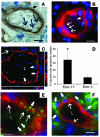
Figure 1
Figure 1. Kim-1–expressing tubule epithelial cells bind and internalize apoptotic bodies and necrotic debris in rat kidneys following ischemic injury.
(A) By light microscopy, necrotic cellular debris (seen by differential interference contrast [DIC]) binds to apically located Kim-1 (dark brown) of surviving tubule epithelial cells (large arrows). An apoptotic body is seen in 1 Kim-1–positive epithelial cell (small arrow). (B) By fluorescence microscopy many DAPI-positive (blue) apoptotic bodies (large arrows) can be seen binding to the surface of Kim-1–positive (red) epithelial cells, and Kim-1–expressing cells have processes (red, small arrow) internalizing an apoptotic body (blue). In addition, apoptotic bodies have been internalized by Kim-1–positive epithelial cells (blue, arrowhead). (C) Confocal image of apoptotic cells (blue) localized in phagocytic cups (small arrows) on the Kim-1–positive (red) apical surface of tubular cells. An internalized apoptotic cell is indicated by a large arrow. L denotes the tubule lumen. (D) The proportion of Kim-1–positive tubules containing internalized apoptotic bodies was greater than adjacent Kim-1–negative tubules in the postischemic kidney. Neither Kim-1 nor apoptotic bodies were identified in the normal kidneys. *P = 0.01 (error bars indicate SD). (E) Kim-1–positive epithelial cells (red) binding many TUNEL-positive apoptotic bodies (green, large arrows) and internalizing other small apoptotic bodies (green, small arrow). (F) Kim-1–positive epithelial cells (red, large arrows) are surrounded by CD68-positive macrophages (green, small arrows), which do not express Kim-1 and are found only in the interstitium. Scale bar: 10 μm.
Figure 2
Figure 2. Primary cultured kidney epithelial cells express Kim-1 and phagocytose apoptotic cells by a Kim-1–dependent mechanism.
(A) Following coculture with apoptotic thymocytes, Kim-1–positive (Kim-1+) (red with blue nuclei [N]) but not Kim-1–negative (Kim-1–) (blue nuclei only [n]) epithelial cells show avid binding (arrowheads) and internalization of fluorescently labeled apoptotic thymocytes (green and blue) (arrows). Note marked ring enhancement of phagosomes with Kim-1, and Kim-1 at the phagocytic cup of bound apoptotic cells. (B) Image of primary cultured rat epithelial cells all expressing cytokeratin (green) but showing heterogenous expression of Kim-1 (red). Scale bars: 10 μm. (C) The number of apoptotic cells bound or phagocytosed per 100 Kim-1–positive or 100 Kim-1–negative epithelial cells following coculture with labeled apoptotic cells and washing to remove bound cells. Note Kim-1–positive cells show avid phagocytosis. **P < 0.001. (D) Phagocytic index (number apoptotic cells/100 phagocytes) of Kim-1–positive primary epithelial cell cultures pretreated with monoclonal anti-rat Kim-1 affinity purified antibodies (15 μg/ml) followed by coculture with labeled apoptotic cells. Epithelial cells were lifted from plates and single epithelial cells in suspension scored for phagocytic index. Note that anti–Kim-1 antibodies directed at the extracellular domain block phagocytosis when compared with cells preincubated with isotype control antibodies. *P < 0.01.
Figure 3
Figure 3. KIM-1–expressing kidney epithelial cell lines avidly bind and phagocytose apoptotic and necrotic material.
(A) KIM-1 (red) in a KIM1-PK1 cell (left panel) is expressed at high levels (arrows) at the point of binding of multiple apoptotic thymocytes (green and blue) and is part of the initial phagocytic cup (arrowhead). Scale bar: 5 μm. At later time points (right panel), KIM-1 remains associated with the internalized apoptotic cell, resulting in ring enhancement (arrow) of the apoptotic body. The cell border is highlighted by broken lines. Scale bar: 10 μm. (B) Multiple apoptotic thymocytes labeled with CMFDA (green) were localized intracellularly in KIM-1–expressing cells after coculture. Internalized apoptotic thymocytes are visualized in the confocal plane of cortical actin filaments (red) in this confocal image confirming internalization. Cell nuclei (N) are highlighted. Scale bar: 10 μm. (C) DIC with fluorescence (green) microscopic images of KIM1-PK1 cells confirm ingestion of CMFDA-labeled (green) apoptotic LLC-PK1 cells (left panel) or sonicated LLC-PK1 cell debris (middle panel). pCDNA-PK1 cells in the same experiment showing no phagocytosis of apoptotic cells (right panel). These microscopic studies confirm internalization of fluorescent apoptotic or necrotic cell debris (arrowheads). Original magnification, ×60.
Figure 4
Figure 4. Quantitative analysis of KIM-1–mediated apoptotic cell and necrotic material phagocytosis.
Flow cytometric plots of green fluorescence against side scatter (SSC) for KIM1-PK1 and pcDNA-PK1 epithelial cells that have ingested fluorescently labeled (CMFDA) apoptotic thymocytes (A), apoptotic LLC-PK1 cells (B), or necrotic debris (necrotic LLC-PK1 cells) (C) in a phagocytosis assay. Percentages represent the proportion of epithelial cells that have ingested fluorescently labeled material. KIM1-PK1 cells that have not ingested apoptotic cells or debris were used to define the gated area. Without coculture with necrotic cells, only 0.93% of KIM-1–expressing epithelial cells were identified in the gated area. (D) Flow cytometric plots of green fluorescence against side scatter for KIM1–tet-off MDCK epithelial cells that have ingested fluorescently labeled (CMFDA) apoptotic LLC-PK1 in a phagocytosis assay. MDCK cells were either treated with doxycycline (100 ng/ml) to inhibit expression of the KIM-1 (left panel) or no doxycycline was used (5 days), permitting high-level expression of KIM-1 (right panel). Values represent the percentage of epithelial cells that have ingested fluorescently labeled apoptotic cells. KIM1–tet-off MDCK cells that have not ingested apoptotic cells or debris were used to define no ingestion.
Figure 5
Figure 5. KIM-1 mediates phagocytosis of apoptotic necrotic cells but not other phagocytotic targets, zymosan or latex beads.
(A) Graph showing percentage of KIM1-PK1 or pcDNA-PK1 cells that have internalized fluorescent apoptotic LLC-PK1 cells after 1 hour incubation with apoptotic fluorescent green labeled cells at 37°C or 4°C (on ice). At 4°C, binding but not internalization occurs. KIM1-PK1 cells showed much less fluorescence at 4°C than at 37°C (**P = 0.006; n = 3 per condition; error bars indicate SD). (B) Graph showing phagocytosis (black bars) by KIM1-PK1 cells as assessed by flow cytometry (left axis; percentage fluorescent cells) or binding plus phagocytosis (white bars) as assessed by spectrophotometry (right axis; relative fluorescence intensity). Labeled apoptotic thymocytes were incubated with KIM1-PK1 cells that had been pretreated with cytochalasin D (30 μM), nocodazole (30 μM), or vehicle. Total (bound plus phagocytosed) thymocytes were equivalent in each group; however, phagocytosis was inhibited by cytochalasin D and nocodazole. (C) Fluorescence images of KIM1-PK1 cells following coincubation with fluorescence-labeled zymosan particles (left, 0.5 mg/ml), latex beads (center, Fluorosphere; 1:800 dilution), or heparin (right, 25 μg/ml) for 1 hour at 37°C. Note no uptake of any of these particles. (D) Preincubation of KIM1-PK1 cells with anti–Kim-1 antibodies (AWE2), but not IgG, reduced phagocytic index (number of apoptotic LLC-PK1 cells/KIM1-PK1 phagocyte) as assessed by flow cytometry (50 μg/ml, left panel). Right panels show photomicrographs of KIM1-PK1 cells after internalization and binding of CMFDA-labeled apoptotic LLC-PK1 cells that had been pretreated (1 hour) with soluble mKIM1-Fc (bottom right panel, 0.8 μg/ml) or IgG-treated (top right panel). Pretreatment with mKIM1-Fc inhibited phagocytosis. Original magnification, ×40 (C); ×10 (D).
Figure 6
Figure 6. The KIM-1 ectodomain binds specifically to the surface of apoptotic epithelial cells and binds specifically to PS and PE.
(A) Flow cytometric histogram plots of fluorescence of normal live LLC-PK1 epithelial cells (left panel) or apoptotic LLC-PK1 epithelial cells (right panel), labeled with KIM1-Fc followed by anti–hIgG-FITC (green), anti–hIgG-FITC alone (red), or no reagents (blue). Note an approximately 50-fold increase in binding of KIM1-Fc to apoptotic cells. (B) Representative photomicrographs of an apoptotic LLC-PK1 cell labeled with KIM1-Fc (green, right panel) and a normal, live LLC-PK1 cell labeled identically (left panel). Nuclei were faintly stained with DAPI (blue) in both cells. Original magnification, ×60. (C) Graph of mean peak fluorescence for binding of KIM1-Fc to apoptotic cells in the absence or presence of the calcium chelators EDTA/EGTA, assessed by flow cytometry. KIM1-Fc binding was abolished by calcium chelators, and binding was restored by the addition of an excess of calcium to the chelators (*P = 0.006). (D) Graph of phagocytosis inhibition by PS liposomes. Pretreatment of KIM-1–expressing cells with PS liposomes almost completely abolished KIM-1–mediated phagocytosis of apoptotic cells (A.C.) (**P = 0.007), while equimolar PC liposomes had no effect. Error bars indicate SD. (E) In vitro binding curves for purified KIM1-Fc binding to equimolar phospholipid coated ELISA plates. KIM1-Fc binding was detected by anti-human IgG — HRP conjugated antibody followed by a colorimetric assay. Note KIM1-Fc binds to aminophospholipids PS and PE but not PC or anionic phosphatidic acid (PA), whereas control Fc proteins, human IgG and c-Ret–Fc do not bind.
Figure 7
Figure 7. Macrophages do not express Kim-1/Tim-1.
RT-PCR (left panel) for Kim1 mRNA from day 7 mouse bone marrow macrophages cultured with LPS or dexamethasone. Emr1 (F4/80 antigen) and Gapdh were used as controls, and mouse kidney cDNA 48 hours following ischemia was used as a positive control. Immunoblot (right panel) for Kim-1 in protein lysates from cultured macrophages and postischemic mouse kidney. Note that mouse bone marrow–derived macrophages generate neither kim1 transcript nor Kim-1 protein, in quiescent or activated states. BMMf, bone marrow–derived macrophages.
Figure 8
Figure 8. KIM-1–expressing epithelial cells bind and internalize ox-LDL, and the KIM-1 ectodomain binds specifically to ox-LDL.
(A) Graph showing internalization of DiI-labeled ox-LDL or native LDL by KIM1-PK1 cells or pcDNA-PK1 cells over 1 hour at 37°C, as quantified by spectrofluorometry of lysed cells. (B) Photomicrographs of KIM1-PK1 cells and pcDNA-PK1 cells showing internalized ox-LDL or native LDL in intracellular vesicles. Original magnification, ×40. (C) Graph showing the effect of a 40-fold excess of unlabeled ox-LDL on internalization of DiI-labeled ox-LDL by KIM1-PK1 cells or pcDNA-PK1 cells incubated at 37°C for 1 hour. Uptake of fluorescent lipoprotein was quantified by spectrofluorometry of lysed cells. (D) Graph showing the effect of doxycycline on KIM1–tet-off MDCK cells’ capacity to internalize DiI-labeled ox-LDL and DiI-labeled native LDL. In the presence of doxycycline (DOX+), KIM-1 expression is suppressed. In these conditions, there is little uptake of labeled lipoprotein after 1 hour. In the absence of doxycycline (DOX-), KIM-1 expression is not suppressed, and there is marked uptake of both lipoproteins. (E) In vitro binding curves for purified KIM1-Fc, human IgG1, or c-Ret–Fc proteins to ox-LDL coated ELISA plates. KIM1-Fc binding was detected by anti-human IgG – HRP-conjugated antibody followed by a colorimetric assay. Note KIM1-Fc strongly binds to ox-LDL but not uncoated plastic, whereas control proteins human IgG and c-Ret–Fc do not bind ox-LDL or plastic. (F) Graph showing the effect of pretreatment of KIM1-PK1 cells with either 40 or 50 μg/ml of ox-LDL on phagocytosis of fluorescently labeled apoptotic LLC-PK1 cells as assessed by flow cytometry. (*P = 0.0009 [40 μg/ml ox-LDL]; **P = 0.0003 [50 μg/ml ox-LDL] compared with no ox-LDL pretreatment. Error bars indicate SD).
Figure 9
Figure 9. KIM1-PK1 cells but not pcDNA-PK1 cells bind and internalize gram negative (E. coli) and gram positive (S. aureus) bacteria.
KIM-1 increases the capacity of epithelial cells to phagocytose both E. coli and S. aureus bacteria. % Phagocytosis, percentage of cells with internalized bacteria. Error bars indicate SD.

References

    1. Bonventre J.V. Dedifferentiation and proliferation of surviving epithelial cells in acute renal failure. J. Am. Soc Nephrol. 2003;14:S55–S61. doi: 10.1097/01.ASN.0000067652.51441.21. - DOI - PubMed
    1. Kelly K.J., et al. Intercellular adhesion molecule-1 deficient mice are protected against renal ischemia. J. Clin. Invest. 1996;97:1056–1063. doi: 10.1172/JCI118498. - DOI - PMC - PubMed
    1. Thadhani R., Pascual M., Bonventre J.V. Acute renal failure. N. Engl. J. Med. 1996;334:1448–1460. doi: 10.1056/NEJM199605303342207. - DOI - PubMed
    1. Henson P.M., Hume D.A. Apoptotic cell removal in development and tissue homeostasis. Trends Immunol. 2006;27:244–250. doi: 10.1016/j.it.2006.03.005. - DOI - PubMed
    1. Savill J., Dransfield I., Gregory C., Haslett C. A blast from the past: clearance of apoptotic cells regulates immune responses. Nat. Rev. Immunol. 2002;2:965–975. doi: 10.1038/nri957. - DOI - PubMed

Publication types

MeSH terms