. 2008 May;118(5):1632-44.
doi: 10.1172/JCI29226.

ROCK1 mediates leukocyte recruitment and neointima formation following vascular injury

Affiliations

ROCK1 mediates leukocyte recruitment and neointima formation following vascular injury

Kensuke Noma et al. J Clin Invest. 2008 May.

Abstract

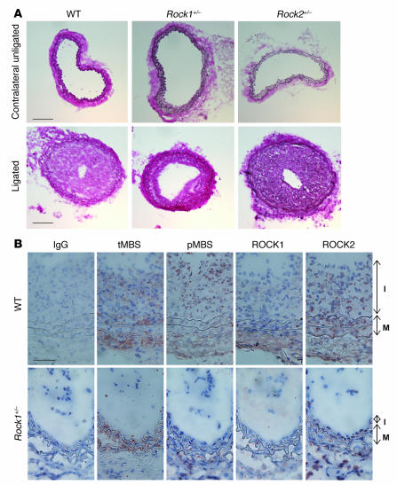
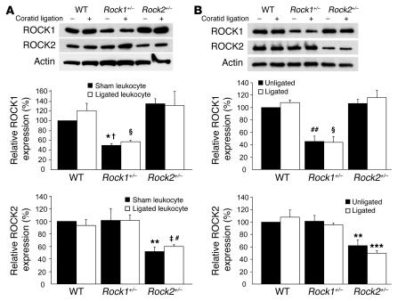
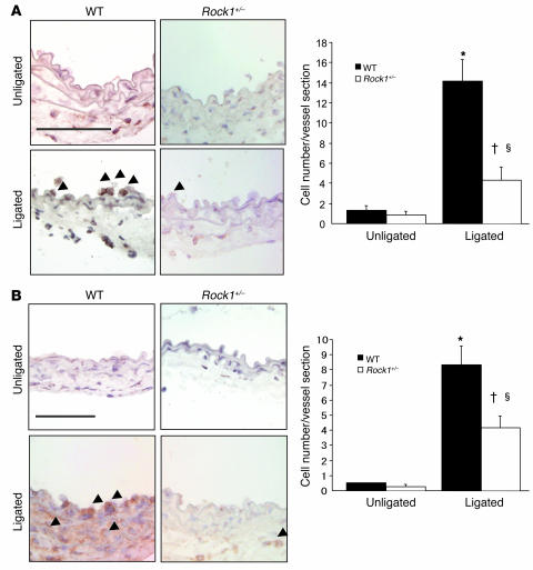
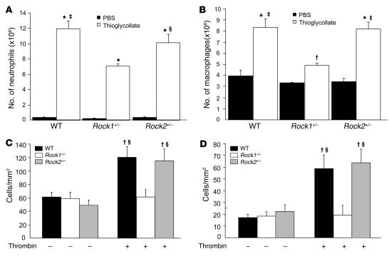
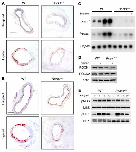
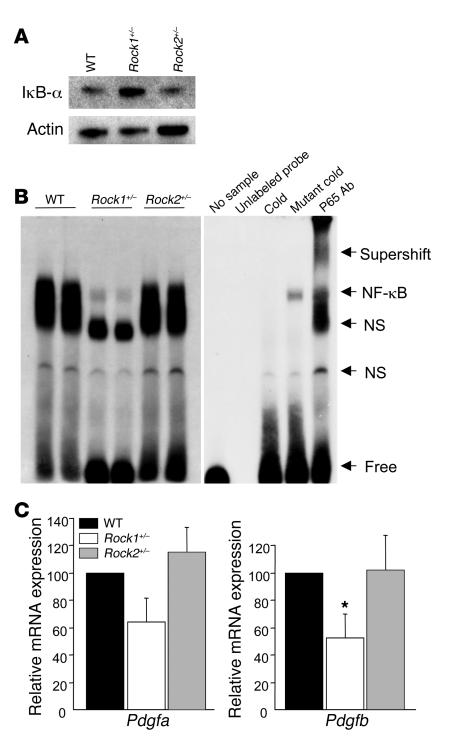
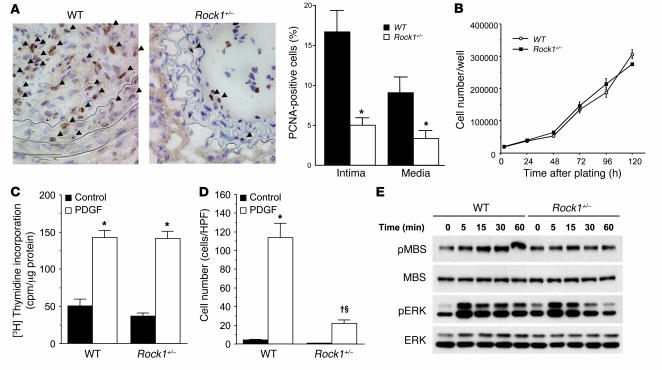
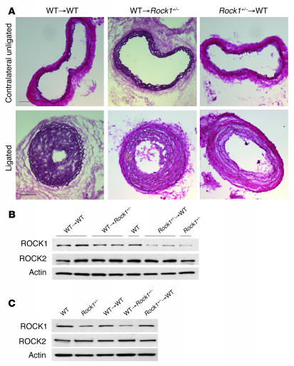
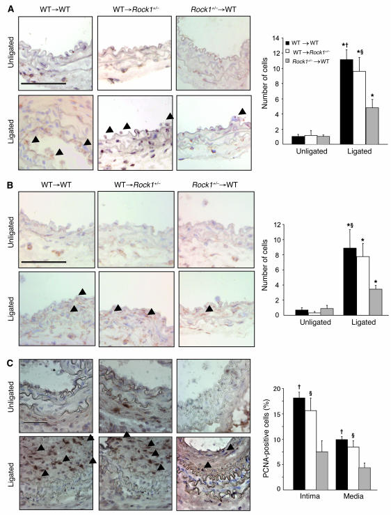
Although Rho-associated kinase (ROCK) activity has been implicated in cardiovascular diseases, the tissue- and isoform-specific roles of ROCKs in the vascular response to injury are not known. To address the role of ROCKs in this process, we generated haploinsufficient Rock1 (Rock1(+/-)) and Rock2 (Rock2(+/-)) mice and performed carotid artery ligations. Following this intervention, we found reduced neointima formation in Rock1(+/-) mice compared with that of WT or Rock2(+/-) mice. This correlated with decreased vascular smooth muscle cell proliferation and survival, decreased levels proinflammatory adhesion molecule expression, and reduced leukocyte infiltration. In addition, thioglycollate-induced peritoneal leukocyte recruitment and accumulation were substantially reduced in Rock1(+/-) mice compared with those of WT and Rock2(+/-) mice. To determine the role of leukocyte-derived ROCK1 in neointima formation, we performed reciprocal bone marrow transplantation (BMT) in WT and Rock1(+/-) mice. Rock1(+/-) to WT BMT led to reduced neointima formation and leukocyte infiltration following carotid ligation compared with those of WT to WT BMT. In contrast, WT to Rock1(+/-) BMT resulted in increased neointima formation. These findings indicate that ROCK1 in BM-derived cells mediates neointima formation following vascular injury and suggest that ROCK1 may represent a promising therapeutic target in vascular inflammatory diseases.


Figures

Figure 1
Figure 1. Decreased neointima formation after carotid artery ligation in Rock1+/– mice.
(A) Representative cross sections of contralateral unligated and ligated carotid arteries in WT, Rock1+/–, and Rock2+/– mice at 28 days after ligation. Scale bar: 100 μm. (B) Representative immunohistochemical analysis of ROCK expression and activity in carotid arteries from WT and Rock1+/– mice at 14 days after ligation. Vessels were stained with nonspecific antibody (IgG), and antibodies were directed at total MBS (tMBS), phosphorylated MBS (pMBS), ROCK1, and ROCK2. The I and M indicate the intima and media, respectively. Scale bar: 50 μm.
Figure 2
Figure 2. ROCK expression in leukocytes and carotids of Rock1+/– and Rock2+/– mice.
(A) Expression of ROCK isoforms in leukocytes from WT, Rock1+/–, and Rock2+/– mice with and without carotid ligation. Representative western blot (upper panel). Quantification of ROCK1 expression (middle panel). Mean ± SEM; n = 3. Quantification of ROCK2 expression (lower panel). Mean ± SEM; n = 3. (B) Expression of ROCK isoforms in unligated and ligated carotid arteries from WT, Rock1+/–, and Rock2+/– mice. Representative western blot (upper panel). Quantification of ROCK1 expression (middle panel). Mean ± SEM; n = 4. Quantification of ROCK2 expression (lower panel). Mean ± SEM; n = 4. *P < 0.05 versus unligated WT mice; P < 0.01 versus unligated Rock2+/– mice; ΧP < 0.01 versus ligated WT and Rock2+/– mice; **P < 0.01 versus unligated WT and Rock1+/– mice; P < 0.01 versus ligated WT mice; #P < 0.05 versus ligated Rock1+/– mice; ##P < 0.01 versus unligated WT and Rock2+/– mice; ***P < 0.01 versus WT and Rock1+/– mice.
Figure 3
Figure 3. Decreased leukocyte recruitment to ligated vessels of Rock1+/– mice.
(A) Representative histological sections from carotid arteries in WT and Rock1+/– mice stained with CD45 Ab for leukocytes. Arrowheads indicate CD45-positive cells. Scale bar: 50 μm (left panel). Quantitative analysis of the number of CD45-positive leukocytes adherent to the unligated (n = 3–4 in each group) and ligated vessel walls (n = 14 in each group) at 3 days after ligation (right panel). (B) Representative histological sections from carotid arteries in WT and Rock1+/– mice stained with MOMA-2 Ab for macrophage. Arrowheads indicate MOMA-2–positive cells. Scale bar: 50 μm (left panel). Quantitative analysis of the number of MOMA-2–positive macrophages infiltrated into the unligated (n = 3–4 in each group) and ligated vessel (n = 12–13 in each group) at day 3. *P < 0.01 versus unligated WT mice; P < 0.05 versus unligated Rock1+/– mice; ΧP < 0.05 versus ligated WT mice.
Figure 4
Figure 4. Decreased recruitment and adhesion of leukocytes in Rock1+/– mice.
Neutrophil and macrophage recruitment was induced by i.p. injection of thioglycollate. (A) Neutrophil recruitment 4 hours after i.p. injection of PBS (control) or 3% thioglycollate into WT, Rock1+/–, and Rock2+/– mice (n = 10, 5, and 5, respectively). (B) Macrophage recruitment 4 days after i.p. injection of PBS (control) or 3% thioglycollate into WT, Rock1+/–, and Rock2+/– mice (n = 9, 8, and 6, respectively). Confluent monolayers of ECs isolated from WT and Rock1+/– mice were incubated with PBS (control, n = 4–6 in each group) or 10 units/ml of thrombin for 10 minutes (n = 9–10 in each group). They were then placed under laminar flow at 0.5 (C) or 1.0 dynes/cm2 (D). U937 monocytes, stably expressed human L selectin (U937-LAM), were perfused over the EC monolayers, and adherent cells were quantified after 3 minutes. *P < 0.01 versus each control group; P < 0.05 versus each control group; P < 0.01 versus Rock1+/– mice with thioglycollate or Rock1+/– ECs with thrombin; ΧP < 0.05 versus Rock1+/– mice with thioglycollate or Rock1+/– ECs with thrombin.
Figure 5
Figure 5. Decreased expression of endothelial adhesion molecules in Rock1+/– mice.
Representative histological sections from unligated and ligated carotid arteries at 7 days after ligation in WT and Rock1+/– mice stained for ICAM-1 (A) and VCAM-1 (B). Scale bars: 50 μm. (C) Representative result of northern blot analysis of Icam1 and Vcam1 mRNA expression in ECs from WT and Rock1+/– mice. ECs were stimulated with 5 U/ml of thrombin for 6 hours. Gapdh mRNA expression was used as an internal control. (D) Representative result of western blot analysis of ROCK1 and ROCK2 expressions in ECs from WT and Rock1+/– mice with or without thrombin stimulation for 5 minutes. Actin was used as an internal control. (E) Representative result of western blot analysis of ROCK and ERK activities in ECs from WT and Rock1+/– mice. ECs were stimulated with 5 U/ml of thrombin for the indicated time periods.
Figure 6
Figure 6. Decreased expression of PDGF-B, degradation of IκB-α, and activation of NF-κB in macrophages from Rock1+/– mice.
(A) Representative western blot analysis of IκB-α expression in peritoneal macrophages from WT, Rock1+/–, and Rock2+/– mice. Actin was used as an internal control. (B) Representative electrophoretic mobility shift assay of NF-κB in WT, Rock1+/–, and Rock2+/– macrophages (left panel). Specificity of NF-κB binding activity was analyzed by the addition of unlabeled probe, by the pretreatment with excess unlabeled probe (cold) or mutant probe (mutant cold), and by anti-p65 Ab (p65 Ab) supershift gel assay (right panel). NF-κB binding band (NF-κB), nonspecific binding band (NS), free probe (free), and the raised bands supershifted by Ab (supershift) are indicated on the right. (C) Expression of Pdgfa and -b were analyzed by quantitative real-time PCR. Total RNA was extracted from thioglycollate-induced peritoneal macrophages in WT, Rock1+/–, and Rock2+/– mice (n = 6–8). *P < 0.05 versus WT and Rock2+/– mice.
Figure 7
Figure 7. Decreased cell proliferation in the neointima of Rock1+/– mice.
(A) Representative histological sections from carotid arteries in WT and Rock1+/– mice stained for PCNA at 14 days after ligation. Arrowheads indicate PCNA-positive cells. Scale bars: 50 μm (left panels). Quantitative analysis of the ratio of PCNA-positive cells to total cell number in the intima and the media (n = 10–16) (right panel). *P < 0.01 versus WT mice. (B) Cell proliferation in response to serum of VSMCs from WT and Rock1+/– mice. Experiments were performed 6 times in triplicates. (C) DNA synthesis in response to PDGF of VSMCs from WT and Rock1+/– mice (n = 12). *P < 0.01 versus without PDGF (control). (D) Cell migration in response to PDGF of VSMCs from WT and Rock1+/– mice (n = 8­–9). *P < 0.01 versus without PDGF (control); P < 0.01 versus control; ΧP < 0.01 versus WT. (E) Representative western blot analysis of ROCK and ERK activities in VSMCs of WT and Rock1+/– mice. VSMCs were stimulated with 10 ng/ml of PDGF for the indicated time periods.
Figure 8
Figure 8. ROCK1 in BM-derived cells mediates to neointima formation.
(A) Representative cross sections of contralateral unligated and ligated carotid arteries in WT to WT, WT to Rock1+/–, and Rock1+/– to WT BMT mice at 28 days after ligation. Scale bars: 100 μm. Representative western blot analysis of ROCK expression in leukocytes (B) and ECs (C) from WT and Rock1+/– BMT mice. Actin was used as an internal control.
Figure 9
Figure 9. BM-derived cells contribute to leukocyte recruitment and cell proliferation in Rock1+/– to WT BMT mice.
(A) Representative histological sections from carotid arteries in WT and Rock1+/– BMT mice stained with CD45 Ab for leukocytes. Arrowheads indicate CD45-positive cells. Scale bar: 50 μm (left panel). Quantitative analysis of the number of CD45-positive leukocytes adherent to the unligated (n = 3–4) and ligated vessel wall at 3 days after ligation (n = 8–12). (B) Representative histological sections from carotid arteries in WT and Rock1+/– BMT mice stained with MOMA-2 Ab for macrophages. Arrowheads indicate MOMA-2–positive cells. Scale bar: 50 μm (left panel). Quantitative analysis of the number of MOMA-2–positive macrophages infiltrated into the unligated (n = 3–4) and ligated vessel wall at days 3 (n = 7–8). (C) Representative histological sections from carotid arteries in WT and Rock1+/– BMT mice stained for PCNA at 14 days after ligation. Arrowheads indicate PCNA-positive cells. Scale bar: 50 μm (left panel). Quantitative analysis of the ratio of PCNA-positive cells to total cell number in the intima and the media (n = 8–9). *P < 0.05 versus unligated mice; P < 0.01, ΧP < 0.05 versus Rock1+/– to WT mice.

References

    1. Pasterkamp G., Galis Z.S., de Kleijn D.P. Expansive arterial remodeling: location, location, location. Arterioscler. Thromb. Vasc. Biol. 2004;24:650–657. doi: 10.1161/01.ATV.0000120376.09047.fe. - DOI - PMC - PubMed
    1. Bobik A., Tkachuk V. Metalloproteinases and plasminogen activators in vessel remodeling. Curr. Hypertens. Rep. 2003;5:466–472. doi: 10.1007/s11906-003-0054-5. - DOI - PubMed
    1. Serrano C.V., Jr., et al. Coronary angioplasty results in leukocyte and platelet activation with adhesion molecule expression. Evidence of inflammatory responses in coronary angioplasty. J. Am. Coll. Cardiol. 1997;29:1276–1283. doi: 10.1016/S0735-1097(97)00070-3. - DOI - PubMed
    1. Tanaka H., et al. Sustained activation of vascular cells and leukocytes in the rabbit aorta after balloon injury. Circulation. 1993;88:1788–1803. - PubMed
    1. Libby P. Inflammation in atherosclerosis. Nature. 2002;420:868–874. doi: 10.1038/nature01323. - DOI - PubMed

Publication types

MeSH terms