

Characterization of antibodies that selectively detect alpha-synuclein in pathological inclusions
- PMID: 18414880
- PMCID: PMC2664556
- DOI: 10.1007/s00401-008-0375-1
Characterization of antibodies that selectively detect alpha-synuclein in pathological inclusions
Abstract
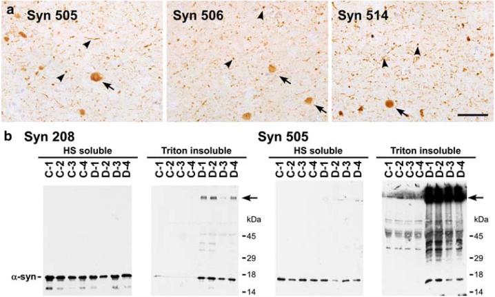
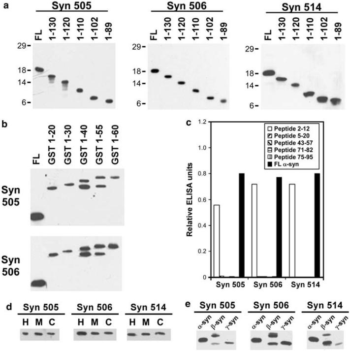
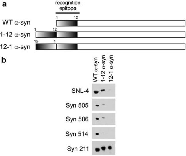
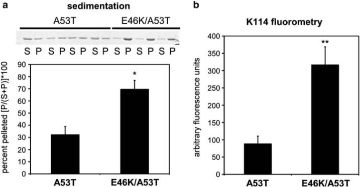
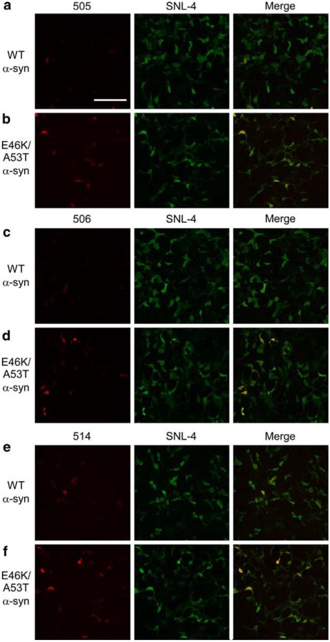
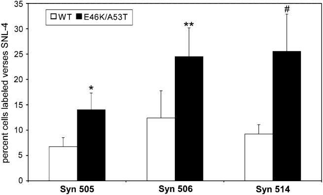
Sensitive detection of alpha-synuclein (alpha-syn) pathology is important in the diagnosis of disorders like Parkinson's disease, dementia with Lewy bodies, and multiple system atrophy and in providing better insights into the etiology of these diseases. Several monoclonal antibodies that selectively react with aggregated alpha-syn in pathological inclusions and reveal extensive and underappreciated alpha-syn pathology in the brains of diseased patients were previously reported by Duda et al. (Ann Neurol 52:205-210, 2002). We sought to characterize the specificity of some of these antibodies (Syn 505, Syn 506 and Syn 514); using C-terminal and N-terminal truncations of alpha-syn, all three antibodies were determined to require N-terminal epitopes that minimally comprise amino acids 2-4, but possibly extend to amino acid 12 of alpha-syn. The selectivity of these antibodies was further assessed using biochemical analysis of human brains and reactivity to altered recombinant alpha-syn proteins with duplication variants of amino acids 1-12. In addition, by expressing wild-type or a double mutant (E46K/A53T) of alpha-syn in cultured cells and by comparing their immunoreactivities to another antibody (SNL-4), which has a similar primary epitope, it was determined that Syn 505, Syn 506 and Syn 514 recognize conformational variants of alpha-syn that is enhanced by the presence of the double mutations. These studies indicate that antibodies Syn 505, Syn 506 and Syn 514 preferentially recognize N-terminal epitopes in complex conformations, consistent with the dramatic conformational change associated with the polymerization of alpha-synuclein into amyloid fibrils that form pathological inclusions.
Figures
Similar articles
-
Generation and characterization of novel conformation-specific monoclonal antibodies for α-synuclein pathology.Neurobiol Dis. 2015 Jul;79:81-99. doi: 10.1016/j.nbd.2015.04.009. Epub 2015 Apr 30. Neurobiol Dis. 2015. PMID: 25937088
-
Production of a monoclonal antibody, against human α-synuclein, in a subpopulation of C57BL/6J mice, presenting a deletion of the α-synuclein locus.J Neurosci Methods. 2010 Oct 15;192(2):268-76. doi: 10.1016/j.jneumeth.2010.08.010. Epub 2010 Aug 13. J Neurosci Methods. 2010. PMID: 20709102
-
Progression of phosphorylated α-synuclein in Macaca fuscata.Brain Pathol. 2021 Sep;31(5):e12952. doi: 10.1111/bpa.12952. Epub 2021 Mar 22. Brain Pathol. 2021. PMID: 33754430 Free PMC article.
-
Antibodies against alpha-synuclein: tools and therapies.J Neurochem. 2019 Sep;150(5):612-625. doi: 10.1111/jnc.14713. Epub 2019 Jun 25. J Neurochem. 2019. PMID: 31055836 Review.
-
Neuropathology of synuclein aggregates.J Neurosci Res. 2000 Jul 15;61(2):121-7. doi: 10.1002/1097-4547(20000715)61:2<121::AID-JNR1>3.0.CO;2-4. J Neurosci Res. 2000. PMID: 10878583 Review.
Cited by
-
Viral expression of ALS-linked ubiquilin-2 mutants causes inclusion pathology and behavioral deficits in mice.Mol Neurodegener. 2015 Jul 8;10:25. doi: 10.1186/s13024-015-0026-7. Mol Neurodegener. 2015. PMID: 26152284 Free PMC article.
-
Abrogation of α-synuclein-mediated dopaminergic neurodegeneration in LRRK2-deficient rats.Proc Natl Acad Sci U S A. 2014 Jun 24;111(25):9289-94. doi: 10.1073/pnas.1403215111. Epub 2014 Jun 9. Proc Natl Acad Sci U S A. 2014. PMID: 24927544 Free PMC article.
-
Robust Central Nervous System Pathology in Transgenic Mice following Peripheral Injection of α-Synuclein Fibrils.J Virol. 2017 Jan 3;91(2):e02095-16. doi: 10.1128/JVI.02095-16. Print 2017 Jan 15. J Virol. 2017. PMID: 27852849 Free PMC article.
-
Propagation of alpha-synuclein pathology: hypotheses, discoveries, and yet unresolved questions from experimental and human brain studies.Acta Neuropathol. 2016 Jan;131(1):49-73. doi: 10.1007/s00401-015-1485-1. Epub 2015 Oct 7. Acta Neuropathol. 2016. PMID: 26446103 Free PMC article. Review.
-
A novel, high-efficiency cellular model of fibrillar alpha-synuclein inclusions and the examination of mutations that inhibit amyloid formation.J Neurochem. 2010 Apr;113(2):374-88. doi: 10.1111/j.1471-4159.2010.06592.x. Epub 2010 Feb 2. J Neurochem. 2010. PMID: 20132485 Free PMC article.
References
-
- Chartier-Harlin MC, Kachergus J, Roumier C, Mouroux V, Douay X, Lincoln S, Levecque C, Larvor L, Andrieux J, Hulihan M, Waucquier N, Defebvre L, Amouyel P, Farrer M, Destee A. Alpha-synuclein locus duplication as a cause of familial Parkinson's disease. Lancet. 2004;364:1167–1169. - PubMed
-
- Choi W, Zibaee S, Jakes R, Serpell LC, Davletov B, Crowther RA, Goedert M. Mutation E46K increases phospholipid binding and assembly into filaments of human alpha-synuclein. FEBS Lett. 2004;576:363–368. - PubMed
-
- Conway KA, Harper JD, Lansbury PT. Accelerated in vitro fibril formation by a mutant alpha-synuclein linked to early-onset Parkinson disease. Nat Med. 1998;4:1318–1320. - PubMed
-
- Conway KA, Harper JD, Lansbury PT. Fibrils formed in vitro from alpha-synuclein and two mutant forms linked to Parkinson's disease are typical amyloid. Biochemistry. 2000;39:2552–2563. - PubMed
Publication types
MeSH terms
Substances
Grants and funding
LinkOut - more resources
Full Text Sources
Other Literature Sources
Medical
Molecular Biology Databases
Miscellaneous
