. 2008 Dec;57(12):1871-8.
doi: 10.1007/s00262-008-0517-2. Epub 2008 Apr 16.

RhoC a new target for therapeutic vaccination against metastatic cancer

Affiliations

RhoC a new target for therapeutic vaccination against metastatic cancer

Lynn Wenandy et al. Cancer Immunol Immunother. 2008 Dec.

Abstract

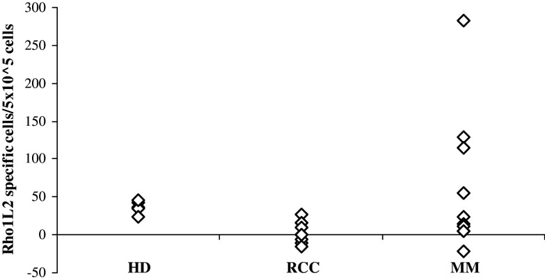
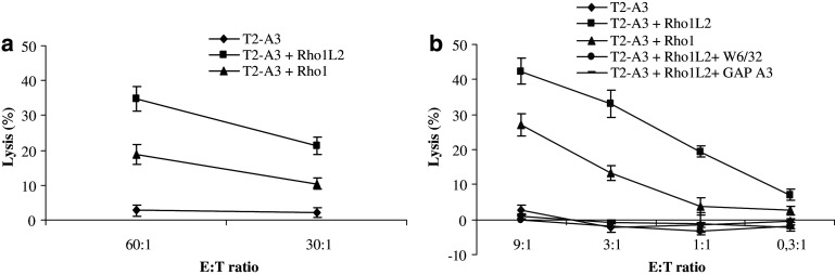
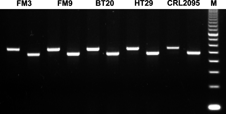
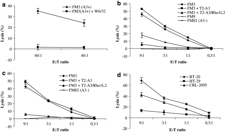
Most cancer deaths are due to the development of metastases. Increased expression of RhoC is linked to enhanced metastatic potential in multiple cancers. Consequently, the RhoC protein is an attractive target for drug design. The clinical application of immunotherapy against cancer is rapidly moving forward in multiple areas, including the adoptive transfer of anti-tumor-reactive T cells and the use of "therapeutic" vaccines. The over-expression of RhoC in cancer and the fact that immune escape by down regulation or loss of expression of this protein would reduce the morbidity and mortality of cancer makes RhoC a very attractive target for anti-cancer immunotherapy. Herein, we describe an HLA-A3 restricted epitope from RhoC, which is recognized by cytotoxic T cells. Moreover, RhoC-specific T cells show cytotoxic potential against HLA-matched cancer cells of different origin. Thus, RhoC may serve as an important and widely applicable target for anti-cancer immunotherapeutic strategies.


Figures

Fig. 1
Fig. 1
HLA-A3 restricted T-cell responses against Rho1L2 as measured by IFN-γ ELISPOT The average number of peptide specific IFN-γ spots formed in response to Rho1L2 among 5 × 105 in vitro stimulated PBMC from five HLA-A3+ healthy donors (HD), PBL from ten renal cell carcinoma patients (RCC), and ten melanoma patients (MM). Measurements were made in triplicates
Fig. 2
Fig. 2
T cell antigen specificity and cross reactivity. a Cytotoxicity by 51Cr-release assay of a bulk culture stimulated with Rho1L2-loaded autologous DC/autologous PBL. Specific lysis of T2-A3 cells with no peptide or pulsed with Rho1L2 (10 μM) or Rho1 (10 μM). E:T ratio = 60:1, 30:1. b Specificity of a T cell clone (clone 9) assayed by 51Cr-release assay. Lysis of T2-A3 cells with no peptide, pulsed with Rho1L2 (10 μM), Rho1 (10 μM) and of Rho1L2-pulsed T2–A3 cells with addition of the HLA-class I specific antibody W6/32 or HLA-A3 specific antibody GAP A3 at different E:T ratios (9:1; 3:1; 1:1; 0,3:1). All measurements were made in duplicates. Error bars indicate the standard deviation
Fig. 3
Fig. 3
PCR products electrophoresed in agarose gel and stained with EtBr. The image represents five different cancer cell lines, all being positive for RhoC. For each cancer cell line the left band represents GAPDH and the right band represents RhoC
Fig. 4
Fig. 4
Functional capacity of RhoC-specific T cells. a Cytotoxicity of a bulk culture stimulated with Rho1L2-loaded autologous DC/autologous PBL. Specific lysis of the HLA-A3+ melanoma cell line FM3 without and with the addition of the HLA-class I specific antibody W6/32. E:T ratio = 60:1, 40:1. b Lysis by a Rho1L2-specific clone (clone 9) of the HLA-A3+ melanoma cell line FM3, cell lysis with addition of unlabeled T2-A3 cells pulsed with Rho1L2 (10 μM) or no peptide (inhibitor to target ratio = 20:1), and cell lysis of the HLA-A3+ melanoma cell line FM9 and the HLA-A3- melanoma cell line FM82. Measurements were made in duplicates for all E:T ratios. c Repetition of the experiments shown in b) (without inclusion of FM9). d Lysis by a Rho1L2-specific clone of the HLA-A3+ breast cancer cell line BT-20, colon cancer cell line HT-29 and head and neck cancer cell line CRL-2095. All measurements were made in duplicates. Error bars indicate the standard deviation

Similar articles

Cited by

References

    1. Andersen MH, Becker JC, Straten P. Regulators of apoptosis: suitable targets for immune therapy of cancer. Nat Rev Drug Discov. 2005;4:399–409. doi: 10.1038/nrd1717. - DOI - PubMed
    1. Andersen MH, Bonfill JE, Neisig A, Arsequell G, Sondergaard I, Valencia G, Neefjes J, Zeuthen J, Elliott T, Haurum JS. Phosphorylated peptides can be transported by tap molecules, presented by class I MHC molecules, and recognized by phosphopeptide-specific CTL. J Immunol. 1999;163:3812–3818. - PubMed
    1. Andersen MH, Pedersen LO, Becker JC, thor Straten P. Identification of a cytotoxic T lymphocyte response to the apoptose inhibitor protein survivin in cancer patients. Cancer Res. 2001;61:869–872. - PubMed
    1. Andersen MH, Pedersen LO, Capeller B, Brocker EB, Becker JC, thor Straten P. Spontaneous cytotoxic T-cell responses against survivin-derived MHC class I-restricted T-cell epitopes in situ as well as ex vivo in cancer patients. Cancer Res. 2001;61:5964–5968. - PubMed
    1. Andersen MH, Svane IM, Kvistborg P, Nielsen OJ, Balslev E, Reker S, Becker JC, Thor SP. Immunogenicity of Bcl-2 in cancer patients. Blood. 2005;15:728–734. doi: 10.1182/blood-2004-07-2548. - DOI - PubMed

Publication types

MeSH terms