Pituitary tumors in childhood: update of diagnosis, treatment and molecular genetics
- PMID: 18416659
- PMCID: PMC2743125
- DOI: 10.1586/14737175.8.4.563
Pituitary tumors in childhood: update of diagnosis, treatment and molecular genetics
Abstract
Pituitary tumors are rare in childhood and adolescence, with a reported prevalence of up to one per 1 million children. Only 2-6% of surgically treated pituitary tumors occur in children. Although pituitary tumors in children are almost never malignant and hormonal secretion is rare, these tumors may result in significant morbidity. Tumors within the pituitary fossa are mainly of two types: craniopharyngiomas and adenomas. Craniopharyngiomas cause symptoms by compressing normal pituitary, causing hormonal deficiencies and producing mass effects on surrounding tissues and the brain; adenomas produce a variety of hormonal conditions such as hyperprolactinemia, Cushing disease and acromegaly or gigantism. Little is known about the genetic causes of sporadic lesions, which comprise the majority of pituitary tumors, but in children, more frequently than in adults, pituitary tumors may be a manifestation of genetic conditions such as multiple endocrine neoplasia type 1, Carney complex, familial isolated pituitary adenoma and McCune-Albright syndrome. The study of pituitary tumorigenesis in the context of these genetic syndromes has advanced our knowledge of the molecular basis of pituitary tumors and may lead to new therapeutic developments.
Figures

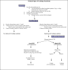
aberrant cAMP signaling (primary initiating event for the polyclonal hyperplasia and/or initial adenoma formation, as evidenced by GNAS and PRKAR1A involvement)
cell-cycle dysregulation and aneuploidy (may initiate or augment growth of a monoclonal pituitary tumor)
menin downregulation, methylation of certain target genes, aneuploidy and/or disruption of genomic integrity in a greater scale (may lead to a growing pituitary adenoma still responsive to medical and/or surgical treatment,depending on the type)
PTTG overexpression and/or additional growth factor upregulation and increased angiogenesis (may lead to aggressive tumors).


References
-
- Kawamura K, Kouki T, Kawahara G, Kikuyama S. Hypophyseal development in vertebrates from amphibians to mammals. Gen Comp Endocrinol. 2002;126(2):130–135. - PubMed
-
- Moore ES, Ward RE, Jamison PL, Morris CA, Bader PI, Hall BD. The subtle facial signs of prenatal exposure to alcohol: an anthropometric approach. J Pediatr. 2001;139(2):215–219. - PubMed
-
- Faglia G, Spada A. Genesis of pituitary adenomas: state of the art. J Neurooncol. 2001;54(2):95–110. *Excellent review of pathogenesis of pituitary adenomas. - PubMed
-
- Zhu X, Gleiberman AS, Rosenfeld MG. Molecular physiology of pituitary development: signaling and transcriptional networks. Physiol Rev. 2007;87(3):933–963. *Excellent review of pathogenesis of pituitary adenomas. - PubMed
-
- Bunin GR, Surawicz TS, Witman PA, Preston-Martin S, Davis F, Bruner JM. The descriptive epidemiology of craniopharyngioma. J Neurosurg. 1998;89(4):547–551. - PubMed
Publication types
MeSH terms
Grants and funding
LinkOut - more resources
Full Text Sources
Medical
