. 2008 Jun 27;283(26):18355-64.
doi: 10.1074/jbc.M800725200. Epub 2008 Apr 16.

Unique Asn-linked oligosaccharides of the human pathogen Entamoeba histolytica

Affiliations

Unique Asn-linked oligosaccharides of the human pathogen Entamoeba histolytica

Paula Magnelli et al. J Biol Chem. .

Abstract

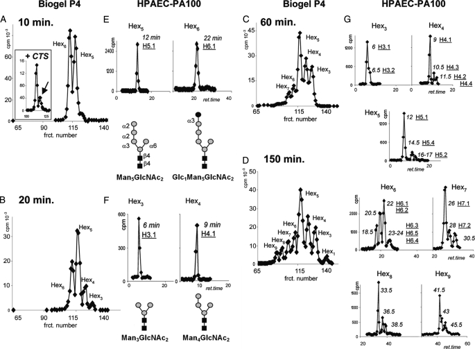
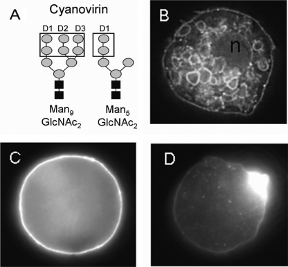
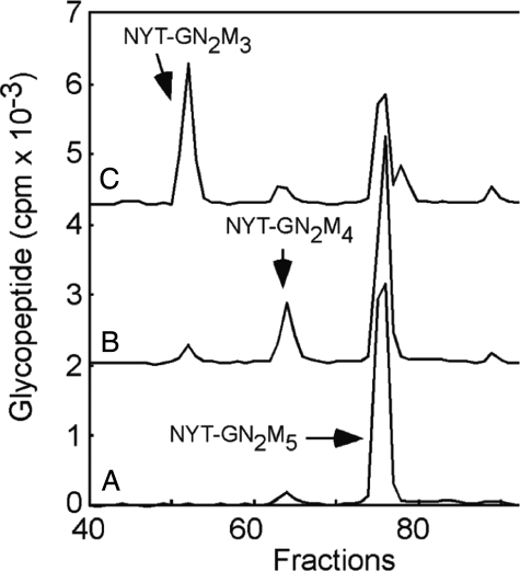
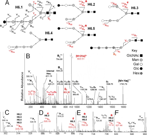
N-Glycans of Entamoeba histolytica, the protist that causes amebic dysentery and liver abscess, are of great interest for multiple reasons. E. histolytica makes an unusual truncated N-glycan precursor (Man(5)GlcNAc(2)), has few nucleotide sugar transporters, and has a surface that is capped by the lectin concanavalin A. Here, biochemical and mass spectrometric methods were used to examine N-glycan biosynthesis and the final N-glycans of E. histolytica with the following conclusions. Unprocessed Man(5)GlcNAc(2), which is the most abundant E. histolytica N-glycan, is aggregated into caps on the surface of E. histolytica by the N-glycan-specific, anti-retroviral lectin cyanovirin-N. Glc(1)Man(5)GlcNAc(2), which is made by a UDP-Glc: glycoprotein glucosyltransferase that is part of a conserved N-glycan-dependent endoplasmic reticulum quality control system for protein folding, is also present in mature N-glycans. A swainsonine-sensitive alpha-mannosidase trims some N-glycans to biantennary Man(3)GlcNAc(2). Complex N-glycans of E. histolytica are made by the addition of alpha1,2-linked Gal to both arms of small oligomannose glycans, and Gal residues are capped by one or more Glc. In summary, E. histolytica N-glycans include unprocessed Man(5)GlcNAc(2), which is a target for cyanovirin-N, as well as unique, complex N-glycans containing Gal and Glc.


Figures

FIGURE 1.
FIGURE 1.
E. histolytica trophozoites make numerous N-glycans from the truncated Man5GlcNAc2 precursor transferred by the OST (3, 8). The most abundant N-glycan in each size range is listed first (e.g. H3.1, H4.1, etc.). Structures were identified by multiple techniques including in vivo and in vitro labeling, size separation on Bio-Gel P-4 and HPAEC, glycosylhydrolase digestions, monosaccharide analysis, and mass spectrometry. An asterisk marks the linkage between Glc and Gal, which could not be determined experimentally.
FIGURE 2.
FIGURE 2.
The complexity of N-glycans made by E. histolytica trophozoites increases dramatically with time. E. histolytica trophozoites were incubated with [2-3H]Man for 10, 20, 60, and 150 min, and N-glycans were released with PNGase F and separated by Bio-Gel P-4 filtration (A-D), where Hexn indicates their size. Isomers with the same number of hexoses were further isolated on an HPAEC-PA100 column (E-H), with the retention time (ret.; min) shown in italic and the identifier name from Fig. 1 (e.g. H5.1) underlined below. After 10-min labeling (A and E), the predominant E. histolytica N-glycans were unmodified Man5GlcNAc2 and the glucosylated product of UDP-Glc:glycoprotein glucosyltransferase (Glc1Man5GlcNAc2) (H6.1). As shown in the inset in A, Glc1Man5GlcNAc2 is by far the most abundant N-glycan in the presence of the glucosidase II inhibitor castanospermine (CTS). After 20-min labeling (B and F), two mannosidase digestion products are apparent: Man4GlcNAc2 (H4.1) and Man3GlcNAc2 (H3.1). After 60-min labeling (C), complex N-glycans are apparent in Hex6 and Hex7 pools. After 150-min labeling (D and G), novel, complex N-glycans are present in all of the pools (see further characterization in Fig. 4). frct., fraction.
FIGURE 3.
FIGURE 3.
The abundance of Glc in complex N-glycans of E. histolytica trophozoites increases with their length. A, E. histolytica N-glycans labeled with [U-14C]Glc and separated by Bio-Gel P-4 filtration. The number of hexoses in each glycomer is indicated (Hexn), according to calibrated standards prepared from Man5GlcNAc2 digested with jack bean α-mannosidase and A. saitoi α1,2-mannosidase. B, relative abundance (weight percentage) of neutral sugars for each peak after acid hydrolysis, as determined by HPAEC chromatography. fract., fraction.
FIGURE 4.
FIGURE 4.
Characterization of the E. histolytica N-glycans by in vivo labeling and glycosylhydrolase digestions. E. histolytica N-glycans, which were prepared as in Fig. 2, were treated with glycosylhydrolases, and digestion products were identified according to retention times (r.t.) of known standards (e.g. Man3GlcNAc2). For isomers H6.2, H6.5, and H7.1, the dashed line enclosing the Manα1,6-arm indicates what corresponds to the residual fragment 11.5, which was fully characterized in a separate [U-14C]Glc-labeling experiment. Note that jack bean α-mannosidase (JBAM) removes an exposed Manα1,3- (as in isomer H4.3), but it does not remove the Manα1,6-unless the Manα1,3-arm is digested first. Hence, isomer H4.2 becomes susceptible to jack beanα-mannosidase digestion only after removal of Galα1,2- by α-galactosidase. The mode of action of jack bean α-mannosidase (37) supports our assumption that the different sensitivities of H4.2 and H4.3 are due to accessibility of α-linked Man residues. In accordance with this, the underlying structures of other isomers were inferred as well. ASAM, A. saitoi α1,2-mannosidase.
FIGURE 5.
FIGURE 5.
The anti-retroviral lectin cyanovirin-N, which recognizes high Man N-glycans (15, 16), binds to the surface E. histolytica trophozoites and forms caps. A, cyanovirin-N binds to terminal α1,2-linked Man, which is present on each of the three arms of Man9GlcNAc2 and is also present on the single arm of Man5GlcNAc2. B, cyanovirin-N binds to the surface and to vesicular membranes of fixed and permeabilized E. histolytica. n, nucleus. C, cyanovirin-N is evenly distributed on the surface of a live E. histolytica incubated with the lectin in the cold, so it cannot cap. D, cyanovirin-N is capped on the surface of a live E. histolytica that is allowed to warm up to 37 °C for 10 min. The negative control was scytovirin (a lectin that binds to an intact D3 arm, which is absent in E. histolytica N-glycans). E. histolytica incubated with scytovirin were not labeled (data not shown).
FIGURE 6.
FIGURE 6.
Synthesis of complex N-glycans in vitro by incubating E. histolytica membranes with UDP-[3H]Glc and UDP[3H]Gal. A, a Bio-Gel P-4 filtration chromatogram of N-glycans is shown. B, isomer separation by HPAEC in PA100 is shown. ret., retention. C, identification of the transferred monosaccharide after total hydrolysis by HPAEC in MA1 is shown. The presence of Gal in the Hex4 isomers and in Hex5 eluting at 17 min is consistent with the α-galactosidase digests in F. In D-F, untreated glycans are marked with closed circles, and the products of glycosylhydrolases are marked with open circles. D, one Gal1Man3GlcNAC2 isomer (H4.3, at 10.5 min) is digested with jack bean α-mannosidase (JBAM), whereas the other isomer (H4.2, at 11.5 min) is not. E, Glc1Man5GlcNAC2 (H6.1, at 22 min) is digested to a disaccharide (asterisk) by endomannosidase. F, both Gal1Man3GlcNAC2 isomers (H4.2. and H4.3 at 10.5 and 11.5 min), as well as Gal1Man4GlcNAC2 (H5.2 at 17 min), digest with α-galactosidase. In contrast, Glc1Gal1Man3GlcNAC2 (H5.6 at 19 min) is not digested by α-galactosidase. G, the table summarizes these results. Isomer H5.2 was fully characterized using complementary data acquired in a separate [23H]Man-labeling experiment (Fig. 2). frct., fraction; Endomann, endomannosidase; comp, composition; ident, identity.
FIGURE 7.
FIGURE 7.
E. histolytica membranes contain a swainsonine-sensitive α-mannosidase. Glycopeptides produced by incubating intact E. histolytica membranes with a radiolabeled tripeptide acceptor (NYT) were captured on concanavalin A and resolved by HPLC. HPLC profiles show glycopeptides from assays incubated in the presence (A) or absence (B and C) of swainsonine for 2 min (B) or 30 m in (A and C). Standards include Man5GlcNAc2-NYT, Man9GlcNAc2-NYT, and a glycopeptide mix, Man3-9GlcNAc2-NYT.
FIGURE 8.
FIGURE 8.
MS2 collision-induced dissociation spectrum of the Hex3HexNAc2 isolated at m/z 597. 33, [M+2Na]2+. A, the diagram shows the origin of fragments from the two isomers present in this pool (see Fig. 2G). A secondary fragment, Y/Y/B3 at m/z 458.21 (B), is consistent with archetypal biantennary Man3GlcNAc2. Cross-ring fragments 0,4A2, m/z 301.14 and 3,5A2, m/z 329.16 (C) define the Manα1,6-arm. A low abundance 1,3A2 fragment at m/z 315.16 (C) indicates the presence of an isomer bearing a terminal 1,2-linked Gal (H3.2). A Y3/0,2A4 fragment ion seen at m/z 375.17 (B) is consistent with the core region of a linear isomer. No signal is seen at m/z 505.2 or 533.2 (for a substituted Manα1,6-). Therefore, the structure for H3.2 indicates a missing Manα1,6-arm, with the remaining Manα1,3-arm extended with Galα1,2-. These results are consistent with the glycosylhydrolase digestion data (Fig. 4).
FIGURE 9.
FIGURE 9.
MS2 collision-induced dissociation spectrum of the Hex6 GlcNAc2 [M+2Na]2+ isolated at m/z 903. 51. A, the diagram shows the origin of fragments from the isomers present in this pool (see Fig. 2G). Asterisk and pound symbols indicate the origin of cross-ring fragments of the same denomination but different m/z. Diagnostic ions for isomers H6.1 and H6.4 are as follows. Fragments 0,4A5 and 3,5 A5, m/z 301.14 and 329.16 (C) define the unsubstituted Manα1,6-arm, whereas fragment 1,3A4, m/z 723.35 (E) indicates the four hexoses of the Manα1,3-arm. Fragment Y/Y/B6, m/z 458.20 (B) reveals a biantennary structure. The spectrum supports evidence for Gal extensions. An ion at m/z 315.16 (C) indicates a 1,3A2x fragment. The single isomer predicted to contain terminal Manα1,2- (H6.2) is just ∼1% of the pool; therefore, the majority of the 1,3A2x fragment originates from the terminal Galα1,2- of the more abundant isomers H6.3, H6.4, and H6.5. Evidence for extensions on the Manα1,6- was found as well. Fragments 0,4A4 and 3,5 A4 at m/z 505.19 and 533.21 (D) and fragments 0,4A4 and 3,5 A4 at m/z 709.28 and 737.30 (E) indicate Manα1,6- with one or two hexoses substitutions. Fragment 1,3A5 at m/z 927.44 (F) indicates an α1,2 with a four-hexose extension. These data, together with results from glycosylhydrolase digestions, define isomers H6.3, H6.2, and H6.5.

References

    1. Haque, R., Huston, C. D., Hughes, M., Houpt, E., and Petri, W. A., Jr. (2003) N. Engl. J. Med. 348 1565-1573 - PubMed
    1. Stanley, S. L. (2003) Lancet 361 1025-1034 - PubMed
    1. Samuelson, J., Banerjee, S., Magnelli, P., Cui, J., Kelleher, D. J., Gilmore, R., and Robbins, P. W. (2005) Proc. Natl. Acad. Sci. U. S. A. 102 1548-1553 - PMC - PubMed
    1. Helenius, A., and Aebi, M. (2004) Annu. Rev. Biochem. 73 1019-1049 - PubMed
    1. Trombetta, E. S., and Parodi, A. J. (2003) Annu. Rev. Cell Dev. Biol. 19 649-676 - PubMed

Publication types

LinkOut - more resources