Comparative Study
. 2008 Jul;82(13):6098-108.
doi: 10.1128/JVI.02121-07. Epub 2008 Apr 16.

Cell-type-specific tyrosine phosphorylation of the herpes simplex virus tegument protein VP11/12 encoded by gene UL46

Affiliations
Comparative Study

Cell-type-specific tyrosine phosphorylation of the herpes simplex virus tegument protein VP11/12 encoded by gene UL46

George Zahariadis et al. J Virol. 2008 Jul.

Abstract

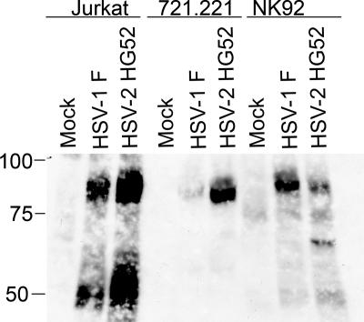
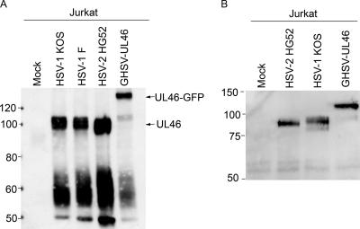
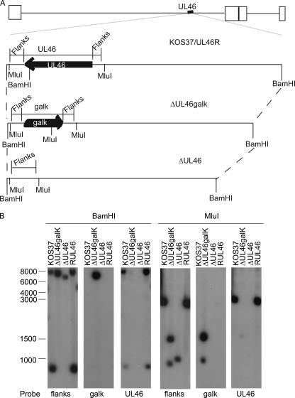
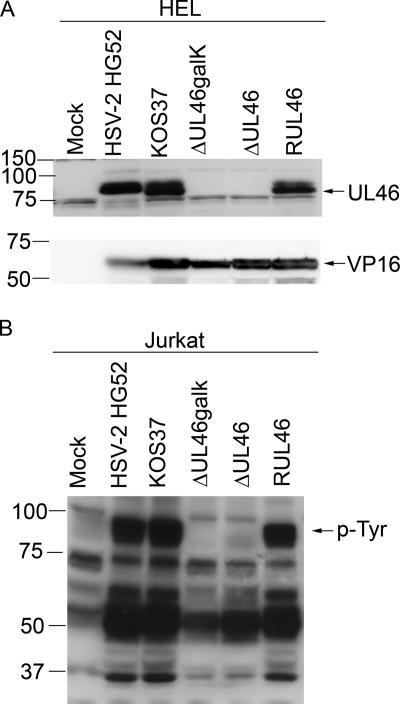
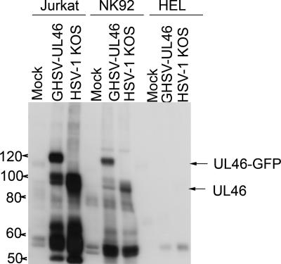
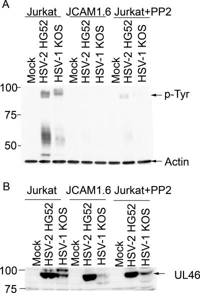
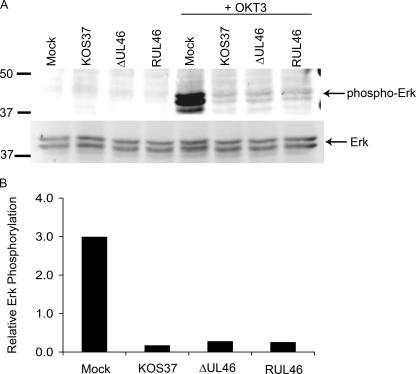
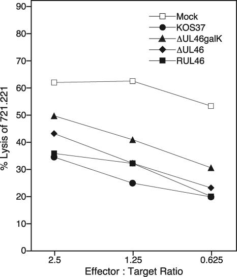
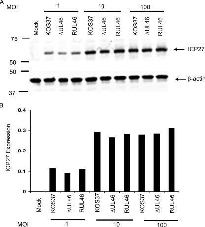
Cytotoxic T lymphocytes (CTL) and natural killer (NK) cells play key roles in limiting herpesvirus infections; consequently, many herpesviruses, including herpes simplex virus (HSV), have evolved diverse strategies to evade and/or disarm these killer lymphocytes. Previous studies have shown that CTL and NK cells are functionally inactivated following contact with HSV-infected fibroblasts. During studies of the mechanisms involved, we discovered that HSV-inactivated NK-92 NK cells and Jurkat T cells contain a strikingly prominent, novel, ca. 90-kDa tyrosine-phosphorylated protein that we identified as the HSV tegument protein VP11/12. Inasmuch as VP11/12 produced in fibroblasts and epithelial cells is not obviously tyrosine phosphorylated, these data suggested that VP11/12 serves as the substrate of a cell-type-specific protein tyrosine kinase. Consistent with this hypothesis, VP11/12 was also tyrosine phosphorylated in B lymphocytes, and this modification was severely reduced in Jurkat T cells lacking the lymphocyte-specific Src family kinase Lck. These findings demonstrate that HSV tegument proteins can be differentially modified depending on the cell type infected. Our data also raise the possibility that VP11/12 may modulate one or more lymphocyte-specific signaling pathways or serve another lymphocyte-specific function. However, HSV type 1 mutants lacking the UL46 gene retained the ability to block signaling through the T-cell receptor in Jurkat cells and remained competent to functionally inactivate the NK-92 NK cell line, indicating that VP11/12 is not essential for lymphocyte inactivation. Further studies are therefore required to determine the biological function of tyrosine-phosphorylated VP11/12.


Figures

FIG. 1.
FIG. 1.
Analysis of tyrosine-phosphorylated proteins in lymphocytes after exposure to HSV-infected fibroblasts. The indicated lymphocytes were incubated for 4 h with HEL fibroblasts that had been either mock infected or infected with HSV-1 F or HSV-2 HG52 12 h previously. The lymphocytes were then removed, and extracts were analyzed by Western blotting with an antiphosphotyrosine antibody. The mobilities of protein markers are indicated on the left.
FIG. 2.
FIG. 2.
Tyrosine phosphorylation of the HSV tegument protein VP11/12, encoded by gene UL46. (A). VP11/12 is tyrosine phosphorylated. HEL fibroblasts were either mock infected or infected with the indicated viruses for 12 h. Jurkat T cells were then coincubated with the fibroblasts for 4 h, and extracts of the lymphocytes were prepared. Cell lysates were analyzed by Western blotting with an antiphosphotyrosine antibody as for Fig. 1. The prominent ca. 90- and 120-kDa bands, presumed to correspond to wild-type VP11/12 (UL46) and the VP11/12-GFP fusion protein encoded by GHSV-UL46 (UL46-GFP), respectively, are indicated by arrows. The mobilities of protein markers are indicated on the left. (B) VP11/12 is immunoprecipitated by an antiphosphotyrosine antibody. Jurkat cells were exposed to HEL fibroblasts as described for panel A and were then harvested 9 h later. Extracts were immunoprecipitated with the antiphosphotyrosine antibody, and precipitated UL46-related proteins were detected by Western blotting with a polyclonal antibody directed against HSV-2 VP11/12. The mobilities of protein markers are indicated on the left.
FIG. 3.
FIG. 3.
Verification of the UL46 mutations produced by recombineering of the KOS-37 BAC. (A) Genomic structures of wild-type and UL46 mutant viruses. Below the schematic diagram of the HSV-1 KOS-37 genome is an expanded view of the UL46 locus. The location and orientation of the UL46 ORF are indicated, along with the locations of cleavage sites for BamHI and MluI. “Flanks” indicates the probe that detects the UL46 5′ and 3′ flanking sequences. ΔUL46galK bears a deletion/substitution mutation that replaces the UL46 ORF with the E. coli galK gene; ΔUL46 bears a precise deletion of the UL46 ORF; and RUL46 is a UL46 repair virus derived from ΔUL46galK. (B) Southern blot analysis of viral DNA. Total cellular DNA extracted from Vero cells infected with the indicated viruses was cleaved with BamHI or MluI and analyzed by Southern blot hybridization using the indicated probes. Mobilities of molecular weight markers (in nucleotides) are indicated.
FIG. 4.
FIG. 4.
Analysis of UL46 and tyrosine-phosphorylated proteins in cells infected with UL46 mutants. (A). HEL fibroblasts were either mock infected or infected with wild-type HSV (HSV-2 HG52, HSV-1 KOS-37), the UL46-null viruses (ΔUL46galk, ΔUL46), or the UL46 repair virus (RUL46). Cell extracts prepared 16 h postinfection were then analyzed by Western blotting using a rabbit polyclonal antiserum directed against HSV-2 strain 186 UL46 (top panel) and monoclonal antibody LP1, directed against VP16 (bottom panel). (B) Jurkat cells were exposed as before to HEL fibroblasts that had been either mock infected or infected by wild-type HSV (HSV-2 HG52, HSV-1 KOS-37), a UL46-null virus (ΔUL46galk, ΔUL46), or the UL46 repair virus (RUL46). The Jurkat cells were then removed, and extracts were analyzed by Western blotting with an antiphosphotyrosine antibody. The mobility of the prominent 90-kDa tyrosine-phosphorylated protein is indicated (p-Tyr).
FIG. 5.
FIG. 5.
Lymphocyte-specific tyrosine phosphorylation of VP11/12. HEL fibroblasts were infected with 10 PFU/cell of the indicated viruses. Twelve hours later, Jurkat T cells or NK-92 cells were added to some of the cultures. Four hours later, the lymphocytes were removed, and extracts were analyzed for tyrosine-phosphorylated proteins as before. Extracts prepared at the same time from HEL cells that had not been exposed to lymphocytes were analyzed in parallel. The mobilities of the prominent ca. 90-kDa and 120-kDa tyrosine-phosphorylated proteins produced by HSV-1 KOS and GHSV-UL46 are indicated (UL46 and UL46-GFP, respectively).
FIG. 6.
FIG. 6.
Src family kinase activity is required for efficient tyrosine phosphorylation of VP11/12 in Jurkat T cells. (A) HEL fibroblasts were either mock infected or infected with HSV-2 HG52 or HSV-1 KOS for 12 h in the absence of PP2. Jurkat cells or cells of the Lck-deficient Jurkat derivative JCaM 1.6 were then added to the cultures, and extracts prepared 4 h later were analyzed for tyrosine-phosphorylated proteins and β-actin by Western blotting as before. Where indicated, PP2 was added at the same time as the Jurkat cells. The mobility of the 90-kDa tyrosine-phosphorylated protein is indicated. (B) The Western blot described for panel A was stripped and reprobed with antisera raised against the HSV-2 186 UL46 protein.
FIG. 7.
FIG. 7.
UL46 is not required for HSV-induced inhibition of TCR signaling. (A) Jurkat T cells were either mock infected or infected with 20 PFU/cell of the indicated virus for 8 h. Each culture was then divided in half and incubated on ice for 15 min with or without 10 μg/ml of an anti-CD3 antibody (OKT3). The anti-CD3 antibody was then cross-linked by incubation on ice for an additional 15 min with goat anti-mouse IgG2a (15 μg/ml). Samples were then incubated for 10 min at 37°C, followed by Western blot analysis using antibodies that detect total and phosphorylated ERK1/2. (B) The levels of phosphorylated ERK present in OKT3-treated cells (see panel A) were quantified using an Odyssey infrared imager and normalized to total ERK levels. Values are expressed in arbitrary units.
FIG. 8.
FIG. 8.
UL46 is not required for HSV-induced inhibition of NK-92 cells. HEL fibroblasts were either mock infected or infected with the indicated virus for 12 h. NK-92 cells were exposed to the fibroblasts from 12 to 16 h postinfection; then they were removed and analyzed for lytic activity against 721.221 target cells in a chromium release assay at various effector-to-target cell ratios. Data points are means from quadruplicate samples within a single experiment.
FIG. 9.
FIG. 9.
Effect of UL46 on the expression of the IE ICP27 gene in lymphocytes. Jurkat T cells were either mock infected or infected with the indicated viruses at a multiplicity of infection (MOI) of 1, 10, or 100 PFU/cell. At 4 h postinfection, the accumulation of the IE protein ICP27 was assessed by Western blotting. (A) ICP27 and host β-actin were simultaneously detected with fluorescently labeled secondary antibodies using an Odyssey infared imager. (B) ICP27 signal intensities normalized to β-actin levels are displayed.

References

    1. Ambagala, A. P., J. C. Solheim, and S. Srikumaran. 2005. Viral interference with MHC class I antigen presentation pathway: the battle continues. Vet. Immunol. Immunopathol. 1071-15. - PubMed
    1. Arnon, T. I., G. Markel, and O. Mandelboim. 2006. Tumor and viral recognition by natural killer cells receptors. Semin. Cancer Biol. 16348-358. - PubMed
    1. Aubert, M., E. M. Krantz, and K. R. Jerome. 2006. Herpes simplex virus genes Us3, Us5, and Us12 differentially regulate cytotoxic T lymphocyte-induced cytotoxicity. Viral Immunol. 19391-408. - PubMed
    1. Benedict, C. A., P. S. Norris, and C. F. Ware. 2002. To kill or be killed: viral evasion of apoptosis. Nat. Immunol. 31013-1018. - PubMed
    1. Blaho, J. A., C. S. Zong, and K. A. Mortimer. 1997. Tyrosine phosphorylation of the herpes simplex virus type 1 regulatory protein ICP22 and a cellular protein which shares antigenic determinants with ICP22. J. Virol. 719828-9832. - PMC - PubMed

Publication types

LinkOut - more resources