Comparative Study
. 2008 Jul;82(13):6259-71.
doi: 10.1128/JVI.00409-08. Epub 2008 Apr 16.

Henipavirus V protein association with Polo-like kinase reveals functional overlap with STAT1 binding and interferon evasion

Affiliations
Comparative Study

Henipavirus V protein association with Polo-like kinase reveals functional overlap with STAT1 binding and interferon evasion

Louise E Ludlow et al. J Virol. 2008 Jul.

Abstract

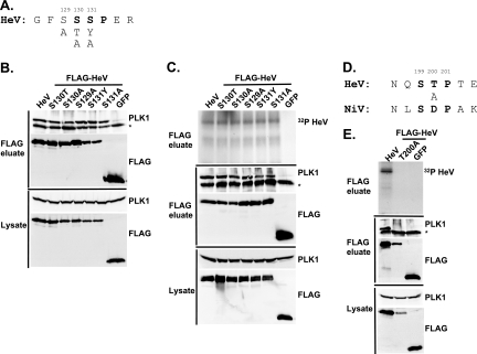
Emerging viruses in the paramyxovirus genus Henipavirus evade host antiviral responses via protein interactions between the viral V and W proteins and cellular STAT1 and STAT2 and the cytosolic RNA sensor MDA5. Polo-like kinase (PLK1) is identified as being an additional cellular partner that can bind to Nipah virus P, V, and W proteins. For both Nipah virus and Hendra virus, contact between the V protein and the PLK1 polo box domain is required for V protein phosphorylation. Results indicate that PLK1 is engaged by Nipah virus V protein amino acids 100 to 160, previously identified as being the STAT1 binding domain responsible for host interferon (IFN) signaling evasion, via a Thr-Ser-Ser-Pro motif surrounding residue 130. A distinct Ser-Thr-Pro motif surrounding residue 199 mediates the PLK1 interaction with Hendra virus V protein. Select mutations in the motif surrounding residue 130 also influenced STAT1 binding and innate immune interference, and data indicate that the V:PLK1 and V:STAT complexes are V mediated yet independent of one another. The effects of STAT1/PLK1 binding motif mutations on the function the Nipah virus P protein in directing RNA synthesis were tested. Remarkably, mutations that selectively disrupt the STAT or PLK1 interaction site have no effects on Nipah virus P protein-mediated viral RNA synthesis. Therefore, mutations targeting V protein-mediated IFN evasion will not alter the RNA synthetic capacity of the virus, supporting an attenuation strategy based on disrupting host protein interactions.


Figures

FIG. 1.
FIG. 1.
Henipavirus P, V, and W proteins associate with PLK1. (A) Schematic representation of the Nipah virus P, V, and W proteins (NiP, NiV, and NiW, respectively) illustrating the STAT1 binding domain, the cysteine-rich CTD, and the nuclear localization signal (NLS). (B) Nipah virus V and Hendra virus V (HeV) but not measles virus V (MeV) proteins copurify with endogenous PLK1. 293T cells were transfected to express FLAG-tagged measles virus V, Nipah virus V, Hendra virus V, or GFP, followed by immunoprecipitation with FLAG affinity gel and elution with FLAG peptide. The FLAG eluate and lysate samples were subjected to immunoblot analysis to detect endogenous PLK1 or FLAG-V protein. The asterisk indicates the immunoglobulin G (IgG) heavy chain, which migrates near PLK1. (C) Expressed HA-tagged PLK1 copurifies with Nipah virus V and Hendra virus V but not measles virus V. 293T cells were transfected to express HA-PLK1 together with either FLAG-tagged measles virus V, Nipah virus V, Hendra virus V, or GFP, followed by HA immunoprecipitation and elution with HA peptide. The HA immune complexes and lysate samples were subjected to immunoblotting with the FLAG antibody to detect V or the anti-HA antibody to detect HA-PLK1. (D) Nipah virus P and W proteins copurify with endogenous PLK1. 293T cells were transfected to express FLAG-tagged Nipah virus P, Nipah virus V, Nipah virus W, or GFP, followed by immunoprecipitation with FLAG affinity gel and elution with FLAG peptide. The FLAG eluate and lysate samples were subjected to immunoblot analysis to detect endogenous PLK1 or FLAG-V protein. (E) Purified HA-PLK1 phosphorylates recombinant Henipavirus V proteins in vitro. (i) Purification of PLK1. 293T cells were transfected to express HA-PLK1, followed by HA immunoprecipitation (I.P.) and elution with HA peptide. Purified PLK1 and the input samples were subjected to SDS-PAGE and silver staining. (ii) Bacterial expression of recombinant Henipavirus GST-V proteins. Recombinant GST-FLAG-Nipah virus V, GST-FLAG-Hendra virus V, and control GST-FLAG were purified from bacteria and analyzed by SDS-PAGE and Coomassie blue staining. (iii) Henipavirus V proteins are phosphorylated by purified HA-PLK1. Approximately 1, 5, or 10 ng of purified PLK1 was incubated with 5 μg of bacterially expressed Henipavirus V protein or dephosphorylated bovine casein in the presence of [γ32-P]ATP. Reaction mixtures were resolved by SDS-PAGE and visualized by autoradiography.
FIG. 2.
FIG. 2.
PLK1 binds Nipah virus V protein via the STAT1 binding domain. (A) Selective PLK1, STAT1, and STAT2 binding to Nipah virus V (NiV) fragments. 293T cells were transfected to express FLAG-tagged Nipah virus V, GFP control, and Nipah virus V truncation constructs (37), followed by FLAG immunoprecipitation and elution with FLAG peptide. The FLAG eluate and lysate samples were analyzed by immunoblotting using antibodies to detect endogenous PLK1, STAT1, STAT2, and FLAG-V proteins. (B) Interaction of Nipah virus V protein with PLK1, STAT1, and STAT2. 293T cells were transfected to express the Rev1.4-GFP fusion vectors as indicated (37), and lysates were immunoprecipitated (IP) with antisera to GFP. Immune complexes were probed for PLK1, STAT1, STAT2, and GFP. (C) Schematic representation of Nipah virus V protein illustrating the PLK1 and STAT1 interaction domains. The consensus S-[pS/pT]-P motifs for Nipah virus and Hendra virus V (HeV) proteins are indicated by boldface type and double underlining.
FIG. 3.
FIG. 3.
The PLK1 PBD is necessary and sufficient to mediate Henipavirus V interactions. (A) Illustration of PLK1 domain structure. The PLK1 N-terminal kinase domain containing residue K82, which is required for ATP binding, and the C-terminal PBD containing residues W414, H538, and K540, which are required for contact with phosphopeptide substrates, are overlined. (B) The PBD mediates Henipavirus V protein interactions. 293T cells were cotransfected to express HA-PLK1, the HA-PLK1 N-terminal kinase domain (residues 1 to 344), or the C-terminal PBD (residues 345 to 603) together with either FLAG-tagged Nipah virus V (NiV) (i), Hendra virus V (HeV) (ii), or GFP (iii), followed by FLAG immunoprecipitation and elution. The FLAG immune complexes and lysate samples were subjected to immunoblot analysis to detect FLAG-V, HA-PLK1, or HA-PLK1 fragments. (C) PBD contact residues mediate V protein interactions. 293T cells were cotransfected to express HA-PLK1 or HA-PLK1 point mutations together with either FLAG-tagged Nipah virus V (i), Hendra virus V (ii), or GFP (iii), followed by FLAG immunoprecipitation and elution. The FLAG immune complexes and lysate samples were subjected to immunoblotting to detect FLAG-V, HA-PLK1, or HA-PLK1 point mutations.
FIG. 4.
FIG. 4.
Nipah virus V protein PLK1 interaction and phosphorylation require a consensus motif surrounding serine 130. (A) Selected residues of the consensus TSSP motif were mutated as indicated. (B) The TSSP motif is necessary for PLK1 binding to the Nipah virus V (NiV) protein. 293T cells were transfected to express FLAG-tagged Nipah virus V, the GFP control, or Nipah virus V point mutations, followed by FLAG immunoprecipitation and elution with FLAG peptide. The FLAG eluate and lysate samples were analyzed by immunoblotting using antibodies to detect associated PLK1 or FLAG-V protein. (C) Contact-dependent phosphorylation of Nipah virus V. 293T cells were transfected to express FLAG-tagged Nipah virus V, the GFP control, or Nipah virus V mutations, followed by FLAG immunoprecipitation and elution. Immune complexes were incubated in the presence of [γ32-P]ATP, and reaction mixtures were resolved by SDS-PAGE and visualized by autoradiography. The FLAG eluate was further assessed by immunoblotting using antibodies detecting endogenous PLK1 and FLAG-V protein. *, immunoglobulin G heavy chain.
FIG. 5.
FIG. 5.
Hendra virus V protein PLK1 interaction and phosphorylation require an alternate consensus motif surrounding threonine 200. (A) Selected residues of the conserved SSSP motif were mutated as indicated. (B) Hendra virus V (HeV) protein mutations retain PLK1 binding. 293T cells were transfected to express FLAG-tagged Hendra virus V, the GFP control, or Hendra virus V point mutations and analyzed as described in the legend of Fig. 4. (C) Hendra virus V mutations retain phosphorylation. Immune complex kinase assays were carried out as described in the legend of Fig. 4. (D) An STP motif that is not conserved with Nipah virus surrounding T200 in the Hendra virus V protein mediates the PLK1 interaction. T200 was mutated to A as indicated. (E) T200 is required for PLK1 binding to the Hendra virus V protein. Proteins were expressed and analyzed for PLK1 associations and phosphorylation as described above.
FIG. 6.
FIG. 6.
Independence of STAT and PLK1 complexes. (A) PLK1 does not associate with STAT1 and STAT2 in the absence of V protein. 293T cells were transfected to express HA-tagged PLK1, followed by HA immunoprecipitation and elution. The lysate, sequential HA peptide eluates (E1 and E2), and the supernatant following immunoprecipitation (I.P) were assessed by immunoblotting to detect HA-PLK1, STAT1, or STAT2. (B) The PLK1 and V protein interactions are independent of STAT1 or STAT2. Parental 2fTGH cells, STAT1-deficient U3A, or STAT2-deficient U6A derivatives were transfected to express FLAG-tagged V proteins or GFP, followed by FLAG immunoprecipitation. Immune complexes and lysates were analyzed by immunoblotting to detect PLK1, STAT1, STAT2, and FLAG-V. (C) V:STAT and V:PLK1 exist as distinct complexes. 293T cells were transfected to express HA-PLK1 together with either FLAG-tagged Nipah virus V (NiV), Hendra virus V (HeV), or GFP, followed by FLAG immunoprecipitation and elution with FLAG peptide (FLAG eluate). The FLAG immune complexes were then reimmunoprecipitated with HA affinity gel and eluted using HA peptide (HA eluate). Lysate (input), FLAG, and HA eluates were assessed by immunoblot analysis to detect endogenous PLK1, STAT1, or STAT2 and then reprobed to detect HA-PLK1 or FLAG-V.
FIG. 7.
FIG. 7.
The conserved T/S SSP motif is necessary for interaction with STAT1 and STAT2. (A) T/S SSP motifs are required for STAT1 and STAT2 V protein interactions. 293T cells were transfected to express FLAG-tagged Nipah virus V (NiV) (i) or Hendra virus V (HeV) (ii), followed by FLAG immunoprecipitation and elution. The FLAG eluate and lysate samples were analyzed by immunoblotting using antibodies to detect endogenous STAT1, STAT2, or FLAG-V protein. (iii) Control analysis of the FLAG-Hendra virus V T200A point mutation. (B) IFN signaling inhibition correlates with STAT1 and STAT2 binding abilities. 2fTGH cells were transfected with the GAS- or ISRE-luciferase (Luc) reporter gene and Nipah virus V (NiV) (i), Hendra virus V (HeV) (ii), point mutations, or an empty vector (CON), as indicated. Cells were stimulated with IFN-γ (5 ng/ml) or IFN-α (1,000 U/ml) for 10 h or not stimulated (−) prior to lysis and luciferase assays. Data were normalized to cotransfected Renilla luciferase, and the bars indicate the averages (n = 3) ± the standard deviations.
FIG. 8.
FIG. 8.
The conserved T/S SSP motif is essential for STAT1 relocalization and IFN signaling evasion. (A) A subset of mutated V proteins fail to retain STAT1 in the cytoplasm. 2fTGH cells were transfected with FLAG-tagged Nipah virus V (NiV) and point mutations, followed by indirect immunofluorescence to detect FLAG-V and STAT1. WT, wild type. (B) V proteins that fail to bind STAT1 do not disrupt the IFN antiviral state. Pools of 2fTGH cells stably expressing Nipah virus V protein, the Hendra virus V protein (HeV), phosphopeptide binding motif mutations, and the pEF-FLAG control were treated with medium alone or IFN-α for 8 h, followed by VSV-GFP infection. (C) After 22 h, GFP levels were analyzed by flow cytometry, and the percentage of cells expressing GFP was calculated in comparison to uninfected 2fTGH cells. N/A, not applicable.
FIG. 9.
FIG. 9.
Interaction of STAT1 and PLK1 is dispensable for P protein-dependent Nipah virus RNA synthesis. BHK/sr/T7 cells were transfected to express Nipah virus N, Nipah virus P (or Nipah virus P point mutations), Nipah virus L, and the Nipah virus CAT minigenome plasmid. For the negative control (CON), the Nipah virus L plasmid was replaced with an equivalent amount of pTM1 empty vector. The assay was performed three separate times in which each sample was assayed in duplicate. Minigenome replication was assessed using a CAT ELISA system as described previously (16). WT, wild type.

References

    1. Aaronson, D. S., and C. M. Horvath. 2002. A road map for those who don't know JAK-STAT. Science 2961653-1655. - PubMed
    1. Andrejeva, J., K. S. Childs, D. F. Young, T. S. Carlos, N. Stock, S. Goodbourn, and R. E. Randall. 2004. The V proteins of paramyxoviruses bind the IFN-inducible RNA helicase, mda-5, and inhibit its activation of the IFN-beta promoter. Proc. Natl. Acad. Sci. USA 10117264-17269. - PMC - PubMed
    1. Anonymous. 2004. Hendra virus—Australia (Queensland). ProMED archive number 20041214.3307. International Society for Infectious Diseases, Brookline, MA.
    1. Bostik, P., G. L. Dodd, F. Villinger, A. E. Mayne, and A. A. Ansari. 2004. Dysregulation of the polo-like kinase pathway in CD4+ T cells is characteristic of pathogenic simian immunodeficiency virus infection. J. Virol. 781464-1472. - PMC - PubMed
    1. Bradford, M. M. 1976. A rapid and sensitive method for the quantitation of microgram quantities of protein utilizing the principle of protein-dye binding. Anal. Biochem. 72248-254. - PubMed

Publication types

MeSH terms