Comparative Study
. 2008 Jul;82(13):6697-710.
doi: 10.1128/JVI.00212-08. Epub 2008 Apr 16.

A single Banana streak virus integration event in the banana genome as the origin of infectious endogenous pararetrovirus

Affiliations
Comparative Study

A single Banana streak virus integration event in the banana genome as the origin of infectious endogenous pararetrovirus

Philippe Gayral et al. J Virol. 2008 Jul.

Abstract

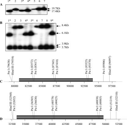
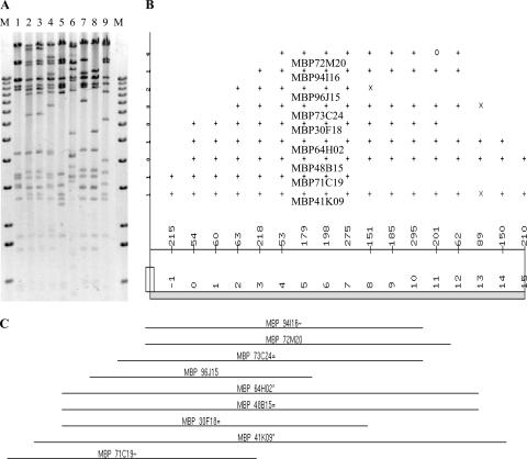
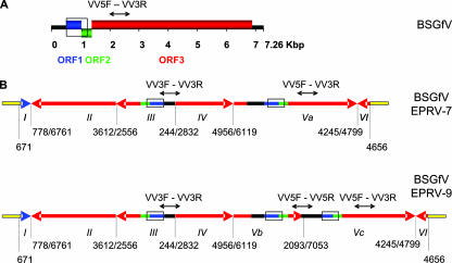
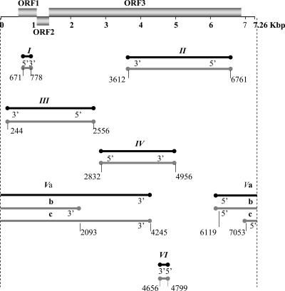
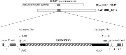
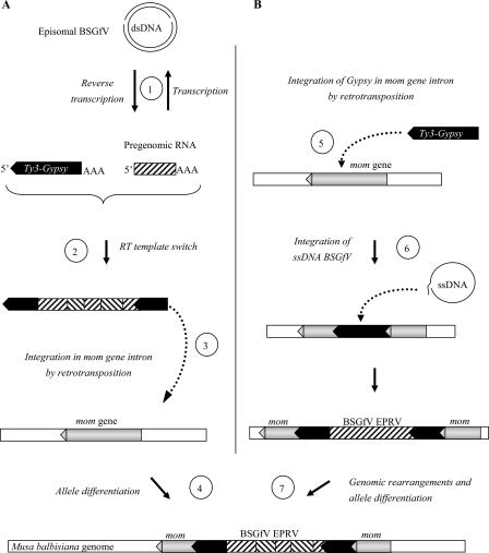
Sequencing of plant nuclear genomes reveals the widespread presence of integrated viral sequences known as endogenous pararetroviruses (EPRVs). Banana is one of the three plant species known to harbor infectious EPRVs. Musa balbisiana carries integrated copies of Banana streak virus (BSV), which are infectious by releasing virions in interspecific hybrids. Here, we analyze the organization of the EPRV of BSV Goldfinger (BSGfV) present in the wild diploid M. balbisiana cv. Pisang Klutuk Wulung (PKW) revealed by the study of Musa bacterial artificial chromosome resources and interspecific genetic cross. cv. PKW contains two similar EPRVs of BSGfV. Genotyping of these integrants and studies of their segregation pattern show an allelic insertion. Despite the fact that integrated BSGfV has undergone extensive rearrangement, both EPRVs contain the full-length viral genome. The high degree of sequence conservation between the integrated and episomal form of the virus indicates a recent integration event; however, only one allele is infectious. Analysis of BSGfV EPRV segregation among an F1 population from an interspecific genetic cross revealed that these EPRV sequences correspond to two alleles originating from a single integration event. We describe here for the first time the full genomic and genetic organization of the two EPRVs of BSGfV present in cv. PKW in response to the challenge facing both scientists and breeders to identify and generate genetic resources free from BSV. We discuss the consequences of this unique host-pathogen interaction in terms of genetic and genomic plant defenses versus strategies of infectious BSGfV EPRVs.


Figures

FIG. 1.
FIG. 1.
Integration patterns of BSGfV EPRVs in cv. PKW. Fingerprint patterns obtained after digestion of BAC clones containing BSGfV EPRVs with HindIII (A) or PstI (B) and hybridization with two BSGfV probes covering the full-length viral genome are shown. Lanes 1 to 9 show the results obtained with BAC clones containing BSGfV inserts: lane 1, MBP 30_F18; lane 2, MBP 41_K09; lane 3, MBP 64_H02; lane 4, MBP 71_C19; lane 5, MBP 72_M20; lane 6, MBP 73_C24; lane 7, MBP 94_I16; lane 8, MBP 48_D15; and lane 9, MBP 96_J15. Asterisks indicate BAC clones with the same restriction pattern. Deduced restriction maps of BSGfV EPRV in BAC clones MBP 71_C19 (C) and MBP 94_I16 (D) are presented. Numbers refer to the position according to BAC annotation. Gray bars indicate the BSGfV EPRV.
FIG. 2.
FIG. 2.
Fingerprint contig building. (A) Restriction patterns of nine XhoI-digested BAC clones containing BSGfV integration analyzed with the software Image, version 3.10 (54). Lane M, 1-kb ladder (Invitrogen); lanes 1 to 9, BAC clones (the lane order is the same as in Fig. 1). (B) Consensus band map displayed in FPC version 4.7.9 (49) from the fingerprint analysis in panel A showing the ordering of clones and their fragments. At the top of the panel, the length of each clone is equal to the number of bands in the clone (total length of 17, ranging from −1 to 15). The points represent partially ordered groups: “+” indicates a match with the bottom band within the tolerance, “×” indicates a match within twice the tolerance, and “○” indicates no match. The middle portion of the panel indicates the consensus band numbers. The parameters used were as follows: tolerance, 7; cutoff, 10e-7. (C) Resulting contig of the nine BAC clones. The suffix symbols “*”, “=”, and “∼” represent the status of each clone: the “*” indicates a parent clone, which shares the same common bands with exact child clones (=) or a percentage of the common bands with approximate child clones (∼).
FIG. 3.
FIG. 3.
BSGfV EPRV structures in cv. PKW. (A) Organization (linear view) of the BSGfV genome (GenBank AY3509). Blue, green, and red boxes indicate the three ORFs of the virus. The intergenic region is shown in black. (B) Structures of BSGfV EPRV-7 (top) and EPRV-9 (bottom) resulting from annotated BAC clones MBP_71C19 and MBP_94I16, respectively. Arrows indicate the orientation of fragments of the BSGfV genome integrated in the Musa genome, shown in yellow. Blue, green, and red (the same code as used in panel A) refer to the different ORFs. Roman numerals identify the fragment. Numbers below each EPRV indicate the position of the fragment in the BSGfV genome. Open boxes indicate the region used in EPRV genotyping by PCR-RFLP DifGf F/R, and black arrows above the fragments indicate the regions amplified by multiplex-PCR with VV3F/R-VV5F/R.
FIG. 4.
FIG. 4.
Positions of EPRV fragments in the BSGfV genome. The genome of BSGfV is represented as in Fig. 3 (top). Lines below the BSGfV genome represent all of the fragments of EPRV-7 (in black) and EPRV-9 (in gray), and the circles indicate the boundaries of each fragment. Fragment names are indicated above each line. Fragments are arranged relative to their position in the BSGfV genome. The annotations 5′ and 3′ indicate the orientation of the fragments relative to the BSGfV genome.
FIG. 5.
FIG. 5.
Musa genomic environment of BSGfV EPRV. The orientation of the mom gene putative exons (gray arrows) and regions of the Ty3/gypsy-like retrotransposon (black arrows) are indicated. mom gene introns are numbered and indicated by thin lines. The 3′ and 5′ LTRs are indicated. TS, target site; PBS, primer binding site; PPT, polypurine tract. The regions of the GAG and POL polyproteins (Ty3/gypsy-encoded RT) in the retrotransposon are indicated.
FIG. 6.
FIG. 6.
PCR analysis of cv. PKW and cv. IDN 110 4x and their F1 progeny, using BSGfV specific primers. (A) Ethidium bromide-stained agarose gel analysis of PCR product (GfF [5′-ACGAACTATCACGACTTGTTCAAGC-3′] and GfR [5′-TCGGTGGAATAGTCCTGAGTCTTC-3′]). (B) Southern blot hybridization of the gel shown in panel A using complete genome probes of BSGfV. Lane M, 1-kb ladder; lane 1, cv. PKW; lane 2, IDN 110 4x; lanes 3 to 6, F1 plants showing no sign of banana streak disease; lanes 7 to 10, F1 plants showing symptoms and BSV particles by immunosorbent electron microscopy as described by Lheureux et al. (30). Hybridization was performed according to the method of Sambrook et al. (45) using the two BSGfV probes (pCR-TOPO [1,262 bp] and pCR-TOPO [6,001 bp]).
FIG. 7.
FIG. 7.
Genotyping of BSGfV EPRV-7 and EPRV-9 (A, B, and C) and detection of recombinant EPRV (C). (A) PCR DifGf F/R-RFLP to genotype BSGfV EPRVs in cv. PKW. Endonuclease TaaI discriminates between EPRV-7 and EPRV-9; amplification products carry two versus one restriction sites, respectively. Lane M, DNA ladder (low molecular weight; Invitrogen). Digestion of PCR product DifGfF/R on clone MBP_94I16 carrying EPRV-9 (lane 1), on clone MBP_71C19 carrying EPRV-7 (lane 2), and on M. balbisiana cv. PKW carrying both EPRVs (lane 3) was performed. No amplification product was seen on M. acuminata cv. IDN 110 4x (data not shown). (B) Multiplex PCR with primers VV3 and VV5 for BSGfV EPRV genotyping. Primers VV3F/R amplify a 376-bp product in both EPRVs, primers VV5F/R amplify a 628-bp product in EPRV-9 only, and primers VV5F/R amplify a 1,012-bp product on both EPRVs and the BSGfV circular genome. Lane M, 1-kb ladder (Invitrogen). Lane 1, BAC MBP_94I16; lane 2, BAC MBP_71C19; lane 3, DNA of M. balbisiana cv. PKW; lane 4, DNA of M. acuminata infected by BSGfV; lane 5, PCR negative control. (C) PCR detection of recombination between the two BSGfV EPRVs. PCR results with Spe7F/R (top) specific to EPRV-7 and Spe9bisF/R (bottom) specific to EPRV-9. Lane M, 1-kb ladder (Invitrogen). Lane 1, negative PCR control (water); lane 2, M. acuminata cv. IDN 110 4x genomic DNA; lane 3, M. balbisiana cv. PKW genomic DNA; lane 4, BAC MBP_94I16; lane 5, BAC MBP_71C19.
FIG. 8.
FIG. 8.
Genotyping of BSGfV viral particles in infected hybrids. (A) IC-multiplex PCR allows specific detection of BSGfV particles (DifGfF/R, 670-bp product) and a monitoring of plant DNA contaminations (Actin1F/R, 420-bp product). Lane M, 1-kb ladder (Invitrogen); lanes 1 to 6, coated plant extracts; lanes 7 to 10, plant total DNA. Lanes 1 to 3 show results for F1 AAB hybrids; lanes 4 to 6 show results for the IC control (lane 4, M. balbisiana cv. PKW; lane 5, M. acuminata cv. Grande Naine infected by BSGfV; lane 6, M. acuminata cv. Grande Naine BSGfV-free). Lanes 7 to 10 show results for the PCR control (lane 7, M. balbisiana cv. PKW; lane 8, M. acuminata cv. Grande Naine infected by BSGfV; lane 9, M. acuminata cv. Grande Naine BSGfV-free; and lane 10, water control). (B) Nested PCR using the internal primers VV1F/GfM2 (642-bp product) and increasing PCR product quantity from diluted DifGfF/R PCR product of infected hybrids. Lanes 1 to 10 show the results for AAB F1 hybrids infected with BSGfV; lane M, 1-kb ladder (Invitrogen). (C) TaaI RFLP test (described in Fig. 7A) to genotype the molecular EPRV signature of the viral BSGfV particle. Lanes 1 to 10 show the results for AAB F1 hybrids infected with BSGfV; lane M shows the results for the 50-bp ladder (NEB).
FIG. 9.
FIG. 9.
Scenarios for BSGfV integration in the M. balbisiana nuclear genome. (A) Hypothesis 1: use of Ty3/gypsy retroelement for BSGfV integration. Viral RT leads to a pregenomic viral RNA (step 1) during episomal BSGfV infection. A template switch of retrotransposon RT between pregenomic viral RNA and a replicating Ty3/gypsy RNA may form a chimeric RNA (step 2). This step leads to the rearrangements of BSGfV EPRV: fragmentation, inversion, and duplication. Complete retrotransposition of the Ty3/gypsy element leads to its integration (step 3) into the genome of M. balbisiana (within intron 5 of the mom gene). Subsequent genomic change such as recombination may lead to the structural differences observed between the two alleles 7 and 9. (B) Hypothesis 2: Ty3/gypsy retroelement integrated in the fifth intron of the mom gene (step 5) during retrotransposition. Double-strand break repair could account for illegitimate recombination with the single-stranded DNA template generated during BSGfV reverse transcription and integration of the BSGfV genome (step 6). A subsequent genomic change, e.g., recombination, may have lead to both the fragmentation, inversion, and duplication observed in BSGfV EPRV and the structural differences between the alleles 7 and 9 (step 7).

References

    1. Altschul, S., W. Gish, W. Miller, E. Myers, and D. Lipman. 1990. Basic local alignment search tool. J. Mol. Biol. 215403-410. - PubMed
    1. Amedeo, P., Y. Habu, K. Afsar, O. M. Scheid, and J. Paszkowski. 2000. Disruption of the plant gene MOM releases transcriptional silencing of methylated genes. Nature 405203-206. - PubMed
    1. Berriman, M., and K. Rutherford. 2003. Viewing and annotating sequence data with Artemis. Brief Bioinform. 4124-132. - PubMed
    1. Capy, P., G. Gasperi, C. Biemont, and C. Bazin. 2000. Stress and transposable elements: co-evolution or useful parasites? Heredity 85101-106. - PubMed
    1. Cocquet, J., A. Chong, G. L. Zhang, and R. A. Veitia. 2006. Reverse transcriptase template switching and false alternative transcripts. Genomics 88127-131. - PubMed

Publication types

LinkOut - more resources