. 2008 Jul;82(13):6288-98.
doi: 10.1128/JVI.00569-08. Epub 2008 Apr 16.

Bovine papillomavirus type 1: from clathrin to caveolin

Affiliations

Bovine papillomavirus type 1: from clathrin to caveolin

Valerie Laniosz et al. J Virol. 2008 Jul.

Abstract

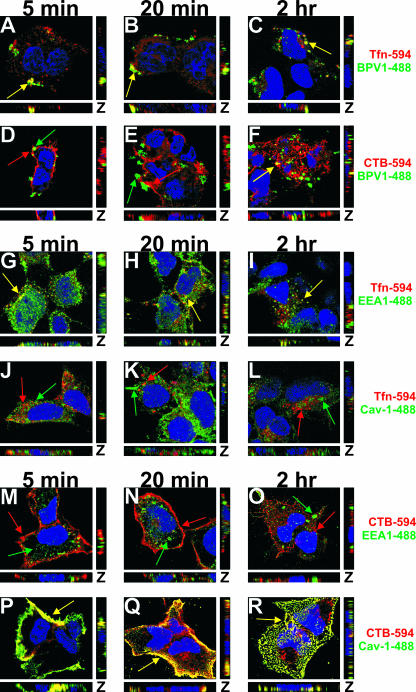
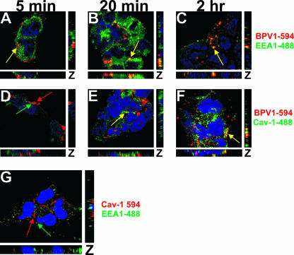
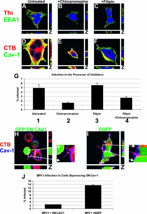
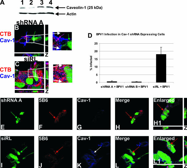
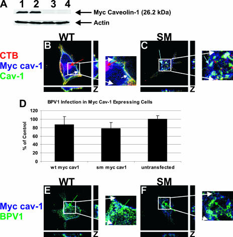
Viruses may infect cells through clathrin-dependent, caveolin-dependent, or clathrin- and caveolin-independent endocytosis. Bovine papillomavirus type 1 (BPV1) entry into cells has been shown to occur by clathrin-dependent endocytosis, a pathway that involves the formation of clathrin-coated pits and fusion to early endosomes. Recently, it has been demonstrated that the closely related JC virus can enter cells in clathrin-coated vesicles and subsequently traffic to caveolae, the organelle where vesicles of the caveolin-dependent pathway deliver their cargo. In this study, we use immunofluorescence staining of BPV1 pseudovirions to show that BPV1 overlaps with the endosome marker EEA1 early during infection and later colocalizes with caveolin-1. We provide evidence through the colocalization of BPV1 with transferrin and cholera toxin B that BPVl trafficking may not be restricted to the clathrin-dependent pathway. Disrupting the entry of caveolar vesicles did not affect BPV1 infection; however, we show that blocking the caveolar pathway postentry results in a loss of BPV1 infection. These data indicate that BPV1 may enter by clathrin-mediated endocytosis and then utilize the caveolar pathway for infection, a pattern of trafficking that may explain the slow kinetics of BPV1 infection.


Figures

FIG. 1.
FIG. 1.
BPV1 PsVs colocalize with Tfn early during infection and later show overlap with CTB. (A to F) BPV1 PsVs were internalized into 293 cells simultaneously with 594-labeled Tfn (A to C, red) or CTB (D to F, red). Virions were labeled with anti-L1 antibody 5B6 (green), and TOPRO-3 was used to stain the nucleus (blue). At 5 and 20 min, the virus overlapped with Tfn (A and B, yellow) but not CTB (D and E). Two hours postinternalization, there was prominent overlap between BPV1 and CTB (F, yellow) and residual overlap with Tfn (C). (G to L) 293 cells were allowed to internalize Tfn (red) for 5 min, 20 min, and 2 h and stained for the early endosome marker EEA1 (G to I, green) and caveolin-1 (Cav-1) (J to L, green). Tfn colocalized with EEA1 at all time points (G to I, yellow). No overlap was seen between Tfn and caveolin-1 at any of the time points (J to L). (M to R) 293 cells internalized CTB (red) for 5 min, 20 min, and 2 h. EEA1 (M to O, green) and caveolin-1 (P to R, green) were labeled in the cells. Colocalization was observed between CTB and caveolin-1 at all time points (P to R, yellow) but not between CTB and EEA1 (M to O). Images shown are z stacks, representing the cell in the x, y, and z planes.
FIG. 2.
FIG. 2.
BPV1 PsVs colocalize with caveolin-1 by 2 h after entry. (A to F) 293 cells were infected with BPV1 PsVs for 5 min, 20 min, and 2 h. Monoclonal antibody 5B6 was used to detect BPV1 (red), the cells were stained for EEA1 (A to C, green) or caveolin-1 (Cav-1) (D to F, green), and the nucleus was stained blue. Five minutes after entry, BPV1 overlapped with EEA1 (A, yellow) but not caveolin-1 (D). At 20 min, colocalization was seen between BPV1 and EEA1 (B, yellow) and to a lesser extent between BPV1 and caveolin-1 (E). Two hours postentry, 5B6 overlapped with EEA1 (C, yellow) and prominently with caveolin-1 (F, yellow). (G) 293 cells were fixed and costained for caveolin-1 (red), EEA1 (green), and nuclei (blue) (TOPRO-3). The markers did not overlap (red and green arrows). Pictures are three-dimensional z stacks.
FIG. 3.
FIG. 3.
Cholesterol sequestration by filipin does not affect BPV1 infection, but a dominant negative caveolin-1 mutant inhibits BPV1 infection. (A to F) 293 cells pretreated for 1 h with chlorpromazine (B and E) or filipin (C and F) were allowed to internalize Tfn (A to C, red) or CTB (D to F, red) for 30 min. The cells were stained for EEA1 (A to C, green) or caveolin-1 (Cav-1) (D to F, green), and the nucleus was stained blue using TOPRO-3. In untreated cells, Tfn colocalized with EEA1 (A, yellow) and CTB overlapped with caveolin-1 (D, yellow). Chlorpromazine treatment resulted in a loss of Tfn detection (B) but normal overlap of CTB with caveolin-1 (E, yellow). Filipin treatment did not affect the colocalization of Tfn with EEA1 (C, yellow) but diminished CTB (F). (G) Inhibitor pretreatment of 293 cells with chlorpromazine decreased infection (bars 2 and 4) compared to filipin treatment (bar 3) or untreated cells (bar 1). Flow cytometry was used to measure GFP expression in infected cells. The experiment was performed in triplicates, and the standard deviations are represented by the error bars. (H to J) The effect of transfecting 293 cells with dominant negative GFP-tagged caveolin-1 (GFP-DN CAV1) (H, green) was compared to that of EGFP expression (I, green). (H and I) CTB (red) was added to transfected cells for 30 min, and cells were stained for endogenous caveolin-1 (blue). In cells expressing dominant negative GFP-tagged caveolin-1 (H, green cell), neither CTB nor caveolin-1 was evident, but an untransfected cell showed internalization of CTB and overlap with caveolin-1 (H, purple arrow). In EGFP-transfected cells (I, green), CTB colocalized with caveolin-1 (I, purple arrow). (J) BPV1 infection of 293 cells transfected with dominant negative caveolin-1 (DN CAV1) or EGFP resulted in a loss of BPV1 infection in cells expressing dominant negative caveolin-1. Transfection was determined by GFP expression, and infection was quantitated by the expression of the DsRed reporter within the pseudovirus. At least 10,000 cells were counted, and bars represent the percentages of transfected cells that were infected (i.e., double positives). Error bars show the standard deviations for data from three experiments.
FIG. 4.
FIG. 4.
Knockdown of caveolin-1 expression results in a loss of BPV1 infection despite normal entry. (A) 293 cells were transfected with shRNA A or B against caveolin-1 (lanes 1 and 2, respectively), infected with siRL (lane 3), or untransfected (lane 4). In cells expressing shRNA A and B, protein levels of caveolin-1 were diminished (lanes 1 and 2). The levels of detection of caveolin-1 were comparable in cells expressing siRL (lane 3) and in untransfected cells (lane 4). The actin loading control is shown. (B and C) CTB (red) endocytosis was tested in cells with caveolin-1 (Cav-1) knockdown (shRNA A) (B, green) and cells expressing the siRL (C, green). Endogenous caveolin-1 was stained blue. In cells transfected with shRNA A, caveolin-1 was not detected, and CTB did not bind or enter (B, green arrow). An untransfected cell showed normal CTB trafficking and overlap with caveolin-1 (shRNA A) (B, purple arrow). Cells expressing siRL (C, green arrow) allowed CTB entry, which colocalized with caveolin-1 (C, purple arrow). (D) 293 cells were transfected with shRNA A or B or infected with siRL. Forty-eight hours later, cells were infected with BPV1 PsVs containing the DsRed reporter plasmid. The percentage of transfected cells that were infected was measured by flow cytometry. The knockdown of caveolin-1 led to an almost total loss of infection (shRNA A + BPV1 and shRNA B + BPV1) compared to that of control cells expressing siRL (siRL + BPV1) (D). (E to L) 293 cells were transfected with shRNA A (E to H, green) or infected with siRL (I to L, green) for 48 h and then infected with BPV1 for 30 min. The virus was detected with anti-L1 monoclonal antibody 5B6 (F and J, red), and endogenous caveolin-1 was stained blue (G and K). Cells expressing shRNA A showed virtually no caveolin-1 staining, indicating the effective knockdown of caveolin-1 (G). In the merge and enlarged images, cells lacking caveolin-1 showed the entry of BPV1 PsVs (H and H1, red arrow, merge and enlarged images, respectively). The siRL control cells had normal levels of caveolin-1 (K, blue), and BPV1 endocytosis was evident (L and L1, red arrow, merged and enlarged images, respectively). Enlarged images depict the z stack, showing staining in the x, y, and z planes.
FIG. 5.
FIG. 5.
A caveolin-1 scaffolding mutant does not affect BPV1 infection or entry. 293 cells were transfected with either WT Myc-tagged caveolin-1 or SM Myc-tagged caveolin-1. (A) Western blot analysis showed the overexpression of WT (lane 1) and SM (lane 2) Myc-caveolin-1 constructs. Control cells were transfected with an empty A3M vector (lane 3) or were not transfected (lane 4) and did not show a band for Myc-tagged caveolin-1. (B and C) 293 cells expressing WT caveolin-1 (Cav-1) (B, green) or SM caveolin-1 (C, green) were allowed to internalize CTB (red) for 30 min. A 647-labeled anti-Myc antibody identified transfected cells (blue), and endogenous caveolin-1 was labeled (green). In cells expressing WT caveolin-1, CTB was internalized and colocalized with caveolin-1 and WT caveolin-1, identified by white seen at the plasma membrane (B, white arrow). (C) Expression of SM caveolin-1 led to the sequestration of caveolin-1 away from the plasma membrane and the failure of CTB to bind and enter the cell. The overlap of endogenous caveolin-1 with the SM was seen within the cell (aqua arrow). (D) Infection of 293 cells expressing WT caveolin-1 or the SM of caveolin-1 was the same as infection of untransfected cells. Ten thousand cells were counted by a flow cytometer, and the error bars represent the standard deviations of the means of data from three experiments. (E and F) 293 cells expressing WT or mutant Myc-caveolin-1 constructs were infected with BPV1 PsVs for 30 min. Transfected cells were labeled blue, and BPV1 was detected using 5B6 (green). BPV1 internalized into cells expressing WT caveolin-1 (E, green arrow) and the caveolin-1 SM (F, green arrow). z-stacked images show the staining pattern in the x, y, and z planes.

Similar articles

Cited by

References

    1. Belnap, D. M., N. H. Olson, N. M. Cladel, W. W. Newcomb, J. C. Brown, J. W. Kreider, N. D. Christensen, and T. S. Baker. 1996. Conserved features in papillomavirus and polyomavirus capsids. J. Mol. Biol. 259249-263. - PMC - PubMed
    1. Berry, J. M., J. M. Palefsky, and M. L. Welton. 2004. Anal cancer and its precursors in HIV-positive patients: perspectives and management. Surg. Oncol. Clin. N. Am. 13355-373. - PubMed
    1. Bosch, F. X., X. Castellsague, N. Munoz, S. de Sanjose, A. M. Ghaffari, L. C. Gonzalez, M. Gili, I. Izarzugaza, P. Viladiu, C. Navarro, A. Vergara, N. Ascunce, E. Guerrero, and K. V. Shah. 1996. Male sexual behavior and human papillomavirus DNA: key risk factors for cervical cancer in Spain. J. Natl. Cancer Inst. 881060-1067. - PubMed
    1. Bossis, I., R. B. Roden, R. Gambhira, R. Yang, M. Tagaya, P. M. Howley, and P. I. Meneses. 2005. Interaction of tSNARE syntaxin 18 with the papillomavirus minor capsid protein mediates infection. J. Virol. 796723-6731. - PMC - PubMed
    1. Bousarghin, L., A. Touze, P. Y. Sizaret, and P. Coursaget. 2003. Human papillomavirus types 16, 31, and 58 use different endocytosis pathways to enter cells. J. Virol. 773846-3850. - PMC - PubMed

Publication types

LinkOut - more resources