

Large arachnoid granulations involving the dorsal superior sagittal sinus: findings on MR imaging and MR venography
- PMID: 18417601
- PMCID: PMC8119153
- DOI: 10.3174/ajnr.A1093
Large arachnoid granulations involving the dorsal superior sagittal sinus: findings on MR imaging and MR venography
Abstract
Background and purpose: Large arachnoid granulations (AG) within the dorsal superior sagittal sinus (SSS) have been incompletely characterized and can be confused with pathology. This report reviews the characteristics of these anatomic structures to establish common imaging features that allow differentiation from pathology.
Materials and methods: Twelve cases of large AG in the dorsal SSS are presented, identified by MR imaging. Signal intensity characteristics, size, location, venographic appearance, and association with adjacent venous and osseous structures were documented.
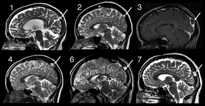
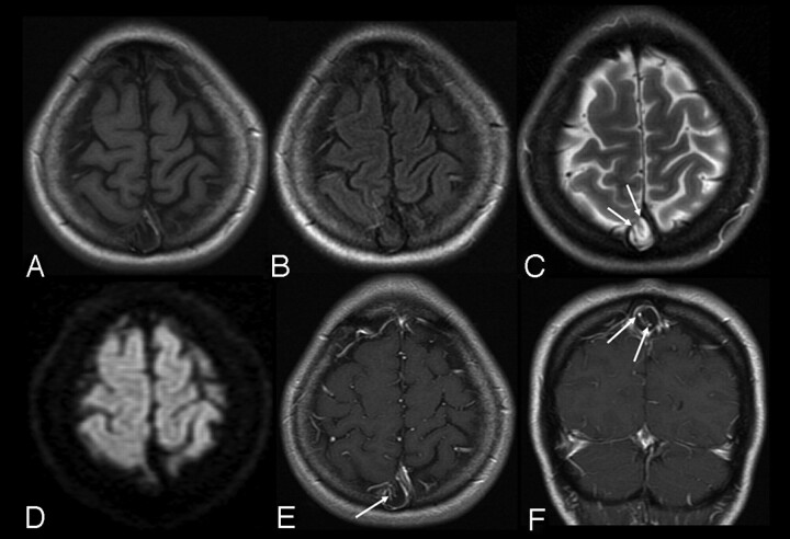
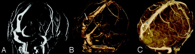
Results: A defect in the dura of the SSS was seen in all of the cases communicating with the subjacent subarachnoid space. The average size of the AG was 8.1 x 9.4 x 10.0 mm (range, 4-19 mm). Ten produced calvarial remodeling, and 11 were in the direct vicinity of the lambda. On T2-weighted images, all were hyperintense to the brain. On T1-weighted images, 8 were hypointense and 4 were hypointense with mixed areas of isointense signal intensity. All of the AGs were associated with cortical venous structures entering the sinus. On MR venography, AGs appeared as focal protrusions into the sinus, displacing, distorting, and narrowing the sinus lumen. Seven patients had headache without other visible cause on MR imaging, and 4 were initially interpreted as thrombosis or tumor.
Conclusion: Large AGs can occur in the dorsal SSS. They are well-defined projections of the subarachnoid space into the sinus, can cause luminal narrowing and calvarial remodeling, and have typical signal intensity characteristics, position, and morphology differentiating them from other pathology. Association with patient symptoms is uncertain.
Figures
References
-
- Leach JL, Fortuna RB, Gaskill-Shipley MG, et al. Imaging of cerebral venous thrombosis. Current techniques, imaging spectrum, diagnostic pitfalls. RadioGraphics 2006;26:S19–43 - PubMed
Publication types
MeSH terms
LinkOut - more resources
Full Text Sources
Medical