Comparative Study
. 2008 Apr 16;28(16):4161-71.
doi: 10.1523/JNEUROSCI.5053-07.2008.

Silencing the Kir4.1 potassium channel subunit in satellite glial cells of the rat trigeminal ganglion results in pain-like behavior in the absence of nerve injury

Affiliations
Comparative Study

Silencing the Kir4.1 potassium channel subunit in satellite glial cells of the rat trigeminal ganglion results in pain-like behavior in the absence of nerve injury

Jean-Philippe Vit et al. J Neurosci. .

Abstract

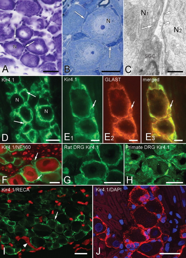
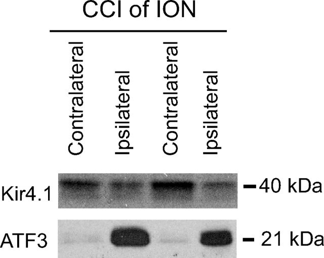
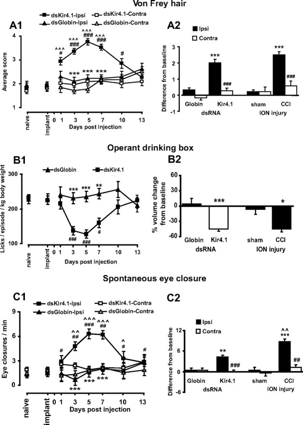
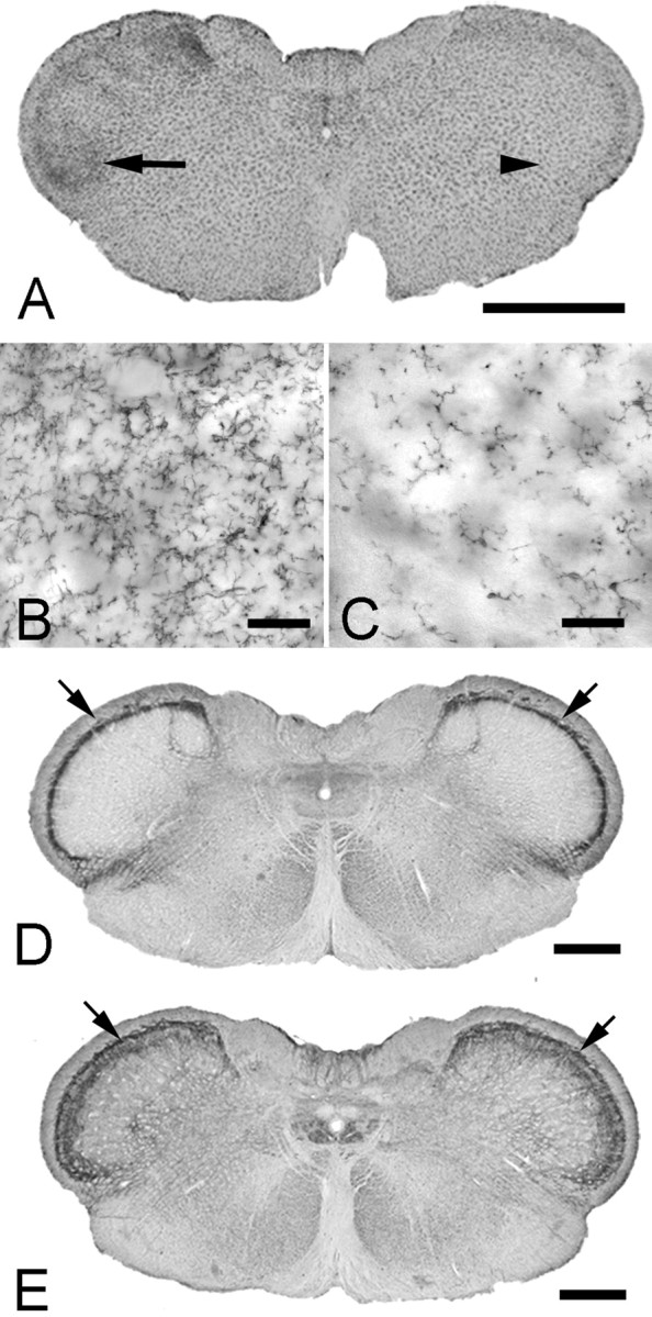
Growing evidence suggests that changes in the ion buffering capacity of glial cells can give rise to neuropathic pain. In the CNS, potassium ion (K+) buffering is dependent on the glia-specific inward rectifying K+ channel Kir4.1. We recently reported that the satellite glial cells that surround primary sensory neurons located in sensory ganglia of the peripheral nervous system also express Kir4.1, whereas the neurons do not. In the present study, we show that, in the rat trigeminal ganglion, the location of the primary sensory neurons for face sensation, specific silencing of Kir4.1 using RNA interference leads to spontaneous and evoked facial pain-like behavior in freely moving rats. We also show that Kir4.1 in the trigeminal ganglion is reduced after chronic constriction injury of the infraorbital nerve. These findings suggests that neuropathic pain can result from a change in expression of a single K+ channel in peripheral glial cells, raising the possibility of targeting Kir4.1 to treat pain in general and particularly neuropathic pain that occurs in the absence of nerve injury.


Figures

Figure 1.
Figure 1.
Morphology and immunohistochemistry of SGCs in sensory ganglia. A, Nissl-stained section showing trigeminal ganglion neurons (N) and the nuclei of surrounding SGCs (arrows). B, Semithin Epon section, 0.1 μm, showing closely packed neurons with surrounding SGCs (arrows). C, Electron micrograph demonstrating that the darkly stained, often attenuated SGC cytoplasm (arrow and arrowheads) closely surrounds the primary sensory neurons (N1, N2). The separation between the neuronal and SGC membranes is ∼20 nm. D, SGCs are immunopositive for Kir4.1. The unstained nuclei of SGCs (arrows) give a characteristic signet-ring appearance. N indicates the cell bodies of sensory neurons. E1–E3, Double immunolabeling with Kir4.1 (green, E1) and GLAST (red, E2) show extensive overlap in the merged image (yellow, E3), confirming that Kir4.1 is confined to SGCs. The arrow indicates the same SGC nucleus in each image. F, Immunolabels for Kir4.1 (green) and the neuronal marker NF160 (red) do not overlap. Arrows indicate unstained SGC nuclei. G, H, SGCs in rat (G) and primate (H) dorsal root ganglion are also Kir4.1 immunopositive. I, Section of control trigeminal ganglion immunostained for Kir4.1 (green, arrow) and blood vessels (RECA-1 antibody, red, arrowheads). There is no overlap of the two labels. J, Control trigeminal ganglion immunostained for Kir4.1 (red) and nuclei (DAPI, blue). The only Kir4.1-immunopositive structures are the SGCs surrounding unstained neurons. Nuclei (arrows) in the surrounding unstained regions belong to Schwann cells, and the outline of unstained myelin sheaths (arrowheads) is visible. Scale bars: A, D, G, H, I, J, 30 μm; B, E, F, 15 μm; C, 5 μm.
Figure 2.
Figure 2.
Changes in Kir4.1 expression after CCI of the ION. Ten days after CCI of the ION, there is a significant decrease in the expression of Kir4.1 in the trigeminal ganglion ipsilateral to the nerve injury and an increase in expression of the cell injury marker ATF3.
Figure 3.
Figure 3.
Silencing Kir4.1 in the trigeminal ganglion leads to pain-like behavior. A1, Rats show increased sensitivity to von Frey hair testing beginning 1 d after Kir4.1 dsRNA injection. This sensitivity reaches a maximum at day 5. A2, When compared with CCI rats, there is no difference between groups. Note that, in all graphs, the values are taken at day 10 after the CCI when nociceptive scores were at the maximum value. B1, Kir4.1 dsRNA reduces the number of licks per episode of drinking. B2, The reduction in drinking behavior is similar to the CCI group. C1, After Kir4.1 dsRNA injection, the number of eye closures on the injected side increases to a maximum on day 5 and then declines thereafter. C2, There is an increase from baseline for both Kir4.1 dsRNA-injected and CCI rats, but, in the CCI group, the eye closure rate is greater than in the Kir4.1 dsRNA group. A1, B1, C1, #p < 0.05, ##p < 0.01, ###p < 0.001 compared with preinjection of dsRNA (implant); **p < 0.01, ***p < 0.001 compared with Kir4.1 dsRNA; ^p < 0.05, ^^ p < 0.01, ^^^p < 0.001 compared with contralateral side (n = 10 per group for Kir4.1 and globin for all comparisons). A2, C2, ##p < 0.01, ###p < 0.001 compared with ipsilateral side. A2, B2, C2, *p < 0.05, **p < 0.01; C2, ***p < 0.001 compared with appropriate control; ^^p < 0.01 compared with Kir4.1 dsRNA (n = 10 per group for Kir4.1 and globin groups and n = 4 per group for CCI and sham groups).
Figure 4.
Figure 4.
Kir4.1 dsRNA injection into the trigeminal ganglion. A1, Injection of Kir4.1 dsRNA into the trigeminal ganglion inhibits the expression of Kir4.1 as shown by reduced SGC immunolabeling (green). A2, A3, Pseudocolor representation of the squares depicted in A1 shows that Kir4.1 inhibition is greatest close (A2) to the injection site indicated by the red fluorescent dye DiI (A1, arrow) when compared with a distant area (A3). B, C, Serial sections from a Kir4.1 dsRNA injection site showing that, after the disappearance of Kir4.1 immunoreactivity (B, green), SGCs are still present as indicated by GLAST labeling (C, green). The asterisk shows the same blood vessel on each section (B, C). D, Kir5.1 immunolabeling (green) is still present in SGCs after Kir4.1 dsRNA injection (arrow shows DiI injection site). E, Immunostaining for SK3 (green) from the region of the trigeminal ganglion injected with Kir4.1 dsRNA indicated by the presence of DiI (red, arrow). There is no reduction of the SGC-specific SK3. F, Kir2.3 immunostaining (green) from the region of the trigeminal ganglion injected with Kir4.1 dsRNA indicated by the presence of DiI (red). There is no reduction of the Kir2.3 immunostaining. G, Section from a Kir4.1 dsRNA injection site (arrows indicate DiI, showing we are in the injection site) on day 14 showing the return of Kir4.1 expression (green). H, Kir4.1 dsRNA injection site showing only a single ATF3-immunopositive neuron (arrow). Inset, Higher magnification of the ATF3-immunolabeled neuron. The asterisk indicates the location of the center of DiI injection. I, Large numbers of ATF3-positive neurons (arrows) are present after CCI of the ION. Scale bars: A, H, 300 μm; B, C, 90 μm; D, I, 30 μm; E, 60 μm; G, I, 180 μm.
Figure 5.
Figure 5.
Changes in Kir4.1 expression 5 d after Kir4.1 dsRNA or globin dsRNA was injected into the trigeminal ganglion. There is a significant decrease in the expression of Kir4.1 in the Kir41. dsRNA-injected ganglia and no change in the globin dsRNA-injected ganglia. I, Ipsilateral ganglion; C, contralateral ganglion.
Figure 6.
Figure 6.
Effect of Kir4.1 dsRNA injection in the trigeminal ganglion. A, B, Nissl-stained sections from Kir4.1 dsRNA-treated (A) and globin dsRNA-treated (B) ganglia. There is no difference in the number of neurons ipsilateral to the injection between Kir4.1 dsRNA (20 ± 1 neurons/100,000 μm2) and globin dsRNA (19 ± 1 neurons/100,000 μm2) groups (ANOVA, F = 2.1, p = 0.180, n = 3). C–H, Neurons (arrows) are immunolabeled for substance-P (C, D, red fluorescence), IB4 (E, F, green fluorescence), or CGRP (G, H, blue fluorescence) in Kir4.1 dsRNA-treated (C, E, G) and globin dsRNA-treated (D, F, H) ganglia. There is no difference in the percentage of substance-P- or IB4-positive neurons ipsilateral to the injection between Kir4.1 dsRNA (substance-P, 12 ± 1%; IB4, 32 ± 1%; CGRP, 34 ± 1%) and globin dsRNA (substance-P, 14 ± 1%; IB4, 33 ± 1%; CGRP, 32 ± 1%) groups (ANOVA; substance-P, F = 2.3, p = 0.204; IB4, F = 0.3, p = 0.815; CGRP, F = 1.1, p = 0.566; n = 3). Scale bars: A–H, 60 μm.
Figure 7.
Figure 7.
Effect of Kir4.1 dsRNA injection in the spinal trigeminal nucleus. A, Kir4.1 dsRNA injection into the trigeminal ganglion results in microglia activation (OX-42 immunostaining) in the brainstem. The arrow and arrowhead indicated the regions shown at high magnification in B and C, respectively. D, E, There is no change in substance-P or NK1 receptor staining in the trigeminal nucleus caudalis after Kir4.1 suppression in the trigeminal ganglion. Sections of the brainstem from a rat with Kir4.1 dsRNA injected into the left trigeminal ganglion immunostained for substance-P (D) or NK1 (E). Densitometry quantification of the immunostaining (arrows) shows no difference in the ipsilateral/contralateral pixel ratio between Kir4.1 dsRNA (substance-P, 1.2 ± 0.1; NK1, 1.0 ± 0.1) and globin dsRNA (substance-P, 0.9 ± 0.1; NK1, 1.0 ± 0.0) groups (ANOVA; substance-P, F = 2.4, p = 0.315; NK1, F = 0.5, p = 0.531). Scale bars: A, 2 mm; B, C, 50 μm; D, E, 1 mm.

References

    1. Alloui A, Zimmermann K, Mamet J, Duprat F, Noel J, Chemin J, Guy N, Blondeau N, Voilley N, Rubat-Coudert C, Borsotto M, Romey G, Heurteaux C, Reeh P, Eschalier A, Lazdunski M. TREK-1, a K+ channel involved in polymodal pain perception. EMBO J. 2006;25:2368–2376. - PMC - PubMed
    1. Anderson CM, Swanson RA. Astrocyte glutamate transport: review of properties, regulation, and physiological functions. Glia. 2000;32:1–14. - PubMed
    1. Banati RB. Brain plasticity and microglia: is transsynaptic glial activation in the thalamus after limb denervation linked to cortical plasticity and central sensitisation? J Physiol (Paris) 2002;96:289–299. - PubMed
    1. Bhangoo SK, Ren D, Miller RJ, Chan DM, Ripsch MS, Weiss C, McGinnis C, White FA. CXCR4 chemokine receptor signaling mediates pain hypersensitivity in association with antiretroviral toxic neuropathy. Brain Behav Immun. 2007;21:581–591. - PMC - PubMed
    1. Bhargava A, Dallman MF, Pearce D, Choi S. Long double-stranded RNA-mediated RNA interference as a tool to achieve site-specific silencing of hypothalamic neuropeptides. Brain Res Brain Res Protoc. 2004;13:115–125. - PubMed

Publication types

MeSH terms

Substances