. 2008 Jul;19(7):1321-30.
doi: 10.1681/ASN.2007121368. Epub 2008 Apr 16.

Endotoxin mediates recruitment of RNA polymerase II to target genes in acute renal failure

Affiliations

Endotoxin mediates recruitment of RNA polymerase II to target genes in acute renal failure

Masayo Naito et al. J Am Soc Nephrol. 2008 Jul.

Abstract

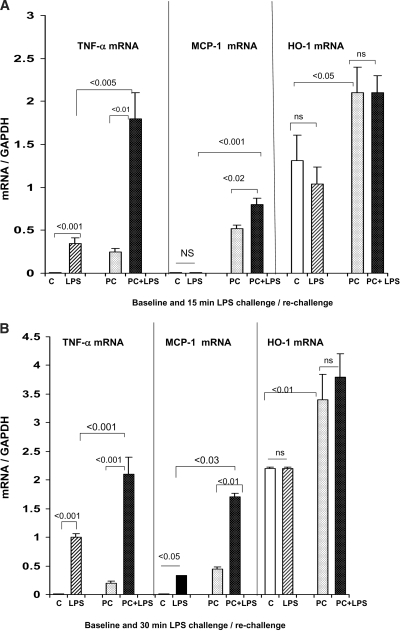
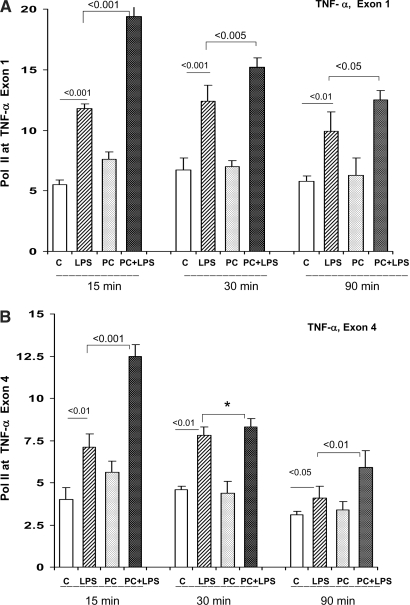
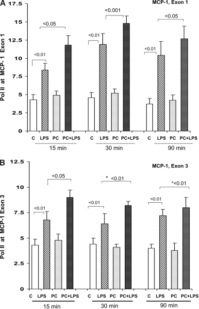
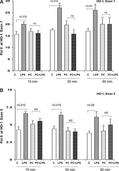
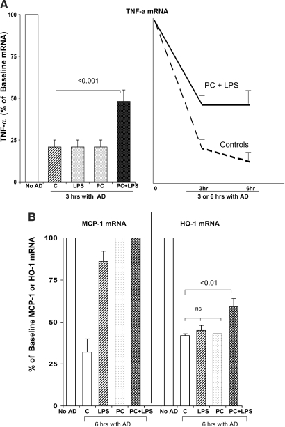
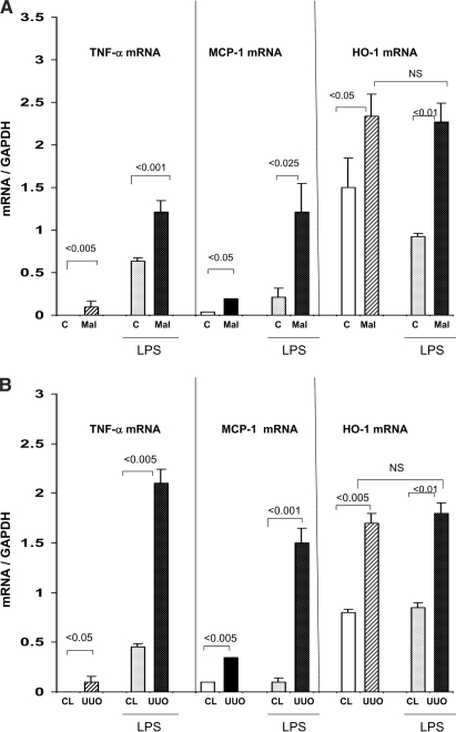
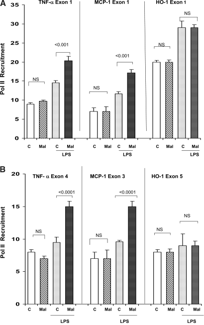
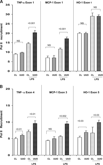
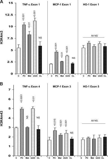
Acute renal failure (ARF) sensitizes the kidney to endotoxin (LPS)-driven production of cytokines and chemokines. This study assessed whether this LPS hyperresponsiveness exists at the genomic level. Three heterogeneous mouse models of ARF were studied: Maleate nephrotoxicity, unilateral ureteral obstruction, and LPS preconditioning. In all cases, LPS was injected approximately 18 h after injury was induced, and over the next 0 to 90 min, RNA polymerase II recruitment to the genome at three LPS-responsive genes (TNF-alpha, monocyte chemoattractant-1 [MCP-1], and heme oxygenase-1 [HO-1]) was assessed by chromatin immunoprecipitation. LPS hyperresponsiveness was noted in each model, measured by exaggerated increases in TNF-alpha and MCP-1 mRNA (approximately two to 10 times higher than LPS-injected controls). Corresponding increases in the recruitment of RNA polymerase II to the TNF-alpha and MCP-1 genes were observed, and increased trimethylation of histone 3 lysine 4 (H3K4m3) at these sites may have played a role in this recruitment. Conversely, recruitment of RNA polymerase II to the HO-1 gene was suppressed ("tolerance"), and no increase in H3K4m3 was observed at HO-1 exons. The ARF-induced changes in mRNA did not correlate with mRNA stability, suggesting the mechanistic importance of RNA polymerase II-mediated transcriptional events. In conclusion, LPS hyperresponsiveness after ARF is likely mediated at the genomic level, possibly by H3K4m3.


Figures

Figure 1.
Figure 1.
(A and B) Renal mRNA responses to LPS injection in control and LPS-PC mice. Mice that were pretreated with LPS 18 h previously had modest but significant TNF-α and MCP-1 mRNA elevations, compared with control (C) mice. When the LPS-PC mice were rechallenged with LPS, significantly greater increases in TNF-α and MCP-1 mRNA were observed, versus those seen in LPS-challenged naive controls. This was true whether the measurements were made at 15 (A) or 30 min (B) after repeat LPS injection. HO-1 mRNA levels were approximately twice as high in the preconditioned versus the control mice (P < 0.01); however, no acute increases were seen in either group at either 15 or 30 min after LPS injection. LPS, 15 or 30 min after LPS injection; PC, 18 h after LPS injection; PC+LPS, preconditioned mice rechallenged with LPS.
Figure 2.
Figure 2.
(A and B) LPS preconditioning: Pol II levels at start and end exons, of the TNF-α gene. Pol II levels at TNF-α exon 1 (A) and exon 4 (B) were assessed at 15, 30, or 90 min after LPS injection in control (C) or LPS-PC mice. In each instance, Pol II recruitment was observed. The degree of this recruitment was significantly greater in the LPS-PC mice, compared with that seen in the LPS-challenged naive controls (*overall statistical comparison at the three time points at exon 4; P < 0.001).
Figure 3.
Figure 3.
(A and B) LPS-PC: Pol II levels at start and end exons (1 and 3) of the MCP-1 gene. The preconditioned and control mice had comparable degrees of Pol II at both MCP-1 exons. When rechallenged with LPS, greater increases were observed in the preconditioned mice, compared with LPS-injected naive controls, particularly at the 15-min time point (*overall statistical analysis for all time points, exon 3, P < 0.01).
Figure 4.
Figure 4.
(A and B) LPS-PC: Pol II levels at start and end exons (1 and 5) of the HO-1 gene. Acute LPS injection into naive controls caused acute, statistically significant Pol II recruitment to both HO-1 exons. This was true at each of the three time points. In striking contrast, when the preconditioned mice were rechallenged with LPS, no significant increase in Pol II levels was observed (at any time point or at either exon). This suggests LPS tolerance vis à vis Pol II recruitment.
Figure 5.
Figure 5.
(A) Assessments of in vitro TNF-α mRNA stability in isolated tubules harvested after in vivo LPS injection. Proximal tubules from control (C) mice and LPS-PC mice (PC) were harvested 30 min after either LPS or vehicle injection, and they were incubated in vitro for 3 or 6 h with or without actinomycin D (AD). Tubules from PC mice that were rechallenged with LPS manifested decreased mRNA degradation, as assessed at either the 3- or 6-h time point. Conversely, neither preconditioning alone nor LPS injection alone altered TNF-α mRNA degradation rates. (B) Assessments of in vitro MCP-1 and HO-1 stability after intravenous LPS injection. LPS induced MCP-1 mRNA stability, whether the assessments were made at 30 min or 18 h (preconditioned mice) after LPS injection. Conversely, only a minimal increase in HO-1 mRNA stability was observed in response to LPS, and this was observed only in preconditioned tubules that were rechallenged with LPS (P < 0.01).
Figure 6.
Figure 6.
(A) Renal cortical mRNA responses to LPS injection in the setting of maleate nephrotoxicity. By 15 to 30 min after LPS injection, significant increases in TNF-α, MCP-1, and HO-1 mRNA were observed in both the control (C) and maleate treatment groups. The degree of these TNF-α and MCP-1 mRNA increases were two- to four-fold greater in the maleate treatment group (compared with their basal values). Conversely, maleate pretreatment did not alter the HO-1 mRNA responses to LPS, given that the post-LPS values were essentially identical between the control and maleate groups. All three mRNAs were somewhat higher at baseline in the maleate versus control groups. (B) Renal cortical mRNA responses 15 to 30 min after LPS injection in the setting of UUO and in CL kidneys. These results mirrored those described for the maleate experiments, as follows: (1) UUO significantly increased TNF-α, MCP-1, and HO-1 mRNA in the absence of LPS; (2) LPS induced dramatic increases in all three mRNAs; (3) the degrees of TNF-α and MCP-1 mRNA increases were significantly greater in UUO versus CL kidneys; and (4) HO-1 mRNA in the UUO kidney did not acutely respond to LPS injection.
Figure 7.
Figure 7.
(A and B) Maleate nephrotoxicity: Pol II expression at start (A) and end (B) exons of TNF-α, MCP-1, and HO-1 genes. Maleate did not independently alter Pol II levels at either the start or end exons of any of the three assessed genes; however, when injected with LPS, Pol II recruitment occurred and was greater at both exons of the TNF-α and MCP-1 genes, compared with the recruitment seen in the controls. Conversely, no preferential recruitment of Pol II was seen at either exon of the HO-1 gene. Thus, these data were highly analogous to the observed corresponding mRNA levels, as presented in Figure 6A.
Figure 8.
Figure 8.
(A and B) UUO: Pol II expression at start (A) and end (B) exons of TNF-α, MCP-1, and HO-1 genes. UUO did not independently alter Pol II levels at exon 1 of any of the assessed genes. It also had either a quantitatively minimal or no impact on Pol II at end exons. When the mice were administered an injection of LPS, significantly greater increases in Pol II recruitment was observed at both start and end exons of the TNF-α and MCP-1 genes. Conversely, no preferential increase was seen at HO-1 exon 1, highly consistent with the previously described HO-1 mRNA data (Figure 6B); however, a modest preferential increase of Pol II was observed at HO-1 exon 5.
Figure 9.
Figure 9.
(A) H3K4m3 at the start of the TNF-α, MCP-1, and HO-1 genes. LPS-PC, maleate nephrotoxicity (Mal), and UUO each induced significant increases in the extent of H3K4m3 at exon 1 of the TNF-α and MCP-1 genes. Conversely, no change in H3K4m3 at HO-1 exon 1 was observed. CL, contralateral kidney in mice subjected to UUO. The CL results did not significantly differ from those observed in normal control (C) kidneys. (B) H3K4m3 at exons 4, 3, and 5 of the TNF-α, MCP-1, and HO-1 genes, respectively. H3K4m3 levels at both TNF-α and MCP-1 end exons were significantly increased with both UUO and LPS (but not maleate) preconditioning. Conversely, no increase in H3K4m3 was observed at HO-1 exon 5. Results in CL kidneys from UUO mice did not differ from normal controls (C).

References

    1. Honda N, Hishida A, Ikuma K, Yonemura K: Acquired resistance to acute renal failure. Kidney Int 31: 1233–1238, 1987 - PubMed
    1. Elliott WC, Houghton DC, Gilbert DN, Baines-Hunter J, Bennett WM: Gentamicin nephrotoxicity. I. Degree and permanence of acquired insensitivity. J Lab Clin Med 100: 501–512, 1982 - PubMed
    1. Zager RA, Baltes LA, Sharma HM, Jurkowitz MS: Responses of the ischemic acute renal failure kidney to additional ischemic events. Kidney Int 26: 689–700, 1984 - PubMed
    1. Nath KA, Balla G, Vercellotti GM, Balla J Jacob HS, Levitt MD, Rosenberg ME: Induction of heme oxygenase in a rapid protective response in rhabdomyolysis in the rat. J Clin Invest 90: 267–270, 1992 - PMC - PubMed
    1. Zager RA, Johnson AC, Hanson SY, Lund S: Ischemic proximal tubular injury primes mice to endotoxin-induced TNF-α generation and systemic release. Am J Physiol 289: F289–F297, 2005 - PubMed

Publication types

MeSH terms

LinkOut - more resources