

Insights into the oncogenic effects of PIK3CA mutations from the structure of p110alpha/p85alpha
- PMID: 18418043
- PMCID: PMC3260475
- DOI: 10.4161/cc.7.9.5817
Insights into the oncogenic effects of PIK3CA mutations from the structure of p110alpha/p85alpha
Abstract
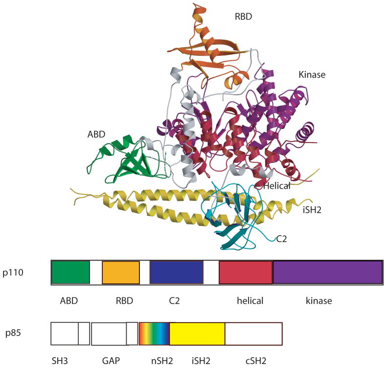
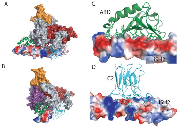
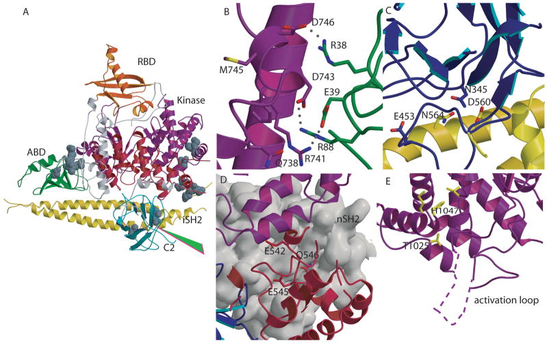
Phosphatidylinositide-3-kinases (PI3K) initiate a number of signaling pathways by recruiting other kinases, such as Akt, to the plasma membrane. One of the isoforms, PI3Kalpha, is an oncogene frequently mutated in several cancer types. These mutations increase PI3K kinase activity, leading to increased cell survival, cell motility, cell metabolism, and cell cycle progression. The structure of the complex between the catalytic subunit of PI3Kalpha, p110alpha, and a portion of its regulatory subunit, p85alpha reveals that the majority of the oncogenic mutations occur at the interfaces between p110 domains and between p110 and p85 domains. At these positions, mutations disrupt interactions resulting in changes in the kinase domain that may increase enzymatic activity. The structure also suggests that interaction with the membrane is mediated by one of the p85 domains (iSH2). These findings may provide novel structural loci for the design of new anti-cancer drugs.
Conflict of interest statement
The authors declare no financial or conflicts of interest in the contents of this paper
Figures
Similar articles
-
Cancer-derived mutations in the regulatory subunit p85alpha of phosphoinositide 3-kinase function through the catalytic subunit p110alpha.Proc Natl Acad Sci U S A. 2010 Aug 31;107(35):15547-52. doi: 10.1073/pnas.1009652107. Epub 2010 Aug 16. Proc Natl Acad Sci U S A. 2010. PMID: 20713702 Free PMC article.
-
Oncogenic mutations weaken the interactions that stabilize the p110α-p85α heterodimer in phosphatidylinositol 3-kinase α.FEBS J. 2015 Sep;282(18):3528-42. doi: 10.1111/febs.13365. Epub 2015 Jul 24. FEBS J. 2015. PMID: 26122737 Free PMC article.
-
Hot-spot mutations in p110alpha of phosphatidylinositol 3-kinase (pI3K): differential interactions with the regulatory subunit p85 and with RAS.Cell Cycle. 2010 Feb 1;9(3):596-600. doi: 10.4161/cc.9.3.10599. Cell Cycle. 2010. PMID: 20009532 Free PMC article.
-
Human tumor mutants in the p110alpha subunit of PI3K.Cell Cycle. 2006 Apr;5(7):675-7. doi: 10.4161/cc.5.7.2605. Epub 2006 Apr 1. Cell Cycle. 2006. PMID: 16627990 Review.
-
Molecular Mechanisms of Human Disease Mediated by Oncogenic and Primary Immunodeficiency Mutations in Class IA Phosphoinositide 3-Kinases.Front Immunol. 2018 Mar 19;9:575. doi: 10.3389/fimmu.2018.00575. eCollection 2018. Front Immunol. 2018. PMID: 29616047 Free PMC article. Review.
Cited by
-
Improved production of class I phosphatidylinositol 4,5-bisphosphate 3-kinase.Protein Expr Purif. 2025 Jan;225:106582. doi: 10.1016/j.pep.2024.106582. Epub 2024 Aug 20. Protein Expr Purif. 2025. PMID: 39173964
-
Cancer-derived mutations in the regulatory subunit p85alpha of phosphoinositide 3-kinase function through the catalytic subunit p110alpha.Proc Natl Acad Sci U S A. 2010 Aug 31;107(35):15547-52. doi: 10.1073/pnas.1009652107. Epub 2010 Aug 16. Proc Natl Acad Sci U S A. 2010. PMID: 20713702 Free PMC article.
-
Somatic mutations in p85alpha promote tumorigenesis through class IA PI3K activation.Cancer Cell. 2009 Dec 8;16(6):463-74. doi: 10.1016/j.ccr.2009.10.016. Cancer Cell. 2009. PMID: 19962665 Free PMC article.
-
PIK3CA hotspot mutations p. H1047R and p. H1047L sensitize breast cancer cells to thymoquinone treatment by regulating the PI3K/Akt1 pathway.Mol Biol Rep. 2022 Mar;49(3):1799-1816. doi: 10.1007/s11033-021-06990-x. Epub 2021 Nov 23. Mol Biol Rep. 2022. PMID: 34816327
-
Multiscale mutation clustering algorithm identifies pan-cancer mutational clusters associated with pathway-level changes in gene expression.PLoS Comput Biol. 2017 Feb 7;13(2):e1005347. doi: 10.1371/journal.pcbi.1005347. eCollection 2017 Feb. PLoS Comput Biol. 2017. PMID: 28170390 Free PMC article.
References
-
- Fruman DA, Meyers RE, Cantley LC. Phosphoinositide kinases. Annu Rev Biochem. 1998;67:481–507. - PubMed
-
- Cantley LC. The phosphoinositide 3-kinase pathway. Science. 2002;296:1655–7. - PubMed
-
- Katso R, Okkenhaug K, Ahmadi K, White S, Timms J, Waterfield MD. Cellular function of phosphoinositide 3-kinases: implications for development, homeostasis, and cancer. Annu Rev Cell Dev Biol. 2001;17:615–75. - PubMed
Publication types
MeSH terms
Substances
Grants and funding
LinkOut - more resources
Full Text Sources
Other Literature Sources
Molecular Biology Databases
Miscellaneous