. 2008 May 1;7(9):1197-204.
doi: 10.4161/cc.7.9.5886. Epub 2008 Mar 10.

Disruption of the circadian clock due to the Clock mutation has discrete effects on aging and carcinogenesis

Affiliations

Disruption of the circadian clock due to the Clock mutation has discrete effects on aging and carcinogenesis

Marina P Antoch et al. Cell Cycle. .

Abstract

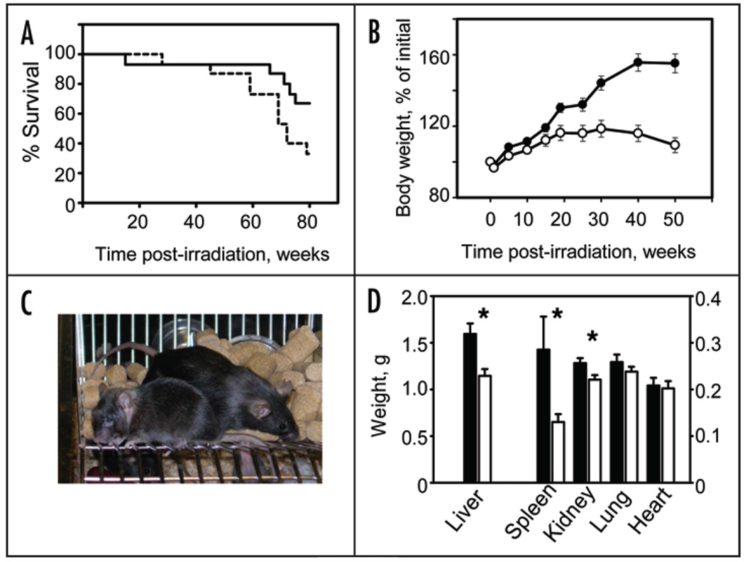
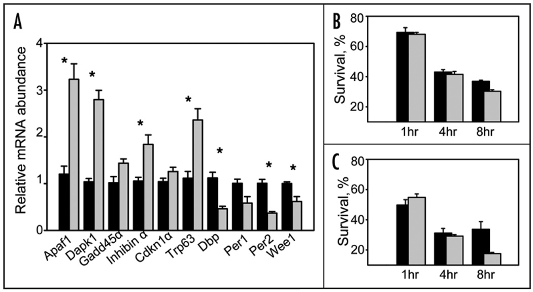
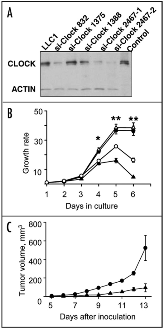
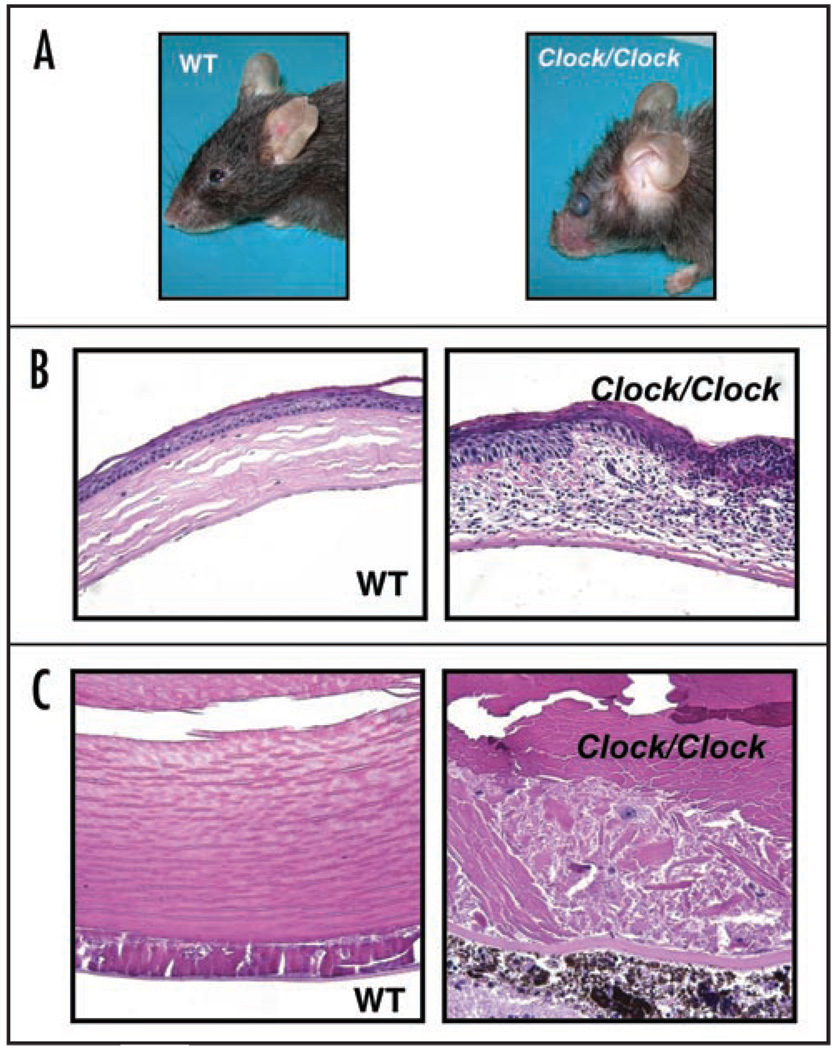
The mammalian circadian system has been implicated in the regulation of various biological processes including those involved in genotoxic stress responses and tumor suppression. Here we report that mice with the functional deficiency in circadian transcription factor CLOCK (Clock/Clock mutant mice) do not display predisposition to tumor formation both during their normal lifespan or when challenged by gamma- radiation. This phenotype is consistent with high apoptotic and low proliferation rate in lymphoid tissues of Clock mutant mice and is supported by the gene expression profiling of a number of apoptosis and cell cycle-related genes, as well as by growth inhibition of cells with CLOCK downregulation. At the same time, Clock mutant mice respond to low-dose irradiation by accelerating their aging program, and develop phenotypes that are reminiscent of those in Bmal1-deficient mice. Taken together, our results demonstrate the dichotomy in biological consequences of the disruption of the circadian clock with respect to ageing and cancer. They also highlight the existence of a complex interconnection between ageing, carcinogenesis and individual components of the circadian clock machinery.


Figures

Figure 1
Figure 1
Increased mortality and body weight loss of Clock/Clock female mice after low-dose irradiation. (A) Kaplan-Meyer survival curve (mean survival for WT and Clock/Clock mice are 73.2 and 63.6 weeks respectively, p = 0.022). Solid line—WT mice, dotted line—Clock/Clock mice. All survived animals were sacrificed 80 weeks post irradiation (B) Changes in body weight of WT and Clock mutant mice; t = 0 corresponds to the time of irradiation. Fifteen WT and fifteen Clock/Clock mice were weighed weekly. Starting at 20 weeks after irradiation Clock mutant mice demonstrate progressive weight loss (p < 0.001). Data shown as mean ± s.e.m. Closed circles—WT; open circles—Clock/Clock. (C) Gross appearance of WT (right) and Clock/Clock (left) female mice 80 weeks after TBI. Note significant reduction in weight, kyphosis and cataract in Clock mutant female. (D) Weight of major organs from female mice after 80 weeks of TBI (mean ± s.e.m.; *p < 0.01; filled bars—WT, open bars—Clock mutant mice. Liver, spleen and kidney of Clock/Clock female mice showed significant reduction when compared to age-matched WT mice.
Figure 2
Figure 2
Major pathological changes in spleens of Clock/Clock and WT mice at 80 weeks after TBI. (A and B) Lymphoma with loss of normal architecture in the white pulp (A, arrow) and numerous mitotic figures (B, arrows). Two wild-type and one Clock/Clock mouse were diagnosed with lymphoma. (C and D). Extensive extramedullary hematopoiesis (EMH, C, arrow) in the red pulp of the spleen. Note large number of megakaryocytes (arrow) as well as myeloid and erythroid precursors, (D) EMH was diagnosed in 9 WT mice, but in only 4 Clock mutants. Calibration Bar: (A and C) 400 μm; (B and D) 50 µm.
Figure 3
Figure 3
The Clock mutation affects the level of radiation-induced apoptosis in radiation-sensitive lymphoid tissues. (A) Upregulation of several key proapoptotic and growth arrest-specific genes in the spleen of Clock/Clock mutant mice. Data presented as mean ± s.e.m., n = 18; *p < 0.001. WT—black bars, Clock/Clock— grey bars. mRNA expression levels were determined by real-time RT-PCR. Expression of the proproliferative gene Wee1 was significantly (p = 0.025) reduced in Clock mice. Consistent with previously reported data, spleens of Clock/Clock mice also showed significantly reduced levels of expression of direct transcriptional targets of the CLOCK protein, Dbp and Per2 (p = 0.002). (B) Percentage of viable (annexin V-negative) CD3- positive cells in thymuses of WT (black bars) and Clock/Clock (grey bars) mice at different times after 8.5 Gy of TBI. Data presented as mean ± s.e.m., n = 4, p = 0.005. (C) Percentage of viable (annexin V-negative) B220-positive cells in spleens of WT (black bars) and Clock/Clock (grey bars) mice after 8.5Gy of TBI. Data presented as mean ± s.e.m., n = 4, p = 0.05.
Figure 4
Figure 4
Suppression of CLOCK protein reduces cell proliferation. (A) CLOCK suppression by siRNA. Western blot of lysates from LLC1 cells infected with four different siClock lentiviral constructs and probed with a CLOCK-specific antibody. Three independently obtained clones (si-Clock-832, siClock-2467-1 and siClock-2467-2) showed significant reduction in CLOCK abundance. (B) CLOCK suppression by siClock-832 (open circles) or 2467-1 (closed triangles) results in a decrease in cell growth rate when compared to parental LLC1 (closed circles) or inactive LLC1-siClock-1375 (open trianges) cell lines, presented as mean ± s.e.m., n = 3, *p < 0.05, **p < 0.01; (C) CLOCK suppression by siClock-2467 reduces the tumor growth rate. LLC1 cells infected by inactive (siClock-1375, circles) or functional (siClock-2467, trianges) lentiviral constructs were inoculated s.c. into C57BL/6J mice. Tumor volumes were measured starting day 5 after inoculation. Data shown as mean ± s.e.m., n = 20, at all days p < 0.01.
Figure 5
Figure 5
Eye pathologies in Clock/Clock mice after 4Gy of TBI. (A) Gross appearance of wild type and Clock/Clock female mice at 80 weeks after TBI. Eye inflammation and opacity of lens can be observed in Clock/Clock animals. (B and C) Representative example of eye histology. (B) Cornea inflammation. Infiltration by immune cells and abundant changes in the structure of cornea in Clock/Clock mice as compared to WT animals. (C) Cataract development. Posterior cataract (this cataract was graded as grade III in Suppl. Fig. 4) developed in the lens of the Clock/Clock mutant mouse. Only minor changes can be observed in posterior zone of wild type lens.

Similar articles

Cited by

References

    1. Schibler U, Sassone Corsi P. A web of circadian pacemakers. Cell. 2002;111:919–922. - PubMed
    1. Panda S, Antoch MP, Miller BH, Su AI, Schook AB, Straume M, Schultz PG, Kay SA, Takahashi JS, Hogenesch JB. Coordinated transcription of key pathways in the mouse by the circadian clock. Cell. 2002;109:307–320. - PubMed
    1. Ko CH, Takahashi JS. Molecular components of the mammalian circadian clock. Human molecular genetics. 2006;15:271–277. - PubMed
    1. Kondratov RV, Gorbacheva VY, Antoch MP. The role of mammalian circadian proteins in normal physiology and genotoxic stress responses. Current topics in developmental biology. 2007;78:173–216. - PubMed
    1. Haus E, Smolensky M. Biological clocks and shift work: circadian dysregulation and potential long-term effects. Cancer Causes Control. 2006;17:489–500. - PubMed

Publication types

MeSH terms

LinkOut - more resources