Comparative Study
. 2008 May 15;7(10):1467-72.
doi: 10.4161/cc.7.10.5883. Epub 2008 Feb 29.

Analysis of DBC1 and its homologs suggests a potential mechanism for regulation of sirtuin domain deacetylases by NAD metabolites

Affiliations
Comparative Study

Analysis of DBC1 and its homologs suggests a potential mechanism for regulation of sirtuin domain deacetylases by NAD metabolites

Vivek Anantharaman et al. Cell Cycle. .

Abstract

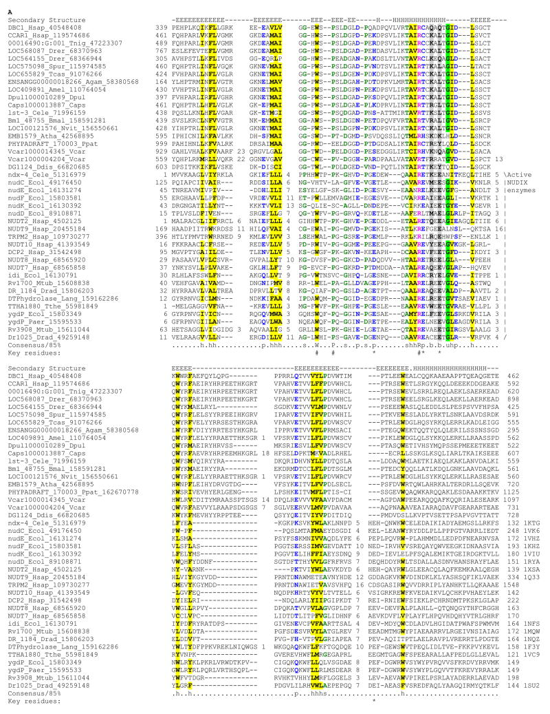
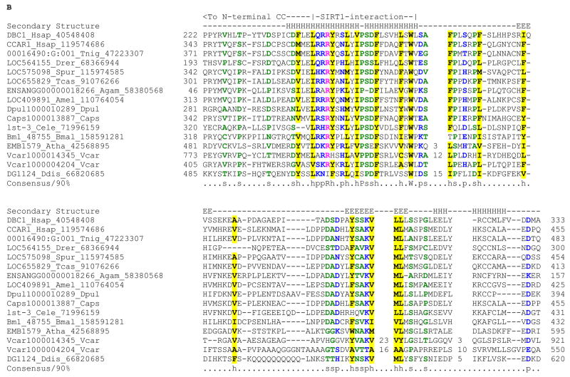
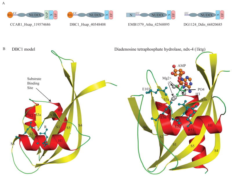
Deleted in Breast Cancer-1 (DBC1) and its paralog CARP-1 are large multi-domain proteins, with a nuclear or perinuclear localization, and a role in promoting apoptosis upon processing by caspases. Recent studies on human DBC1 show that it is a specific inhibitor of the sirtuin-type deacetylase, Sirt1, which deacetylates histones and p53. Using sensitive sequence profile searches and HMM-HMM comparisons we show that the central conserved globular domain present in the DBC1 and it homologs from diverse eukaryotes is a catalytically inactive version of the Nudix hydrolase (MutT) domain. Given that Nudix domains are known to bind nucleoside diphosphate sugars and NAD, we predict that this domain in DBC1 and its homologs binds NAD metabolites such as ADP-ribose. Hence, we propose that DBC1 and its homologs are likely to regulate the activity of SIRT1 or related deacetylases by sensing the soluble products or substrates of the NAD-dependent deacetylation reaction. The complex domain architectures of the members of the DBC1 family, which include fusions to the RNA-binding S1-like domain, the DNA-binding SAP domain and EF-hand domains, suggest that they are likely to function as integrators of distinct regulatory signals including chromatin protein modification, soluble compounds in NAD metabolism, apoptotic stimuli and RNA recognition.


Figures

Fig. 1
Fig. 1. A) Multiple alignment of Nudix domains of the DBC1 family with known representatives
Multiple sequence alignment of the Nudix domain was constructed using Kalign after parsing high-scoring pairs from PSI-BLAST search results. The alignment was refined based on the pairwise alignments produced by the profile-profile searches with the HHpred program against the Nudix profile. The secondary structure from the crystal structures is shown above the alignment with E representing a strand and H a helix. The 85% consensus shown below the alignment was derived using the following amino acid classes: hydrophobic (h: ALICVMYFW, yellow shading); small (s: ACDGNPSTV, green); polar (p: CDEHKNQRST, blue) and its charged subset (c: DEHKR, pink), and big (b: FILMQRWYEK; grey shading). The limits of the domains are indicated by the residue positions, on each end of the sequence. The numbers within the alignment are non-conserved inserts that have not been shown. The key positions involved in stabilizing the substrate-binding cleft and constituting the active site motif of the active version are respectively marked with “#” and “*” symbols. The sequences are denoted by their gene name followed by the species abbreviation and GenBank Identifier (gi). The PDB ids, when, available are shown to the right end of the alignment. The species abbreviations are: Agam : Anopheles gambiae; Amel : Apis mellifera; Atha : Arabidopsis thaliana; Bmal : Brugia malayi; Caps : Capitella sp; Cbri : Caenorhabditis briggsae; Cele : Caenorhabditis elegans; Ddis : Dictyostelium discoideum; Dpul : Daphnia pulex; Drad : Deinococcus radiodurans; Drer : Danio rerio; Ecol : Escherichia coli; Hsap : Homo sapiens; Lang : Lupinus angustifolius; Mtub : Mycobacterium tuberculosis; Nvit : Nasonia vitripennis; Paer : Pseudomonas aeruginosa; Ppat : Physcomitrella patens; Spur : Strongylocentrotus purpuratus; Tcas : Tribolium castaneum; Tnig : Tetraodon nigroviridis; Tthe : Thermus thermophilus; Vcar : Volvox carteri B) Multiple alignment of the N-terminal extension in DBC1 Multiple sequence alignment of the conserved N-terminal extension of the inactive Nudix domains in the DBC1 family was constructed as described above. The helical region that interacts with the SIRT1 in DBC1 is marked. The extended coiled coil region continues N-terminal to this region.
Fig. 2
Fig. 2. A. Domain architectures of the DBC1 family
The domain architectures found in the DBC1 family are shown. Domain architectures are labeled with a representative gene name, the species abbreviation, and the Genebank identifier (GI) number separated by underscores. The N-terminal extension of the Nudix domain is shown as a small ellipse. Domain abbreviations are: S1L – S1-like OB fold domain; EF – EF hand; CC – coiled coil region; and N – N terminal domain specific to plant members of the DBC1 family. B. Model of the DBC1 Nudix domain compared with the crystal structure of the catalytically active version ndx-4 (pdb: 1ktgA). The DBC1 model was constructed using DR1025, a nucleotide pyrophosphatase (PDB: 1SU2), DR1184, a CoA pyrophosphatase (PDB: 1NQZ), both from Deinococcus and ndx-4, a diadenosine tetraphosphate hydrolase(PDB: 1KTG) from Caenorhabditis, as templates. The crystal structure of ndx-4, a diadenosine tetraphosphate hydrolase (1KTG), is shown for comparison. The conserved active-site residues, the substrate binding site, Magnesium ions, phosphate ion and the substrate ADP are shown.

References

    1. Hamaguchi M, Meth JL, von Klitzing C, Wei W, Esposito D, Rodgers L, Walsh T, Welcsh P, King MC, Wigler MH. DBC2, a candidate for a tumor suppressor gene involved in breast cancer. Proc Natl Acad Sci U S A. 2002;99:13647–52. - PMC - PubMed
    1. Sundararajan R, Chen G, Mukherjee C, White E. Caspase-dependent processing activates the proapoptotic activity of deleted in breast cancer-1 during tumor necrosis factor-alpha-mediated death signaling. Oncogene. 2005;24:4908–20. - PubMed
    1. Rishi AK, Zhang L, Yu Y, Jiang Y, Nautiyal J, Wali A, Fontana JA, Levi E, Majumdar AP. Cell cycle- and apoptosis-regulatory protein-1 is involved in apoptosis signaling by epidermal growth factor receptor. J Biol Chem. 2006;281:13188–98. - PubMed
    1. Kim JE, Chen J, Lou Z. DBC1 is a negative regulator of SIRT1. Nature. 2008;451:583–6. - PubMed
    1. Zhao W, Kruse JP, Tang Y, Jung SY, Qin J, Gu W. Negative regulation of the deacetylase SIRT1 by DBC1. Nature. 2008;451:587–90. - PMC - PubMed

Publication types