. 2008 May 21;27(10):1513-24.
doi: 10.1038/emboj.2008.74. Epub 2008 Apr 17.

Topoisomerase IIIalpha is required for normal proliferation and telomere stability in alternative lengthening of telomeres

Affiliations

Topoisomerase IIIalpha is required for normal proliferation and telomere stability in alternative lengthening of telomeres

Nassima Temime-Smaali et al. EMBO J. .

Abstract

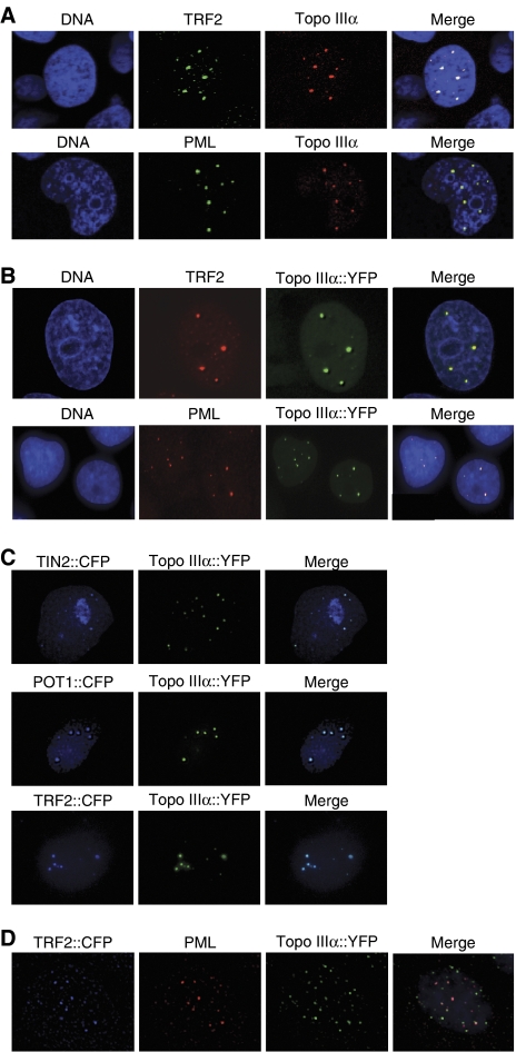
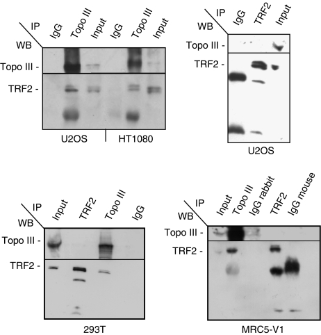
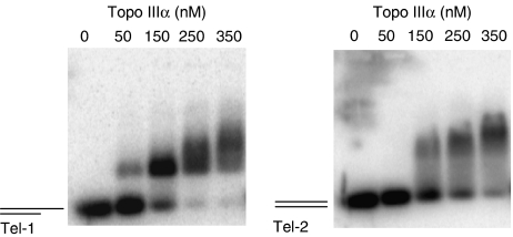
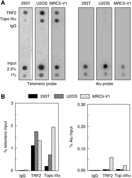
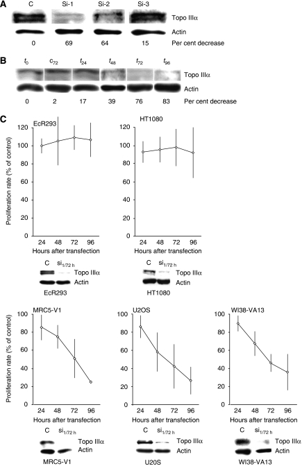
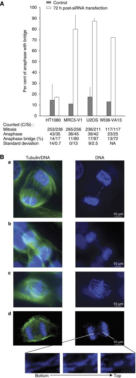
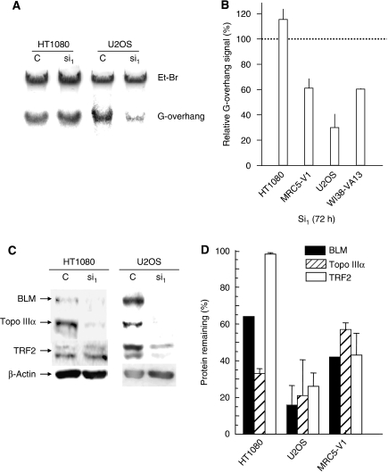
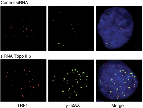
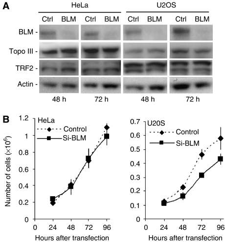
Topoisomerase (Topo) IIIalpha associates with BLM helicase, which is proposed to be important in the alternative lengthening of telomeres (ALT) pathway that allows telomere recombination in the absence of telomerase. Here, we show that human Topo IIIalpha colocalizes with telomeric proteins at ALT-associated promyelocytic bodies from ALT cells. In these cells, Topo IIIalpha immunoprecipitated with telomere binding protein (TRF) 2 and BLM and was shown to be associated with telomeric DNA by chromatin immunoprecipitation, suggesting that these proteins form a complex at telomere sequences. Topo IIIalpha depletion by small interfering RNA reduced ALT cell survival, but did not affect telomerase-positive cell lines. Moreover, repression of Topo IIIalpha expression in ALT cells reduced the levels of TRF2 and BLM proteins, provoked a strong increase in the formation of anaphase bridges, induced the degradation of the G-overhang signal, and resulted in the appearance of DNA damage at telomeres. In contrast, telomere maintenance and TRF2 levels were unaffected in telomerase-positive cells. We conclude that Topo IIIalpha is an important telomere-associated factor, essential for telomere maintenance and chromosome stability in ALT cells, and speculate on its potential mechanistic function.


Figures

Figure 1
Figure 1
Topo IIIα colocalizes with PML and shelterin components at APB in MRC5-V1 ALT cells. The stable expression of a YFP-tagged Topo IIIα protein was also used to determine its localization. (A) Colocalization of Topo IIIα (red, detected by immunofluorescence) with TRF2 or PML (green, detected by immunofluorescence. (B) Representative images of colocalization in MRC5-V1 of Topo IIIα tagged with YFP (Topo IIIα∷YFP, green), with TRF2 or PML (red, detected by immunofluorescence). (C) Representative images of colocalization in MRC5-V1 of Topo IIIα tagged with YFP (Topo IIIα∷YFP, green) with TIN2 tagged with CFP (blue, TIN2∷CFP), POT1 tagged with CFP (blue, POT1∷CFP), or TRF2 tagged with CFP (blue, TRF2∷CFP). (D) Representative images of colocalization in MRC5-V1 of Topo IIIα tagged with YFP (Topo IIIα∷YFP, green), TRF2 tagged with CFP (TRF2∷CFP, blue), and PML (red detected by immunofluorescence).
Figure 2
Figure 2
Topo IIIα/TRF2 complex is detected in telomerase-positive and ALT cells as revealed by immunoprecipitation with D6 Topo IIIα antibody. The antibodies (TRF2, Topo III, or control IgG) used for immunoprecipitation (IP) are listed at the top of each panel and the antibodies (BLM, Topo III, and TRF2) used for western blot analysis (WB) are listed at the left of each panel. Topo IIIα co-immunoprecipitates TRF2 in HT1080 or 293T (telomerase-positive) and in U2OS or MRC5-V1 (ALT) cell lines. Reciprocal immunoprecipitation of Topo IIIα by TRF2 antibody (4A794, mouse) is not detected in 293T, U2OS, or MRC5-V1 and is unreproducible in other cells.
Figure 3
Figure 3
Topo IIIα interacts with telomeric sequences in vitro. EMSA were performed using 5 nM of telomeric template with a 3′ single-stranded extension (Tel-1, left panel) or without the single-stranded region (Tel-2, right panel) (see Materials and methods for details) in the presence of increasing concentrations (50, 150, 250, and 350 nM) of purified recombinant Topo IIIα (Goulaouic et al, 1999). For Tel-1, a Topo IIIα/DNA bandshift was detected at 50 nM, whereas the Tel-2/Topo IIIα bandshift was detected at 150 nM, suggesting a preferential binding to the 3′ single-stranded extension.
Figure 4
Figure 4
Topo IIIα interacts with telomeric sequences in vivo as shown by ChIP. Three different cell lines (telomerase-positive 293T and MRC5-V1 and U2OS ALT cell lines) were evaluated after ChIP with TRF2 or Topo IIIα antibodies. IgG antibodies were used as negative controls. Total input fractions (2.5 and 1%) and antibody-recovered fractions (10% of input) were subjected to Southern blot analysis using telomeric or ALU repeat-specific probes. (A) Representative ChIP experiment. (B) Quantification of radioactivity by Imagequant™ software of the experiment presented in (A). The percentage of precipitated DNA was calculated as a ratio of input (telomeric or Alu) signals and plotted.
Figure 5
Figure 5
Effect of Topo IIIα depletion by RNA interference on telomerase-positive and ALT cells growth. (A) Representative example of effect of three different siRNAs targeting Topo IIIα (Si-1, Si-2, and Si-3; see sequences in Materials and methods) and a control siRNA (C) on the amount of intracellular Topo IIIα protein at 72 h in the MRC5-V1 (ALT) cell line. The loading control was β-actin. The per cent decrease of Topo IIIα compared to treatment with the control is indicated at the bottom. (B) Data from a representative experiment in MRC5-V1 (ALT) cell line with Si-1. Negative control siRNA at 72 h (c72) and the amount of Topo IIIα before treatment (t0) are shown as controls. The per cent decrease of Topo IIIα relative to the control is indicated at the bottom. (C) Effect of Topo IIIα depletion (Si-1, 100 nM) on the growth of telomerase-positive (EcR293 and HT1080) and ALT (MRC5-V1, U2OS, and WI38-VA13) cell lines for up to 96 h after transfection. The results are expressed relative to control siRNA-treated cells, defined as 100%, and correspond to the mean value and standard deviation of three independent experiments. As a control for Topo IIIα depletion, a western blot experiment performed in parallel is presented at the bottom of each growth curve. Protein extracts from Si-1-treated cells at 72 h (Si-1/72 h) or from control siRNA-treated cells (C) were blotted using antibodies against Topo IIIα and β-actin.
Figure 6
Figure 6
Topo IIIα depletion increases anaphase bridge formation in ALT cell lines. (A) Per cent of cells in mitosis harbouring anaphase bridges 72 h after transfection with control siRNA (grey bar) or Si-1 (white bar). Numbers of cells in mitosis and anaphase, percentage of cells with anaphase bridges, and standard deviation are indicated for each cell line for control siRNA (C) and for the siRNA targeting Topo IIIα Si-1 (Si). Experiments were performed in triplicate, except for WI38-VA13 (single determination). NA indicates not applicable. (B) Representative images of the types of anaphase bridges observed: (a) single anaphase bridge, (b) multiple anaphase bridges, (c) abnormal anaphase bridges or chromosome entanglements, and (d) cross-like anaphase bridges. Cells were immunolabelled with α-tubulin antibody (green; see Materials and methods) and DNA was stained with DAPI (blue). Only the merge pictures (tubulin/DNA) and the DAPI stainings (DNA) are shown. Magnification and z-variation of this structure from the bottom to the top of the cell are shown under the main pictures.
Figure 7
Figure 7
Effect of Topo IIIα depletion on G-overhang signal and stability of the Topo IIIα/BLM/TRF2 complex. (A) Representative experiment showing the effect of Topo IIIα depletion on the telomeric G-overhang signal in HT1080 and U2OS cells. DNA was extracted from cells 72 h after transfection with the control siRNA (C) or with Si-1 (si1). The G-overhang signal was evaluated by nondenaturing solution hybridization with a telomeric probe. G-overhang: signal obtained with the (AATCCC)4 probe; EtBr: ethidium bromide DNA staining in the gel. (B) Quantification of the effect of Topo IIIα depletion on the G-overhang signal in four different cell lines. The G-overhang hybridization signal was normalized relative to the EtBr signal. The results are expressed as the percentage of G-overhang signal in control siRNA-transfected cells, defined as 100%. Mean±s.d. is of three independent experiments, including those presented in (A), except for WI38-VA13 (two experiments). (C) Representative experiment showing the effect of Topo IIIα depletion on the stability of the Topo IIIα/BLM/TRF2 complex in HT1080 and U2OS cells by western blot analysis. Cell extracts were prepared 72 h after transfection with the control siRNA (C) or with Si-1 (si1). (D) Quantification of the effect of Topo IIIα depletion on the BLM, TRF2, and Topo IIIα protein levels in HT1080, U20S, and MRC5-V1 cell lines. The results are expressed as the percentage of protein level remaining relative to control siRNA-treated cells. Mean±s.d. is of three independent experiments, including those presented in (C).
Figure 8
Figure 8
DNA damage response at the telomere after Topo IIIα depletion in U2OS cells. U20S cells were treated for 48 h with control siRNA or with Si-1 and examined for γ-H2AX foci (green) that colocalized with TRF1 (red).
Figure 9
Figure 9
Effect of BLM depletion on Topo IIIα and TRF2 protein levels and on the growth of telomerase-positive cells and ALT cells. (A) Western blot analysis of Topo IIIα or TRF2 levels after siRNA-mediated depletion of BLM in HeLa or U2OS cells. Proteins were extracted at the indicated time points after transfection of HeLa or U2OS cells with anti-BLM siRNA (100 nM) or a control siRNA. BLM (upper panel), Topo IIIα, and TRF2 levels were monitored by western blotting using specific antibodies. The membrane was reprobed with anti-β-actin antibody as a loading control (lower panel). (B) Effect of BLM depletion (Si-BLM, 100 nM) on the growth of telomerase-positive (HeLa) and ALT (U2OS) cell lines for up to 96 h after transfection. In brief, cells were harvested and counted every 24 h using one well for each time point. Cells were then lysed and sonicated and BLM depletion was monitored by western blot analysis (data not shown). The values represent the mean of two independent experiments and error bars indicate variation.

References

    1. Ababou M, Dutertre S, Lecluse Y, Onclercq R, Chatton B, Amor-Gueret M (2000) ATM-dependent phosphorylation and accumulation of endogenous BLM protein in response to ionizing radiation. Oncogene 19: 5955–5963 - PubMed
    1. Amiard S, Doudeau M, Pinte S, Poulet A, Lenain C, Faivre-Moskalenko C, Angelov D, Hug N, Vindigni A, Bouvet P, Paoletti J, Gilson E, Giraud-Panis MJ (2007) A topological mechanism for TRF2-enhanced strand invasion. Nat Struct Mol Biol 14: 147–154 - PubMed
    1. Amor-Gueret M (2006) Bloom syndrome, genomic instability and cancer: the SOS-like hypothesis. Cancer Lett 236: 1–12 - PubMed
    1. Cawthon RM (2002) Telomere measurement by quantitative PCR. Nucleic Acids Res 30: e47. - PMC - PubMed
    1. Cesare AJ, Griffith JD (2004) Telomeric DNA in ALT cells is characterized by free telomeric circles and heterogeneous t-loops. Mol Cell Biol 24: 9948–9957 - PMC - PubMed

Publication types

MeSH terms