

Solvent structure and hammerhead ribozyme catalysis
- PMID: 18420140
- PMCID: PMC2610685
- DOI: 10.1016/j.chembiol.2008.03.010
Solvent structure and hammerhead ribozyme catalysis
Abstract
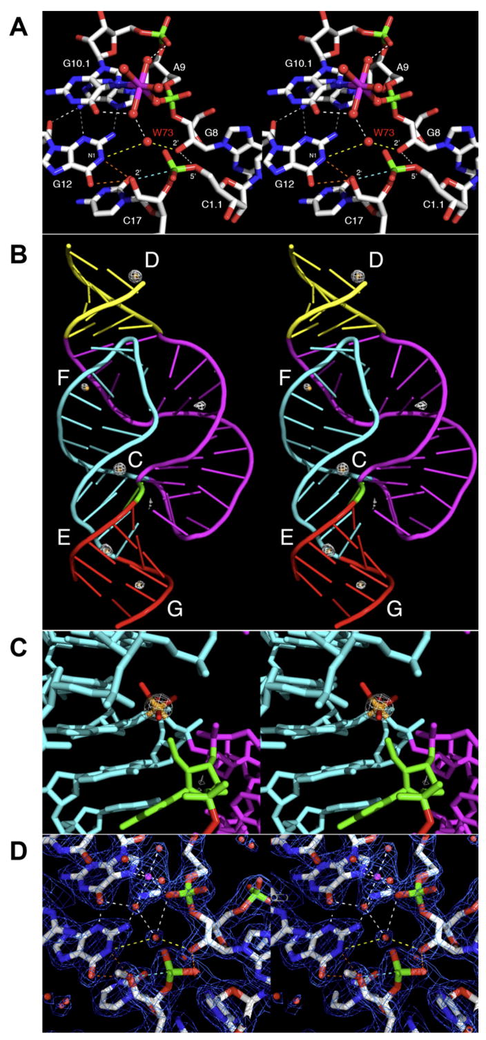
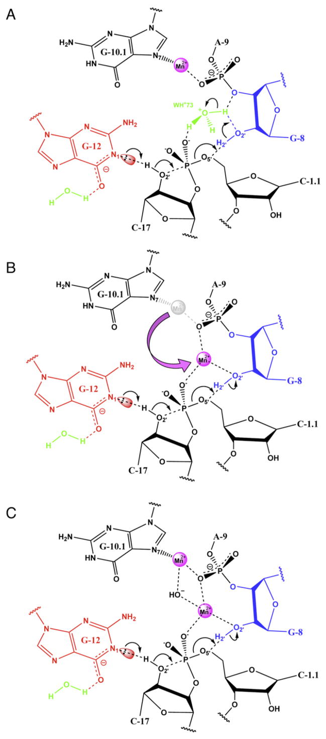
Although the hammerhead ribozyme is regarded as a prototype for understanding RNA catalysis, the mechanistic roles of associated metal ions and water molecules in the cleavage reaction remain controversial. We have investigated the catalytic potential of observed divalent metal ions and water molecules bound to a 2 A structure of the full-length hammerhead ribozyme by using X-ray crystallography in combination with molecular dynamics simulations. A single Mn(2+) is observed to bind directly to the A9 phosphate in the active site, accompanying a hydrogen-bond network involving a well-ordered water molecule spanning N1 of G12 (the general base) and 2'-O of G8 (previously implicated in general acid catalysis) that we propose, based on molecular dynamics calculations, facilitates proton transfer in the cleavage reaction. Phosphate-bridging metal interactions and other mechanistic hypotheses are also tested with this approach.
Figures
Similar articles
-
Structural and catalytic effects of an invariant purine substitution in the hammerhead ribozyme: implications for the mechanism of acid-base catalysis.Acta Crystallogr D Biol Crystallogr. 2014 Sep;70(Pt 9):2256-63. doi: 10.1107/S1399004714010608. Epub 2014 Aug 23. Acta Crystallogr D Biol Crystallogr. 2014. PMID: 25195740 Free PMC article.
-
Two Divalent Metal Ions and Conformational Changes Play Roles in the Hammerhead Ribozyme Cleavage Reaction.Biochemistry. 2015 Oct 20;54(41):6369-81. doi: 10.1021/acs.biochem.5b00824. Epub 2015 Oct 2. Biochemistry. 2015. PMID: 26398724 Free PMC article.
-
Extensive molecular dynamics simulations showing that canonical G8 and protonated A38H+ forms are most consistent with crystal structures of hairpin ribozyme.J Phys Chem B. 2010 May 20;114(19):6642-52. doi: 10.1021/jp1001258. J Phys Chem B. 2010. PMID: 20420375 Free PMC article.
-
Structure-function studies of the hammerhead ribozyme.Curr Opin Chem Biol. 1997 Dec;1(4):532-6. doi: 10.1016/s1367-5931(97)80049-x. Curr Opin Chem Biol. 1997. PMID: 9667883 Review.
-
Structural Simplicity and Mechanistic Complexity in the Hammerhead Ribozyme.Prog Mol Biol Transl Sci. 2018;159:177-202. doi: 10.1016/bs.pmbts.2018.07.006. Epub 2018 Sep 17. Prog Mol Biol Transl Sci. 2018. PMID: 30340787 Review.
Cited by
-
Extension of the Variational Free Energy Profile and Multistate Bennett Acceptance Ratio Methods for High-Dimensional Potential of Mean Force Profile Analysis.J Phys Chem A. 2021 May 20;125(19):4216-4232. doi: 10.1021/acs.jpca.1c00736. Epub 2021 Mar 30. J Phys Chem A. 2021. PMID: 33784093 Free PMC article.
-
Biochemical Studies Provide Insights into the Necessity for Multiple Arabidopsis thaliana Protein-Only RNase P Isoenzymes.J Mol Biol. 2019 Feb 1;431(3):615-624. doi: 10.1016/j.jmb.2018.11.004. Epub 2018 Nov 8. J Mol Biol. 2019. PMID: 30414965 Free PMC article.
-
Hammerhead ribozymes: true metal or nucleobase catalysis? Where is the catalytic power from?Molecules. 2010 Aug 6;15(8):5389-407. doi: 10.3390/molecules15085389. Molecules. 2010. PMID: 20714304 Free PMC article.
-
Molecular simulations of the pistol ribozyme: unifying the interpretation of experimental data and establishing functional links with the hammerhead ribozyme.RNA. 2019 Nov;25(11):1439-1456. doi: 10.1261/rna.071944.119. Epub 2019 Jul 30. RNA. 2019. PMID: 31363004 Free PMC article.
-
Computational identification of RNA functional determinants by three-dimensional quantitative structure-activity relationships.Nucleic Acids Res. 2014;42(17):11261-71. doi: 10.1093/nar/gku816. Epub 2014 Sep 8. Nucleic Acids Res. 2014. PMID: 25200082 Free PMC article.
References
-
- Allen MP, Tildesley DJ. Computer Simulation of Liquids. Oxford: Oxford University Press; 1987.
-
- Auffinger P, Westhof E. RNA hydration: three nanoseconds of multiple molecular dynamics simulations of the solvated tRNAAsp anticodon hairpin. J Mol Biol. 1997;269:326–341. - PubMed
-
- Auffinger P, Westhof E. Water and ion binding around RNA and DNA (C,G) oligomers. J Mol Biol. 2000;300:1113–1131. - PubMed
-
- Bayly CI, Cieplak P, Cornell W, Kollman PA. A well-behaved electrostatic potential based method using charge restraints for deriving atomic charges: the RESP model. J Phys Chem. 1993;97:10269–10280.
-
- Blount KF, Uhlenbeck OC. The structure-function dilemma of the hammerhead ribozyme. Annu Rev Biophys Biomol Struct. 2005;34:415–440. - PubMed
Publication types
MeSH terms
Substances
Associated data
- Actions
Grants and funding
LinkOut - more resources
Full Text Sources
