. 2008 Jul 15;230(2):227-34.
doi: 10.1016/j.taap.2008.02.022. Epub 2008 Mar 4.

Activation of oxidative stress-responsive signaling pathways in early splenotoxic response of aniline

Affiliations

Activation of oxidative stress-responsive signaling pathways in early splenotoxic response of aniline

Jianling Wang et al. Toxicol Appl Pharmacol. .

Abstract

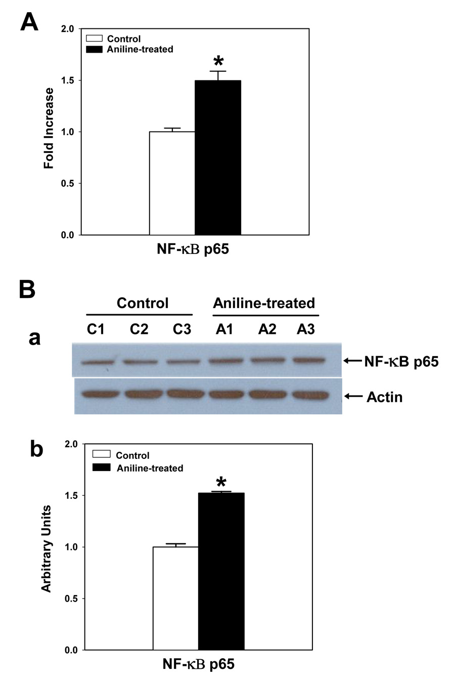
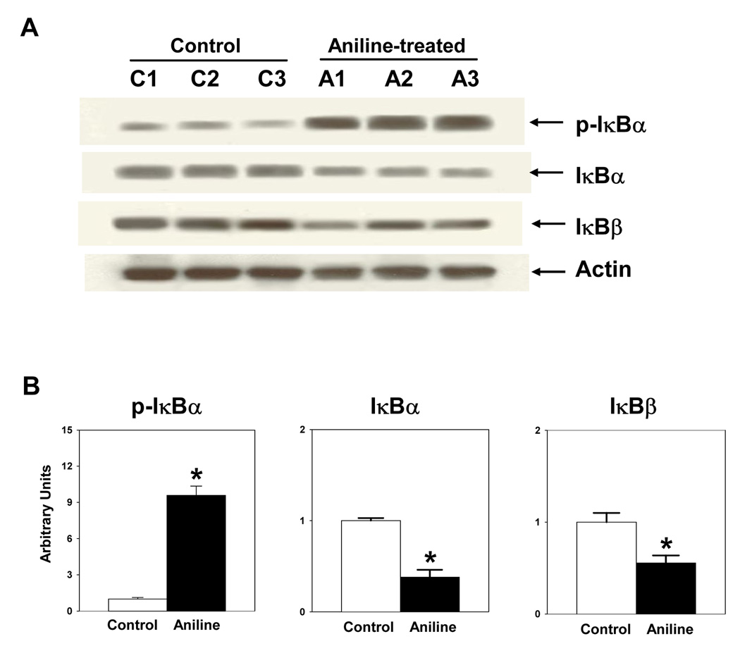
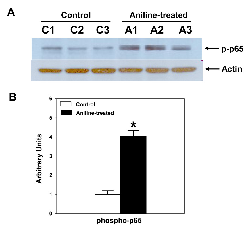
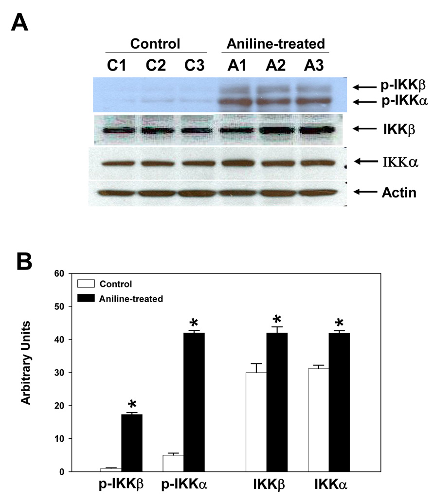
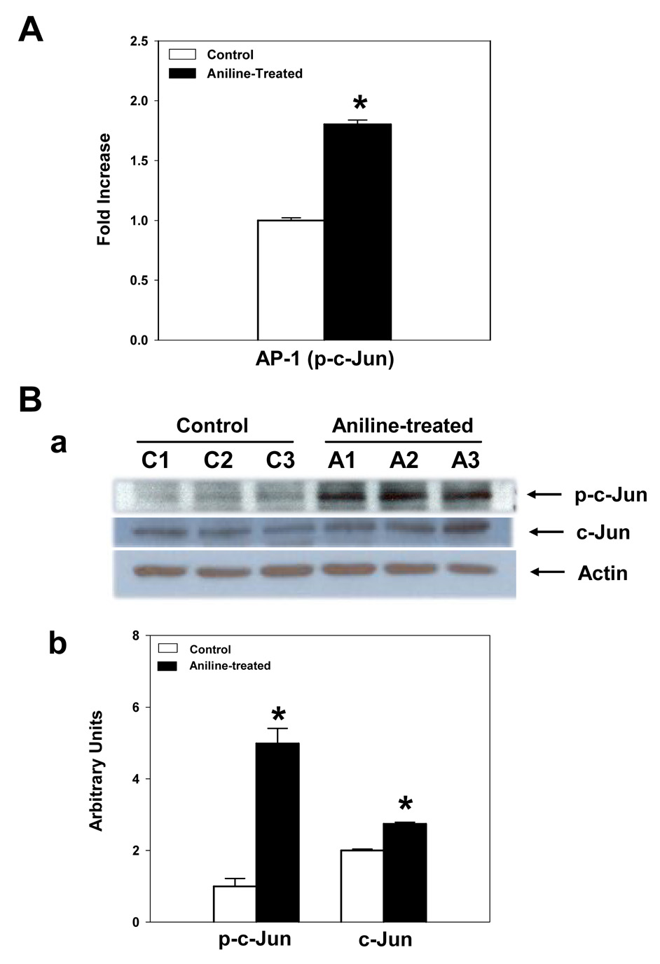
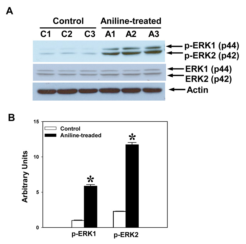
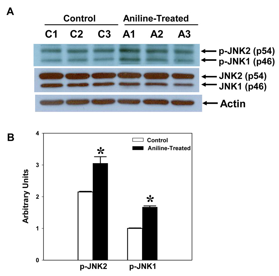
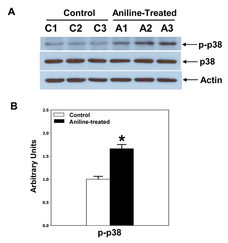
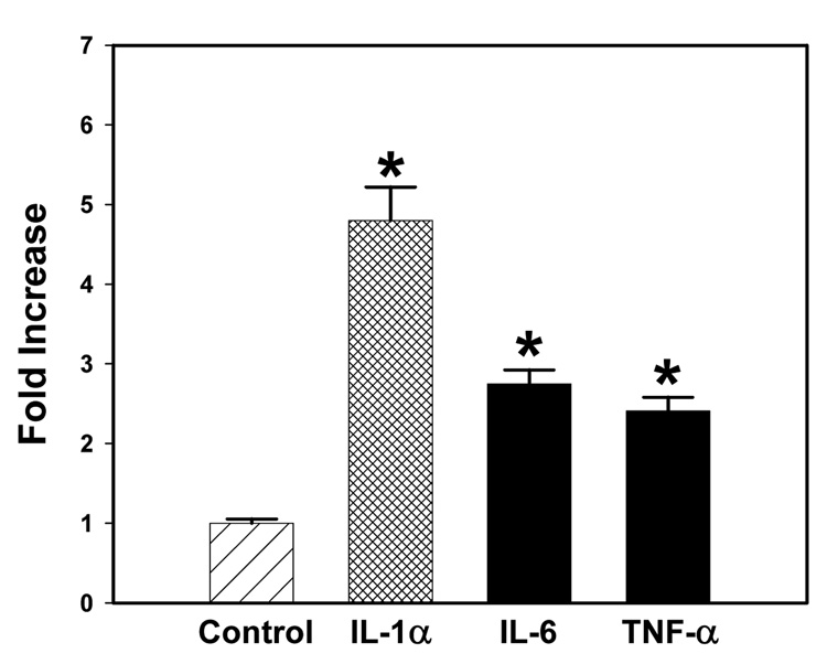
Aniline exposure causes toxicity to the spleen, which leads to a variety of sarcomas, and fibrosis appears to be an important preneoplastic lesion. However, early molecular mechanisms in aniline-induced toxicity to the spleen are not known. Previously, we have shown that aniline exposure results in iron overload and induction of oxidative stress in the spleen, which can cause transcriptional upregulation of fibrogenic/inflammatory cytokines via activation of oxidative stress (OS)-responsive signaling pathways. To test this mechanism, male SD rats were treated with aniline (1mmol/kg/day via gavage) for 7 days, an experimental condition that precedes the appearance of fibrosis. Significant increases in both NF-kappaB and AP-1 binding activity was observed in the nuclear extracts of splenocytes from aniline-treated rats as determined by ELISAs, and supported by Western blot data showing increases in p-IkappaBalpha, p-p65 and p-c-Jun. To understand the upstream signaling events which could account for the activation of NF-kappaB and AP-1, phosphorylation patterns of IkappaB kinases (IKKalpha and IKKbeta) and mitogen-activated protein kinases (MAPKs) were pursued. Our data showed remarkable increases in both p-IKKalpha and p-IKKbeta in the splenocytes from aniline-treated rats, suggesting their role in the phosphorylation of both IkappaBalpha and p65 subunits. Furthermore, aniline exposure led to activation of all three classes of MAPKs, as evident from increased phosphorylation of extracellular-signal-regulated kinase (ERK1/2), c-Jun N-terminal kinase (JNK1/2) and p38 MAPKs, which could potentially contribute to the observed activation of both AP-1 and NF-kappaB. Activation of upstream signaling molecules was also associated with simultaneous increases in gene transcription of cytokines IL-1, IL-6 and TNF-alpha. The observed sequence of events following aniline exposure could initiate a fibrogenic and/or tumorigenic response in the spleen.


Figures

Fig. 1
Fig. 1
(A) NF-κB activation in the splenocytes from control and aniline-treated rats. NF-κB activation was determined in the nuclear extracts of splenocytes using TransAM NF-κB p65 ELISA kit. Values are means ± SD (n=6). *p < 0.05. (B) NF-κB p65 expression in splenocytes following aniline exposure. (a) NF-κB p65 expression was determined in the cell lysates of splenocytes from control and aniline-treated rats by Western blotting using antibody specific for NF-κB p65. (b) Densitometric analysis of NF-κB p65 bands using Eagle Eye II software. Values are means ± SD (n=3). *p < 0.05.
Fig. 2
Fig. 2
Effects of aniline exposure on total IκBα and IκBβ, and phosphorylation of IκBα in rat spleen. (A) Western blot analysis of cell lysates from control and aniline-treated rats using antibodies specific for IκBα, IκBβ and p-IκBα (Ser32/36). (B) Densitometric analysis of protein bands using Eagle Eye II software. Values are means ± SD (n=3). * p < 0.05.
Fig. 3
Fig. 3
Aniline-induced phosphorylation of NF-κB p65 in rat spleen. Splenocytes were isolated from control and aniline-treated rats and phosphorylation of NF-κB p65 was determined in the cell lysates by Western blotting using antibody specific for phosphorylated form of p65 (Ser536). (A) Western blot detection of phospho-p65. (B) Densitometric analysis of the bands. Values are means ± SD (n=3). *p < 0.05.
Fig. 4
Fig. 4
Effect of aniline exposure on IKK signaling in rat spleen. (A) Splenocytes were isolated from control and aniline-treated rats and both phosphorylated and total IKKα and IKKβ were determined in the cell lysates by Western blotting using antibodies specific for p-IKKα (Ser180), p-IKKβ (Ser181), IKKα and IKKβ. (B) Densitometric analysis of p-IKKα/β and total IKKα/β bands. Values are means ± SD (n=3). *p < 0.05.
Fig. 5
Fig. 5
AP-1 activation in the spleen following aniline exposure. (A) AP-1 activation (p-c-Jun) was determined in the nuclear extracts of freshly isolated splenocytes from control and aniline-treated rats using TransAM AP-1 ELISA kit. Values are means ± SD (n=6). *p < 0.05. (B) Western blot analyses of p-c-Jun and total c-Jun in rat spleen. Splenocytes were isolated from control and aniline-treated rats and p-c-Jun and total c-Jun were determined in the cell lysates by Western blotting using antibody specific for phospho-c-Jun (Ser73) and c-Jun. (a) Western blot detection of p-c-Jun and c-Jun. (b) Densitometric analysis of the bands. Values are means ± SD (n=3). *p < 0.05.
Fig. 6
Fig. 6
Aniline-induced phosphorylation of ERK1 (p44) and ERK2 (p42) in rat spleen. Splenocytes were isolated from control and aniline-treated rats and phosphorylation of ERK1/2 was determined in the whole cell lysates by Western blotting using antibodies specific for phospho-p44/42 MAPKs (Tyr204). (A) Western blot detection of phospho-ERK and EKR bands. (B) Densitometric analysis of p-ERK bands (p-ERK1 and p-ERK2). Values are means ± SD (n=3). *p < 0.05.
Fig. 7
Fig. 7
Aniline-induced phosphorylation of JNK1/2 (p46 and p54) in rat spleen. Splenocytes were isolated from control and aniline-treated rats and phosphorylation of JNKs was determined in the cell lysates by Western blotting using antibodies specific for phospho-JNK1/2 (Thr183/Tyr185). (A) Western blot detection of phospho and total JNK. (B) Densitometric analysis of p-JNK bands (p-JNK1 and p-JNK2). Values are means ± SD (n=3). *p < 0.05.
Fig. 8
Fig. 8
Aniline-induced phosphorylation of p38 MAPK in rat spleen. Splenocytes were isolated from control and aniline-treated rats and phosphorylation of p38 were determined in the cell lysates by Western blotting using antibody specific for p38 (Tyr182). (A) Western blot detection of total and p-p38 bands. (B) Densitometric analysis of p-p38 bands. Values are means ± SD (n=3). *p < 0.05.
Fig. 9
Fig. 9
Real-time PCR analysis of cytokine mRNA expression in the spleens of control and aniline-treated rats. Total RNA was extracted from spleen, real-time PCR was performed, and the fold change in mRNA (2−ΔΔCT) expression was determined. Values are means ± SD (n=3). *p < 0.05.

References

    1. Abate C, Patel L, Rauscher FJ, III, Curran T. Redox regulation of fos and jun DNA-binding activity in vitro. Science. 1990;249:1157–1161. - PubMed
    1. Akira S, Kishimoto T. NF-IL6 and NF-κB in cytokine gene regulation. Adv. Immunol. 1997;65:1–46. - PubMed
    1. Angel P, Karin M. The role of Jun, Fos and the AP-1 complex in cell proliferation and transformation. Biochim. Biophys. Acta. 1991;1072:129–157. - PubMed
    1. Baldwin AS. The NF-kappa B and I kappa B proteins: new discoveries and insights. Annu. Rev. Immunol. 1996;14:649–683. - PubMed
    1. Bus JS, Popp JA. Perspectives on the mechanism of action of the splenic toxicity of aniline and structurally related compounds. Food Chem. Toxicol. 1987;25:619–626. - PubMed

Publication types

MeSH terms

LinkOut - more resources