. 2008 Jul 15;230(2):167-74.
doi: 10.1016/j.taap.2008.02.024. Epub 2008 Mar 6.

Intracellular localization and subsequent redistribution of metal transporters in a rat choroid plexus model following exposure to manganese or iron

Affiliations

Intracellular localization and subsequent redistribution of metal transporters in a rat choroid plexus model following exposure to manganese or iron

Xueqian Wang et al. Toxicol Appl Pharmacol. .

Abstract

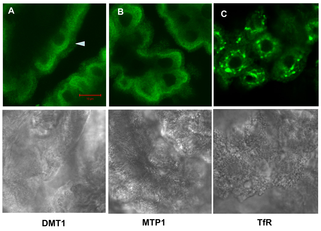
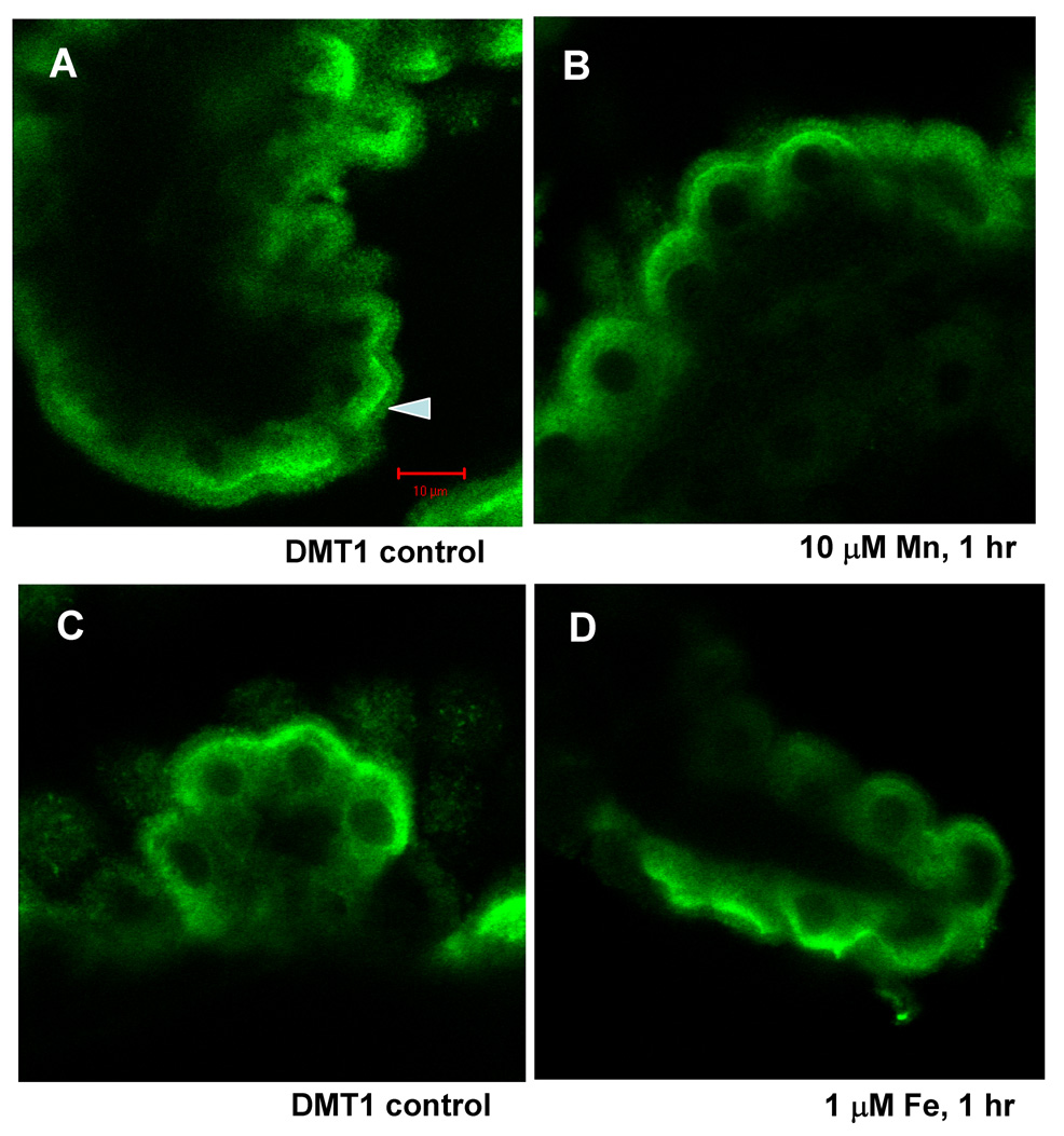
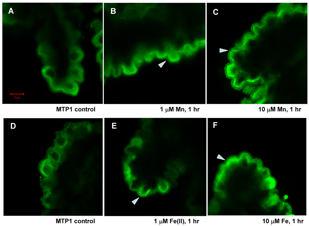
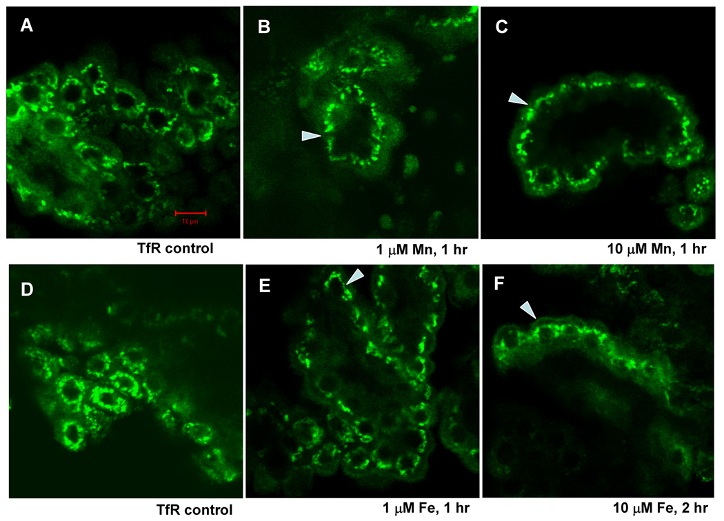
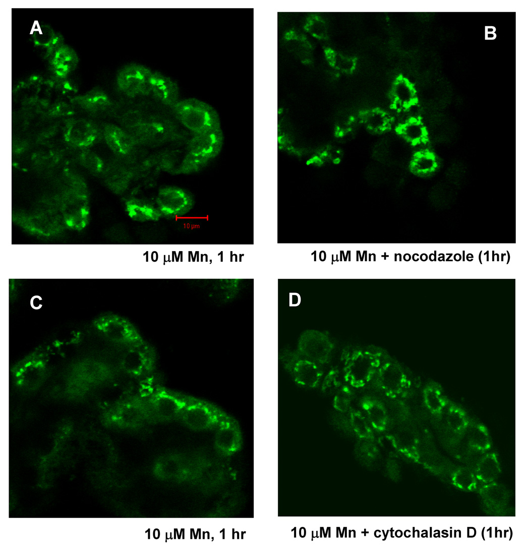
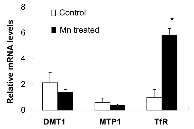
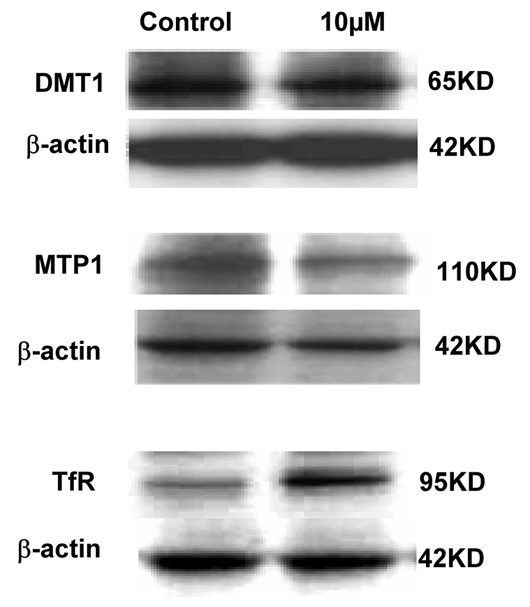
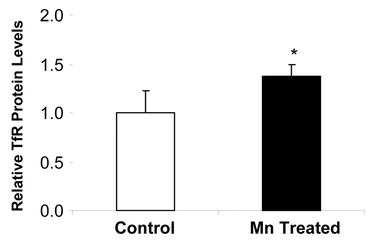
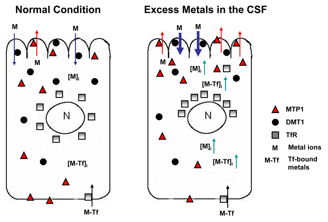
Confocal microscopy was used to investigate the effects of manganese (Mn) and iron (Fe) exposure on the subcellular distribution of metal transporting proteins, i.e., divalent metal transporter 1 (DMT1), metal transporter protein 1 (MTP1), and transferrin receptor (TfR), in the rat intact choroid plexus which comprises the blood-cerebrospinal fluid barrier. In control tissue, DMT1 was concentrated below the apical epithelial membrane, MTP1 was diffuse within the cytosol, and TfR was distributed in vesicles around nuclei. Following Mn or Fe treatment (1 and 10 microM), the distribution of DMT1 was not affected. However, MTP1 and TfR moved markedly toward the apical pole of the cells. These shifts were abolished when microtubules were disrupted. Quantitative RT-PCR and Western blot analyses revealed a significant increase in mRNA and protein levels of TfR but not DMT1 and MTP1 after Mn exposure. These results suggest that early events in the tissue response to Mn or Fe exposure involve microtubule-dependent, intracellular trafficking of MTP1 and TfR. The intracellular trafficking of metal transporters in the choroid plexus following Mn exposure may partially contribute to Mn-induced disruption in Fe homeostasis in the cerebrospinal fluid (CSF) following Mn exposure.


Figures

Fig. 1
Fig. 1
Immunohistochemical staining of DMT1, MTP1 and TfR in rat choroid plexus tissues. DMT1 was cytosol-distributed, much along the apical side as the arrow showed; MTP1 was diffusive in the cytoplasm and some on the brush border; and TfR was localized around nuclei in the form of clusters. None of them existed in the nucleus. Lower panel showed corresponding transmission image, indicating normal morphology of choroid plexus tissues
Fig. 2
Fig. 2
Confocal study of DMT1 cellular distribution in rat choroid plexus tissues as affected by treatment with 0, 10 µM Mn (A and B) or 0, 1 µM Fe (C and D) for 1 hr. Tissues were immunostained with anti-DMT1 antibody. The distribution of DMT1 did not change following Mn or Fe incubation at concentrations tested.
Fig. 3
Fig. 3
Confocal study of MTP1 cellular distribution in rat choroid plexus tissues as affected by treatment with 0, 1 and 10 µM Mn (A, B, C) or Fe (D, E, F) for 1 hr. Tissues were immunostained with anti-MTP1 antibody. Following 1 µM Mn exposure, MTP1 stain moved towards the apical pole of the choroidal epithelia. At 10 µM Mn exposure, MTP1 stain on the brush border was intensified (see the arrow). Fe treatment resulted in the similar, yet less intensive redistribution of MTP1 in comparison to Mn treatment. Arrows indicated MTP1 redistribution of in response to Mn or Fe.
Fig. 4
Fig. 4
Confocal study of TfR cellular distribution in rat choroid plexus tissues as affected by treatment with 0, 1, and 10 µM Mn (A, B, and C) or Fe (D, E and F) for 1 hr or 2 hr. Tissues were immunostained with anti-TfR antibody. With an increase in Mn concentrations, TfR stains moved toward the apical side of the choroidal epithelia, yet still seen closely to the nucleus. Incubation with Fe resulted in the similar redistribution of MTP1. Long exposure (2 hrs) did not move TfR further to the apical side. Arrows indicated TfR redistribution of in response to Mn or Fe.
Fig. 5
Fig. 5
Microtubule inhibitor-nocodazole eliminates TfR redistribution as affected by Mn or Fe treatment in rat choroid plexus tissues, so does microfilament inhibitor-cytochalasin D. Confocal study showed TfR cellular redistribution in rat choroid plexus tissues as affected by treatment with 10 µM Mn (A and C) for 1 hr. Pre-treatment of nocodazole (10µM, 1 hr) and cytochalasin D (1µM, 1 hr) blocked Mn- induced redistribution (B and D).
Fig. 6
Fig. 6
Expression of DMT1, MTP1 or TfR mRNA in rat choroid plexus tissues as affected by Mn exposure. The choroid plexus tissues were incubated with 10 µM Mn for 1hr. The relative mRNA levels of DMT1, MTP1, TfR and GAPDH were quantified by real-time RT-PCR and expressed as the ratio of DMT1/GAPDH, MTP1/GAPDH or TfR/GAPDH. Data represent mean±SE, n=3; *: p<0.01 as compared to control.
Fig. 7
Fig. 7
Expression of DMT1, MTP1 or TfR proteins in rat choroid plexuses as affected by Mn exposure. The choroid plexus tissues were incubated with 10 µM Mn for 1 hr. The Western blots (A) are representative of triplicate experiments. The relative protein levels of TfR (B) were estimated from the corresponding band densities and normalized to those of β-actin. Data represent mean±SE, n=3; p<0.05 as compared to control.
Fig. 7
Fig. 7
Expression of DMT1, MTP1 or TfR proteins in rat choroid plexuses as affected by Mn exposure. The choroid plexus tissues were incubated with 10 µM Mn for 1 hr. The Western blots (A) are representative of triplicate experiments. The relative protein levels of TfR (B) were estimated from the corresponding band densities and normalized to those of β-actin. Data represent mean±SE, n=3; p<0.05 as compared to control.
Fig. 8
Fig. 8
Metal transport by MTP1, DMT1 and TfR in choroidal epithelial cells. Under physiological condition, DMT1 takes up Mn or Fe from the CSF, while MTP1 expels metals to the CSF. Upon metal exposure from the CSF, the uptake of metals by apical DMT1 increases intracellular metal concentrations. MTP1, in response, moves toward apical microvilli, expelling metal ions back to the CSF. Elevated Mn concentration enhances the translational expression of TfR, leading to a TfR-mediated transport of transferrin-bound metals including Fe to the CSF.

References

    1. Abboud S, Haile DJ. A novel mammalian iron-regulated protein involved in intracellular iron metabolism. J Biol Chem. 2000;275:19906–19912. - PubMed
    1. Andrews NC. The iron transporter DMT1. Int J Biochem Cell Biol. 1999;31:991–994. - PubMed
    1. Aschner M, Vrana KE, Zheng W. Manganese uptake and distribution in the central nervous system (CNS) Neurotoxicology. 1999;20:173–180. - PubMed
    1. Bali PK, Zak O, Aisen P. A new role for the transferrin receptor in the release of iron from transferrin. Biochemistry. 1991;30:324–328. - PubMed
    1. Berg D, Gerlach M, Youdim MB, Double KL, Zecca L, Riederer P, Becker G. Brain iron pathways and their relevance to Parkinson's disease. J Neurochem. 2001;79:225–236. - PubMed

Publication types

MeSH terms