. 2008 Jun 13;283(24):16283-92.
doi: 10.1074/jbc.M802240200. Epub 2008 Apr 17.

O-GlcNAc regulates FoxO activation in response to glucose

Affiliations

O-GlcNAc regulates FoxO activation in response to glucose

Michael P Housley et al. J Biol Chem. .

Abstract

FoxO proteins are key transcriptional regulators of nutrient homeostasis and stress response. The transcription factor FoxO1 activates expression of gluconeogenic, including phosphoenolpyruvate carboxykinase and glucose-6-phosphatase, and also activates the expression of the oxidative stress response enzymes catalase and manganese superoxide dismutase. Hormonal and stress-dependent regulation of FoxO1 via acetylation, ubiquitination, and phosphorylation, are well established, but FoxOs have not been studied in the context of the glucose-derived O-linked beta-N-acetylglucosamine (O-GlcNAc) modification. Here we show that O-GlcNAc on hepatic FoxO1 is increased in diabetes. Furthermore, O-GlcNAc regulates FoxO1 activation in response to glucose, resulting in the paradoxically increased expression of gluconeogenic genes while concomitantly inducing expression of genes encoding enzymes that detoxify reactive oxygen species. GlcNAcylation of FoxO provides a new mechanism for direct nutrient control of transcription to regulate metabolism and stress response through control of FoxO1 activity.


Figures

FIGURE 1.
FIGURE 1.
FoxO1 is O-GlcNAcylated. A, Fao cells were infected with Ad-FLAG-FoxO1. FLAG-FoxO1 was immunoprecipitated (IP) and blotted (IB) with the anti-O-GlcNAc antibody CTD110.6 or the terminal GlcNAc-binding lectin succinylated wheat germ agglutinin. Specificity was confirmed by GlcNAc competition and increased antibody or lectin reactivity following treatment with the O-GlcNAcase inhibitor PUGNAc. B, autoradiograph showing endogenous FoxO1 immunoprecipitated from rat liver and labeled with [3H]UDP-galactose using a GlcNAc-specific galactosyltransferase. BSA, bovine serum albumin.
FIGURE 2.
FIGURE 2.
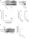
O-GlcNAcylation of FoxO1 is elevated in diabetes. A, FoxO1 was immunoprecipitated from control and STZ-induced diabetic rat livers and blotted with anti-O-GlcNAc antibodies (CTD110.6). Specificity was confirmed by GlcNAc competition. B, UDP-GlcNAc levels were determined from control and STZ-induced diabetic rat livers using capillary electrophoresis (y axis) and plotted against the densitometry analysis from C (x axis). C, RT-PCR analysis of Pepck and G6pc expression show an elevated mRNA expression following STZ-induced diabetes. The dash represents the mean. D, FoxO1 was immunoprecipitated from control and high fat-fed mice and blotted with anti-O-GlcNAc antibodies (CTD110.6). Densitometry analysis reveals elevated GlcNAcylation of FoxO1. IB, immunoblotting.
FIGURE 3.
FIGURE 3.
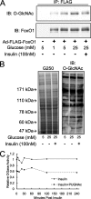
Insulin reduces FoxO1 GlcNAcylation elevated by high glucose. A, FLAG-FoxO1 was immunoprecipitated (IP) from Fao cells that were treated with insulin for 1 h. GlcNAcylation was detected by Western blotting using the CTD110.6 antibody. Glucose increases, whereas insulin decreases, FoxO1 GlcNAcylation. B, insulin treatment does not reduce total cellular GlcNAcylation in Fao cells. C, insulin treatment does not alter O-GlcNAcase activity in Fao cells. IB, immunoblotting.
FIGURE 4.
FIGURE 4.
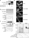
There is a complex interplay between phosphorylation and GlcNAcylation of FoxO1. A, the AKT-insensitive and constitutively nuclear FoxO1 mutant (T24A,S256A,S319A, called 3A) is hyper-GlcNAcylated in Fao cells. B, recombinant FoxO1 was in vitro GlcNAcylated then subsequently in vitro AKT labeled, subjected to SDS-PAGE, and blotted with anti-FoxO1 Ser(P)256, stripped, and then blotted with anti-O-GlcNAc antibodies (CTD110.6). Specificity of the Ser(P)256 antibody was confirmed by alkaline phosphatase treatment. C, recombinant FoxO1 was in vitro AKT-phosphorylated, then subsequently in vitro O-GlcNAc labeled, subjected to SDS-PAGE, and blotted with anti-O-GlcNAc antibodies (CTD110.6), stripped, and then blotted with anti-FoxO1 Ser(P)256 antibodies. Specificity of the Ser(P)256 antibody was confirmed by alkaline phosphatase treatment. In vitro AKT phosphorylation does not block in vitro OGT labeling of FoxO1. D, Fao cells were infected with adenovirus expressing FLAG-tagged FoxO1. 24 h after infection cells were serum-starved in RPMI + 0.5% bovine serum albumin for 4 h and then treated as indicated for an additional 16 h in RPMI + 0.5% bovine serum albumin. FoxO1 was visualized using anti-FoxO1 antibody. PUGNAc (an O-GlcNAcase inhibitor) or 25 mm glucose did not result increased nuclear translocation of FoxO1 under conditions tested. E, the FoxO3 isoform is an in vitro substrate for OGT. [3H]UDP-GlcNAc incorporation was detected by autoradiography following Coomassie G250 staining. The truncated (amino acids 1–525) form incorporated significantly less label, indicating a significant number of sites are in the C-terminal region. The mutant FoxO3 lacking AKT phosphorylation sites (T24A,S215A,S316A, or “triple mutant” or TM) incorporates [3H]UDP-GlcNAc similar to the truncated form, indicating that AKT phosphorylation sites are not significantly in vitro O-GlcNAc labeled. IB, immunoblotting; wt, wild type.
FIGURE 5.
FIGURE 5.
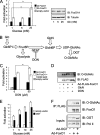
O-GlcNAc increases FoxO-dependent luciferase reporter transcription in HEK293 cells via the HBP. A, luciferase reporter activity is increased by high glucose for both wild type (wt) and 3A FoxO1 (all panels are plotted as relative luciferase activity normalized to β-galactosidase activity; error bars indicate standard errors; *, p < 0.05 by Student's t test). FoxO1 protein levels are unaffected. B, schematic of the UDP-GlcNAc synthesis pathway in which 2–5% of glucose that enters the cells is used for production of the donor sugar nucleotide. C, addition of 50 μm DON, which inhibits the rate-limiting enzyme in UDP-GlcNAc synthesis (glutamine:fructose-6-phosphate amidotransferase, GFAT), reduces high glucose activation of luciferase activity. 10 mm glucosamine (GlcN) rescues activation of FoxO1. D, O-GlcNAc levels on FLAG-FoxO1 in Fao cells following treatment with 10 mm glucosamine and 50 μm DON. E, overexpression of OGT increases FoxO-dependent luciferase activity. F, overexpression of OGT increases FoxO GlcNAcylation. CTD110.6 antibodies were used to detect FLAG-precipitated FoxO1. IP, immunoprecipitation; IB, immunoblotting.
FIGURE 6.
FIGURE 6.
Glucose activates expression of FoxO1 target genes in an OGT-dependent manner. A, RT-PCR analysis shows gluconeogenic enzyme expression (G6pc and Pepck) is elevated by glucose in Fao hepatoma cells (all panels are relative expression normalized to 18 S rRNA; the error bars indicate standard errors; *, p < 0.05 and **, p < 0.005 by Student's t test). B, RT-PCR analysis shows reactive oxygen species detoxification enzyme expression (Catalase, MnSOD) is elevated by glucose. Infection with an adenovirus expressing a dominant negative FoxO1 (Δ256) prevents activation of target genes by high glucose. C, RT-PCR analysis in OGT–/flox MEFs shows that OGT is required for high glucose activation of Catalase and MnSOD. D, RT-PCR analysis in Fao cells following OGT overexpression shows elevated Pepck and G6pc. E, RT-PCR analysis of mouse livers 7 days post-injection of adeno-OGT or GFP viruses. F, electrophoretic mobility shift assay with recombinant GlcNAcylated or naked GST-FoxO1 and a 300-base pair fragment of the IRE-luciferase reporter plasmid (used in Fig. 5) and visualized with ethidum bromide. G, electrophoretic mobility shift assay as in F with either 2 μg of GST + 6 μg of OGT, 2 μg of FoxO1, or 2 μg of FoxO1 + 6 μg of OGT.
FIGURE 7.
FIGURE 7.
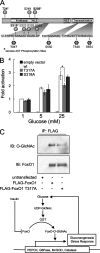
FoxO1 is GlcNAcylated at multiple sites. A, schematic indicating four O-GlcNAc sites mapped using ETD MS/MS on human FoxO1 (Thr317, Ser550, Thr648, and Ser656). B, luciferase reporter assays of point mutants of FoxO1, indicating that mutation of Thr317 to alanine reduces activation by 25 mm glucose (expressed as relative activity normalized to β-galactosidase; error bars represent standard errors; *, p < 0.05 by Student's t test). C, HEK293 cells were transfected with FLAG-FoxO1 vectors containing the indicated site mutations, subjected to SDS-PAGE, and blotted using anti-O-GlcNAc antibodies (CTD110.6). IP, immunoprecipitation; IB, immunoblotting.

References

    1. Buse, M. G. (2006) Am. J. Physiol. 290 E1–E8 - PMC - PubMed
    1. Zhang, W., Patil, S., Chauhan, B., Guo, S., Powell, D. R., Le, J., Klotsas, A., Matika, R., Xiao, X., Franks, R., Heidenreich, K. A., Sajan, M. P., Farese, R. V., Stolz, D. B., Tso, P., Koo, S. H., Montminy, M., and Unterman, T. G. (2006) J. Biol. Chem. 281 10105–10117 - PubMed
    1. Puig, O., and Tjian, R. (2006) Cell Cycle 5 503–505 - PubMed
    1. Lehtinen, M. K., Yuan, Z., Boag, P. R., Yang, Y., Villen, J., Becker, E. B., DiBacco, S., de la Iglesia, N., Gygi, S., Blackwell, T. K., and Bonni, A. (2006) Cell 125 987–1001 - PubMed
    1. Tothova, Z., Kollipara, R., Huntly, B. J., Lee, B. H., Castrillon, D. H., Cullen, D. E., McDowell, E. P., Lazo-Kallanian, S., Williams, I. R., Sears, C., Armstrong, S. A., Passegue, E., DePinho, R. A., and Gilliland, D. G. (2007) Cell 128 325–339 - PubMed

Publication types

MeSH terms