. 2008 Jun 6;283(23):15869-77.
doi: 10.1074/jbc.M709316200. Epub 2008 Apr 17.

The role of CDC48 in the retro-translocation of non-ubiquitinated toxin substrates in plant cells

Affiliations

The role of CDC48 in the retro-translocation of non-ubiquitinated toxin substrates in plant cells

Richard S Marshall et al. J Biol Chem. .

Abstract

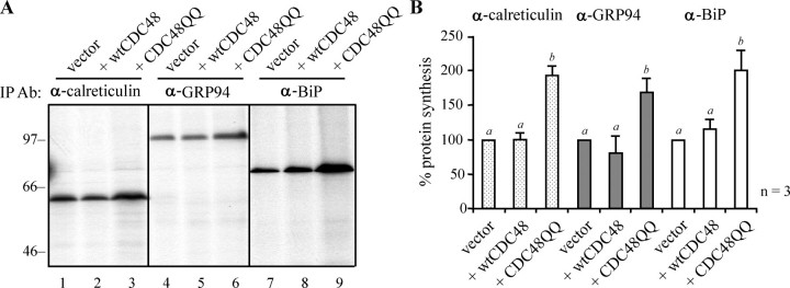
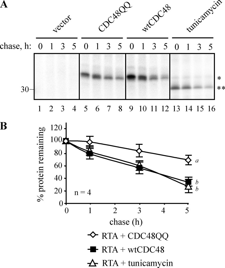
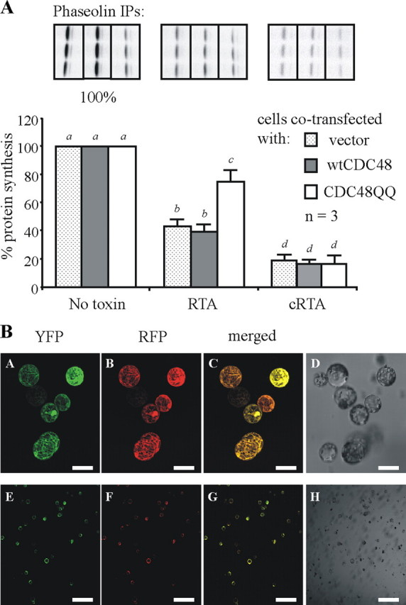
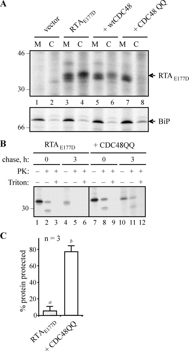
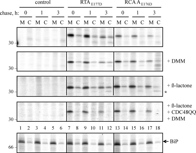
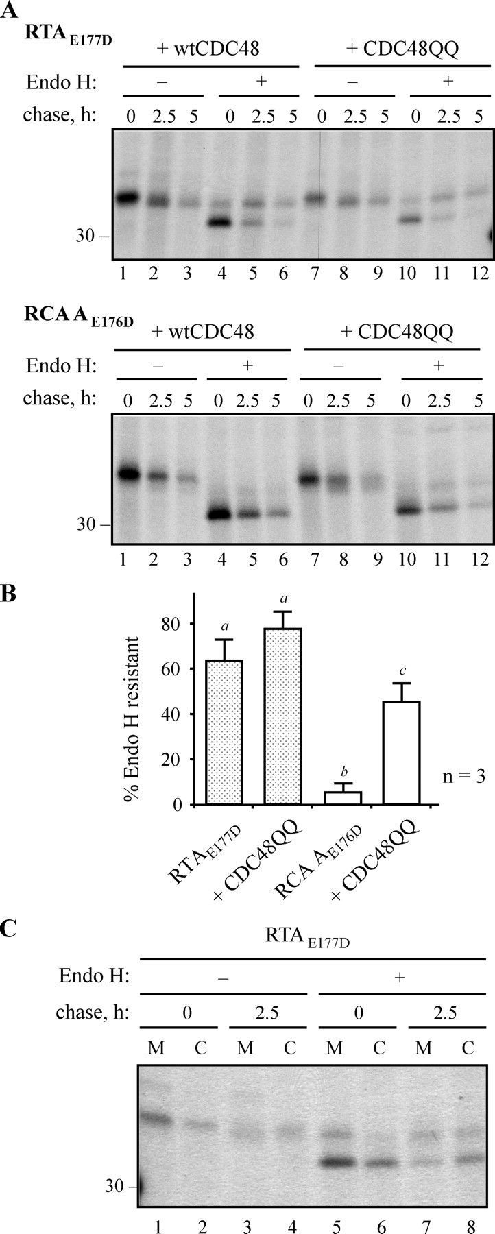
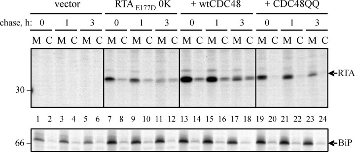
When the catalytic A subunits of the castor bean toxins ricin and Ricinus communis agglutinin (denoted as RTA and RCA A, respectively) are delivered into the endoplasmic reticulum (ER) of tobacco protoplasts, they become substrates for ER-associated protein degradation (ERAD). As such, these orphan polypeptides are retro-translocated to the cytosol, where a significant proportion of each protein is degraded by proteasomes. Here we begin to characterize the ERAD pathway in plant cells, showing that retro-translocation of these lysine-deficient glycoproteins requires the ATPase activity of cytosolic CDC48. Lysine polyubiquitination is not obligatory for this step. We also show that although RCA A is found in a mannose-untrimmed form prior to its retro-translocation, a significant proportion of newly synthesized RTA cycles via the Golgi and becomes modified by downstream glycosylation enzymes. Despite these differences, both proteins are similarly retro-translocated.


Figures

FIGURE 1.
FIGURE 1.
Expression of CDC48QQ promotes an up-regulation of ER chaperones. A, tobacco leaf cells were transfected, by Agrobacterium-mediated stomatal infiltration, with the pGreenII-0029 vector alone (vector) or with plasmids encoding wtCDC48 or CDC48QQ. After 3 days, protoplasts were recovered and pulsed with radioactive amino acids for 1 h. Proteins were immunoselected sequentially from cell homogenates using anti-calreticulin, anti-GRP94, and anti-BiP antisera and analyzed by reducing SDS-PAGE and fluorography. BiP immunoprecipitates were exposed to film for 2 days, whereas the GRP94 and calreticulin immunoprecipitates (IP) were exposed for 10 days. Numbers on the left indicate molecular mass markers in kilodaltons. B, quantitation of A, showing the average values from three independent experiments. The intensity of the immunoselected bands was measured by densitometry and expressed as a percentage of the control (vector). Bars indicate standard deviation and the presence of different letters above the bars indicate a significant difference (p < 0.05).
FIGURE 2.
FIGURE 2.
Expression of mutant CDC48 increases the stability of ER-sequestered ricin A chain. A, protoplasts were transfected with pDHA vector alone (vector) or co-transfected with plasmids encoding ER-targeted ricin A chain with an active site mutation (RTAE177D), and either wtCDC48 or CDC48QQ. Where indicated, protoplasts were preincubated for 1 h with 50 μg/ml tunicamycin. Protoplasts were radiolabeled with 35S-labeled amino acids for 1 h and chased with unlabeled amino acids for the times indicated. RTA was immunoprecipitated from cell homogenates and analyzed by reducing SDS-PAGE and fluorography. The single asterisk indicates the position of glycosylated RTA, whereas the double asterisk marks the position of non-glycosylated RTA. The position of the 30-kDa molecular mass marker is indicated on the left. B, quantitation of the amount of immunoprecipitated RTA, made in the presence of wtCDC48, CDC48QQ, or tunicamycin, as measured by densitometry and expressed as a percentage of the total RTA present at the end of the pulse. The graph shows the average values from four independent experiments. Bars indicate standard deviation and different letters at the end of each series indicate a significant difference (p < 0.05) between treatments at all but the zero time point.
FIGURE 3.
FIGURE 3.
Expression of a mutant CDC48 decreases the toxicity of RTA to ribosomes. A, triplicate preparations of protoplasts were co-transfected with a plasmid encoding a phaseolin protein synthesis reporter along with either pDHA vector alone (no toxin) or a plasmid encoding ER-targeted RTA (RTA) or cytosolic RTA (cRTA). These co-transfections were carried out in combination with either the pamPAT-MCS vector alone (no CDC48), or this plasmid encoding either wtCDC48 or CDC48QQ. Protoplasts were pulsed for 1 h before cell lysis, and immunoprecipitates (IP) were subjected to SDS-PAGE, as described in the legend to Fig. 2. Fluorographs of a representative set of phaseolin immunoprecipitates taken from one of three experimental repeats are shown above the relevant densitometry quantifications. The levels of phaseolin made in the absence of any toxin expression are taken as 100%. The other data are expressed as a percentage of this and represent the average values from three independent experiments. Bars indicate standard deviation and different letters above bars indicate a significant difference (p < 0.05). B, protoplasts were co-transfected with plasmids encoding cytosolic YFP and cytosolic RFP. After overnight expression cells were analyzed by confocal microscopy. Panels A and E show YFP fluorescence (green), panels B and F show RFP fluorescence (red), and panels C and G show merged images. Panels D and H represent the total cell population. YFP was excited at 514 nm, and RFP was excited at 561 nm. All images shown were acquired using the same photo-multiplier gain and offset settings. For each sample, two magnifications are shown. Scale bars represent 50 μm (panels A–D) and 400 μm (panels E–H).
FIGURE 4.
FIGURE 4.
RTA is retained in the membrane fraction when CDC48-dependent retro-translocation is impaired. A, protoplasts were transfected with pDHA vector alone (vector) or the RTAE177D-encoding plasmid plus wtCDC48 or CDC48QQ plasmids. Protoplasts were radiolabeled with [35S]cysteine and [35S]methionine for 6 h, before being homogenized in the absence of detergent and fractionated to yield microsomes (M) and cytosol (C). Proteins were immunoselected sequentially using anti-RTA and anti-BiP antisera, and analyzed by reducing SDS-PAGE and fluorography. Numbers on the left indicate molecular mass markers in kilodaltons. B, protoplasts were transfected with the plasmids shown, before being radiolabeled for 1 h and chased as indicated. The cells were then homogenized as in A, before dividing three ways and incubating in the absence or presence of proteinase K (PK) and detergent (Triton X-100). RTA was immunoselected and resolved as in A. The position of the 30-kDa molecular mass marker is indicated on the left. C, quantitation, from three separate pulse-chase experiments, of the amount of RTA made in the absence or presence of CDC48QQ that was subsequently protected from proteinase K after 3 h. Bars indicate standard deviation and different letters above the bars indicate a significant difference (p < 0.05).
FIGURE 5.
FIGURE 5.
CDC48-dependent retro-translocation does not distinguish between different glycosylated forms. Protoplasts were transfected with pDHA vector alone (control), or constructs encoding ER-targeted RTAE177D or R. communis agglutinin A chain with the equivalent active site point mutation (RCA AE176D). Where indicated, protoplasts were co-transfected with a plasmid expressing CDC48QQ, preincubated for 1 h with 5 mm 1-deoxymannojirimycin (DMM) before radiolabeling, and/or incubated with 80 μm clasto-lactacystin β-lactone during the pulse. Protoplasts were subjected to pulse-chase before being homogenized in the absence of detergent. Homogenates were centrifuged to yield microsomal (M) and cytosol (C) fractions and proteins immunoprecipitated sequentially using anti-RTA and anti-BiP antisera before analysis by reducing SDS-PAGE and fluorography. A representative gel showing the distribution of BiP in the membrane and cytosol fractions is shown in the bottom panel. The asterisk at the right-hand side of the β-lactone panel indicates the likely deglycosylated RTA species that is more clearly observed when proteasomes are inhibited (17). Numbers on the left indicate molecular mass markers in kilodaltons.
FIGURE 6.
FIGURE 6.
A plant ERAD substrate may cycle through the Golgi prior to its CDC48-dependent retro-translocation. A, protoplasts were co-transfected with plasmids encoding RTAE177D or RCA AE176D, and wtCDC48 or CDC48QQ, before pulse-chase analysis. RTA or RCA A were immunoprecipitated, and the immunoprecipitates treated for 16 h in the absence or presence of Endo H before analysis by reducing SDS-PAGE and fluorography. The position of the 30-kDa molecular mass marker is indicated on the left. B, quantitation, from three separate pulse-chase experiments, of the amounts of RTA or RCA A, made in the absence or presence of CDC48QQ, that were resistant to Endo H treatment after 5 h. Bars indicate standard deviation and different letters above the bars indicate a significant difference (p < 0.05). C, protoplasts were transfected to express RTAE177D, radiolabeled as in A, and chased with unlabeled amino acids for 0 and 2.5 h, before being homogenized in the absence of detergent. Homogenates were fractionated into microsomes (M) and cytosol (C) and RTA immunoselected, treated with Endo H, and analyzed as in A. The position of the 30-kDa molecular mass marker is indicated on the left.
FIGURE 7.
FIGURE 7.
CDC48-dependent retro-translocation can be independent of lysyl ubiquitination. Protoplasts were transfected with pDHA vector alone (vector) or with a plasmid encoding RTAE177D lacking its two endogenous lysine residues (RTAE177D 0K), plus a wtCDC48 or CDC48QQ construct where indicated. Protoplasts were subjected to a 1-h pulse and chased as shown. Cell homogenates were centrifuged to yield microsomal (M) and cytosol (C) fractions, and proteins immunoselected sequentially using anti-RTA and anti-BiP antisera before analysis by reducing SDS-PAGE and fluorography. Numbers on the left indicate molecular mass markers in kilodaltons.

References

    1. Vitale, A. (2001) Plant Cell 131260 -1262 - PMC - PubMed
    1. Romisch, K. (2005) Annu. Rev. Cell Dev. Biol. 21435 -456 - PubMed
    1. Meusser, B., Hirsch, C., Jarosch, E., and Sommer, T. (2005) Nat. Cell Biol. 7 766-772 - PubMed
    1. Braun, S., Matuschewski, K., Rape, M., Thoms, S., and Jentsch, S. (2002) EMBO J. 21 615-621 - PMC - PubMed
    1. Ye, Y., Meyer, H. H., and Rapoport, T. A. (2001) Nature 414652 -656 - PubMed

Publication types

MeSH terms